Search Journal-type in search term and press enter
Southwest Pulmonary and Critical Care Fellowships
In Memoriam

Imaging

Last 50 Imaging Postings

(Click on title to be directed to posting, most recent listed first)

May 2024 Medical Image of the Month: Hereditary Hemorrhagic
   Telangiectasia in a Patient on Veno-Arterial Extra-Corporeal Membrane
   Oxygenation
May 2024 Imaging Case of the Month: Nothing Is Guaranteed
April 2024 Medical Image of the Month: Wind Instruments Player Exhibiting
   Exceptional Pulmonary Function
March 2024 Medical Image of the Month: Sputum Cytology in Patients with
   Suspected Lung Malignancy Presenting with Acute Hypoxic Respiratory
   Failure
February 2024 Medical Image of the Month: Pulmonary Alveolar Proteinosis
   in Myelodysplastic Syndrome
February 2024 Imaging Case of the Month: Connecting Some Unusual Dots
January 2024 Medical Image of the Month: Polyangiitis Overlap Syndrome
   (POS) Mimicking Fungal Pneumonia 
December 2023 Medical Image of the Month: Metastatic Pulmonary
   Calcifications in End-Stage Renal Disease 
November 2023 Medical Image of the Month: Obstructive Uropathy
   Extremis
November 2023 Imaging Case of the Month: A Crazy Association
October 2023 Medical Image of the Month: Swyer-James-MacLeod
   Syndrome
September 2023 Medical Image of the Month: Aspergillus Presenting as a
   Pulmonary Nodule in an Immunocompetent Patient
August 2023 Medical Image of the Month: Cannonball Metastases from
   Metastatic Melanoma
August 2023 Imaging Case of the Month: Chew Your Food Carefully
July 2023 Medical Image of the Month: Primary Tracheal Lymphoma
June 2023 Medical Image of the Month: Solitary Fibrous Tumor of the Pleura
May 2023 Medical Image of the Month: Methamphetamine Inhalation
   Leading to Cavitary Pneumonia and Pleural Complications
April 2023 Medical Image of the Month: Atrial Myxoma in the setting of
   Raynaud’s Phenomenon: Early Echocardiography and Management of
   Thrombotic Disease
April 2023 Imaging Case of the Month: Large Impact from a Small Lesion
March 2023 Medical Image of the Month: Spontaneous Pneumomediastinum
   as a Complication of Marijuana Smoking Due to Müller's Maneuvers
February 2023 Medical Image of the Month: Reversed Halo Sign in the
   Setting of a Neutropenic Patient with Angioinvasive Pulmonary
   Zygomycosis
January 2023 Medical Image of the Month: Abnormal Sleep Study and PFT
   with Supine Challenge Related to Idiopathic Hemidiaphragmatic Paralysis
December 2022 Medical Image of the Month: Bronchoesophageal Fistula in
   the Setting of Pulmonary Actinomycosis
November 2022 Medical Image of the Month: COVID-19 Infection
   Presenting as Spontaneous Subcapsular Hematoma of the Kidney
November 2022 Imaging Case of the Month: Out of Place in the Thorax
October 2022 Medical Image of the Month: Infected Dasatinib Induced
   Chylothorax-The First Reported Case 
September 2022 Medical Image of the Month: Epiglottic Calcification
Medical Image of the Month: An Unexpected Cause of Chronic Cough
August 2022 Imaging Case of the Month: It’s All About Location
July 2022 Medical Image of the Month: Pulmonary Nodule in the
   Setting of Pyoderma Gangrenosum (PG) 
June 2022 Medical Image of the Month: A Hard Image to Swallow
May 2022 Medical Image of the Month: Pectus Excavatum
May 2022 Imaging Case of the Month: Asymmetric Apical Opacity–
   Diagnostic Considerations
April 2022 Medical Image of the Month: COVID Pericarditis
March 2022 Medical Image of the Month: Pulmonary Nodules in the
   Setting of Diffuse Idiopathic Pulmonary NeuroEndocrine Cell Hyperplasia
   (DIPNECH) 
February 2022 Medical Image of the Month: Multifocal Micronodular
   Pneumocyte Hyperplasia in the Setting of Tuberous Sclerosis
February 2022 Imaging Case of the Month: Between A Rock and a
   Hard Place
January 2022 Medical Image of the Month: Bronchial Obstruction
   Due to Pledget in Airway Following Foregut Cyst Resection
December 2021 Medical Image of the Month: Aspirated Dental Implant
Medical Image of the Month: Cavitating Pseudomonas
   aeruginosa Pneumonia
November 2021 Imaging Case of the Month: Let’s Not Dance
   the Twist
Medical Image of the Month: COVID-19-Associated Pulmonary
   Aspergillosis in a Post-Liver Transplant Patient
Medical Image of the Month: Stercoral Colitis
Medical Image of the Month: Bleomycin-Induced Pulmonary Fibrosis
   in a Patient with Lymphoma
August 2021 Imaging Case of the Month: Unilateral Peripheral Lung
   Opacity
Medical Image of the Month: Hepatic Abscess Secondary to Diverticulitis
   Resulting in Sepsis
Medical Image of the Month: Metastatic Spindle Cell Carcinoma of the
   Breast
Medical Image of the Month: Perforated Gangrenous Cholecystitis
May 2021 Imaging Case of the Month: A Growing Indeterminate Solitary
   Nodule
Medical Image of the Month: Severe Acute Respiratory Distress
Syndrome and Embolic Strokes from Polymethylmethacrylate
   (PMMA) Embolization
Medical Image of the Month: Pulmonary Aspergillus Overlap Syndrome
   Presenting with ABPA, Multiple Bilateral Aspergillomas
Medical Image of the Month: Diffuse White Matter Microhemorrhages
   Secondary to SARS-CoV-2 (COVID-19) Infection
February 2021 Imaging Case of the Month: An Indeterminate Solitary
   Nodule
Medical Image of the Month: Mucinous Adenocarcinoma of the Lung
   Mimicking Pneumonia
Medical Image of the Month: Superior Vena Cava Syndrome
Medical Image of the Month: Buffalo Chest Identified at the Time of
   Lung Nodule Biopsy
November 2020 Imaging Case of the Month: Cause and Effect?


For complete imaging listings click here

Those who care for patients with pulmonary, critical care or sleep disorders rely heavily on chest radiology and pathology to determine diagnoses. The Southwest Journal of Pulmonary and Critical Care publishes case-based articles with characteristic chest imaging and related pathology. The editor of this section will oversee and coordinate the publication of a core of the most important chest imaging topics. In doing so, they encourage the submission of unsolicited manuscripts. It cannot be overemphasized that both radiologic and pathologic images must be of excellent quality. As a rule, 600 DPI is sufficient for radiographic and pathologic images. Taking pictures of plain chest radiographs and CT scans with a digital camera is strongly discouraged. The figures should be cited in the text and numbered consecutively. The stain used for pathology specimens and magnification should be mentioned in the figure legend. Those who care for patients with pulmonary, critical care or sleep disorders rely heavily on chest radiology and pathology to determine diagnoses. The Southwest Journal of Pulmonary and Critical Care publishes case-based articles with characteristic chest imaging and related pathology. The editor of this section will oversee and coordinate the publication of a core of the most important chest imaging topics. In doing so, they encourage the submission of unsolicited manuscripts. It cannot be overemphasized that both radiologic and pathologic images must be of excellent quality. As a rule, 600 DPI is sufficient for radiographic and pathologic images. Taking pictures of plain chest radiographs and CT scans with a digital camera is strongly discouraged. The figures should be cited in the text and numbered consecutively. The stain used for pathology specimens and magnification should be mentioned in the figure legend.

-------------------------------------------------------------------------------------------  

Wednesday
Jan212015

Medical Image of the Week: Dobhoff Placement in a Patient with Hiatal Hernia

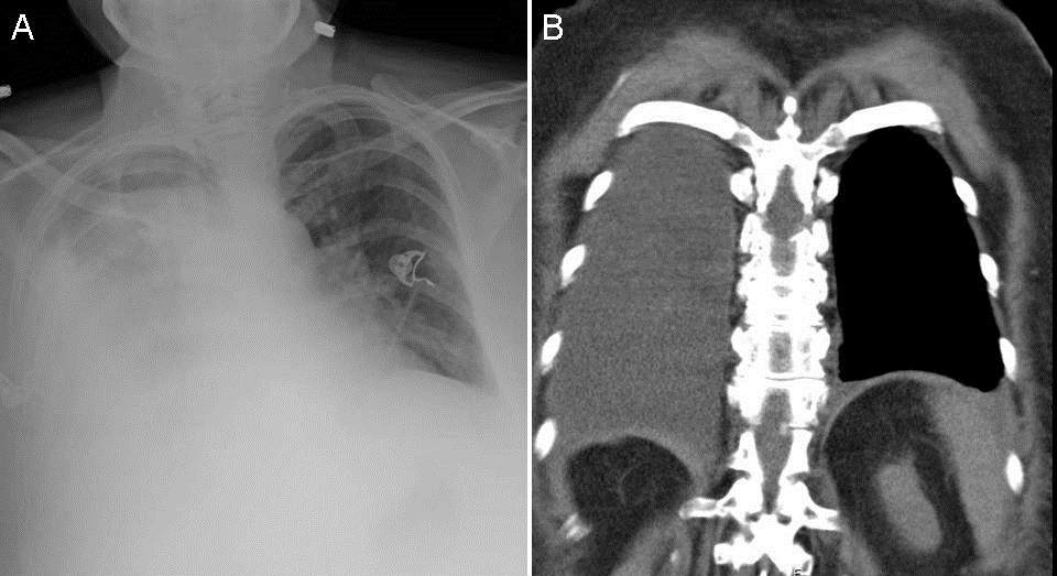
Figure 1. Arrows designate tip of Dobhoff feeding tube (DHT). Panel A: Chest radiograph. DHT appears to follow the left main bronchus into the left lower lobe. Panel B: Abdominal view of DHT placement. Panel C: Chest CT showing degree of hiatal hernia and DHT in the intra-thoracic hernia. Panel D: Follow-up fluoroscopy imaging showing appropriately placed DHT in the duodenum.

A 79 year-old woman with a past medical history of obstructive sleep apnea, chronic obstructive pulmonary disease on home oxygen, obesity hypoventilation syndrome, hypertension, and anxiety presented with a 2 day history of altered mental status and symptoms consistent with a COPD exacerbation, including dyspnea and increased oxygen requirements. She was found to be hypercarbic and did not tolerate a trial of BiPAP due to her altered mentation. She was subsequently intubated. Due to an expected prolonged intubation period, plans for enteral access were made. A Dobhoff naso-duodenal feeding tube (DHT) was inserted. On chest radiograph and a concurrent abdominal radiograph, the DHT appeared to have been inserted into the left mainstem bronchus terminating in the left lower lobe (Figure 1A and 1B). The nursing staff removed and replaced the DHT resulting in a similar radiograph. A third placement was attempted with similar radiographic results. Therefore, a computed tomography (CT) scan of the chest was performed to evaluate tube placement. The CT of the chest showed a large hiatal hernia contained within thoracic cavity (Figure 1C). Upon chart review, previous radiographs mentioned hiatal hernia but it appeared that the degree of herniation had progressed. Fluoroscopy was used to confirm placement of the DHT beyond the herniated gastric contents into the duodenum (Figure 1D) and tube feeds were initiated.

Post-pyloric feeding tubes are often used in place of gastric feeding tubes under the assumption that the risk of aspiration in the intubated patient is reduced. Enteral nutrition is typically started within 36 hours of intubation as this has been shown to decrease mortality in intubated patients (1). There are contraindications to the use of nasogastric or nasoenteric feeding tubes, which include facial trauma, esophageal web, or recent esophagectomy. Hiatal hernias are not a contraindication to nasoenteric feeding tube placement, however, patients with unusual anatomy may benefit from placement under fluoroscopic or endoscopic visualization in order to ensure appropriate positioning (2).

Kawanjit K Sekhon, MD and Ryan Nahapetian, MD, MPH

Department of Internal Medicine

Division of Pulmonary, Allergy, Critical Care, and Sleep Medicine

University of Arizona, Tucson, AZ

References

1. Marik PE, Zaloga GP. Gastric versus post-pyloric feeding: a systematic review. Crit Care. 2003;7(3):R46-51. [CrossRef] [PubMed]

2. Hodin RA, Bordeianou L. Nasogastric and nasoenteric tubes. Uptodate.com. Oct 17, 2013. Dec 20, 2013. Available at: http://www.uptodate.com/contents/nasogastric-and-nasoenteric-tubes?source=machineLearning&search=hiatal+hernia+feeding+tube&selectedTitle=1%7E150&sectionRank=3&anchor=H522922014#H522922014 (requires subscription).

Reference as: Sekhon KK, Nahapetian R. Medical image of the week: Dobhoff placement in a patient with hiatal hernia. Southwest J Pulm Crit Care. 2015;10(1):49-50. doi: http://dx.doi.org/10.13175/swjpcc005-15 PDF

Wednesday
Jan142015

Medical Image of the Week: Hepatic Hydrothorax

Figure 1. Panel A: Chest x-ray showing right pleural effusion. Panel B: Coronal view of the thoracic CT scan in soft tissue windows showing right pleural effusion.


Figure 2. Nuclear scan after intraperitoneal injection of technetium 99mTc albumin aggregated (99mTc-MAA). After less than one hour most of the tracer migrated into the right hemithorax consistent with hepatic hydrothorax.


A 63 year-old woman, with known alcoholic liver cirrhosis, esophageal varices with history of banding presented to an outside hospital with progressive shortness of breath, and was found to have a large right transudative pleural effusion. The patient underwent 2 diagnostic and therapeutic thoracenteses within 3 days, removing 1100 ml and 1500 ml respectively. No ascites was present. At the time of admission the patient had recurrent right effusion (Figure 1). Abdominal ultrasound showed minimal free intrabdominal fluid and she had signs of third spacing on her lower extremities. The patient underwent intraperitoneal injection of Technetium 99mTc albumin aggregated (99mTc-MAA). After less than one hour most of the tracer migrated into the right hemithorax consistent with hepatic hydrothorax (Figure 2).

While the exact mechanism involved in the development of hepatic hydrothorax is incompletely understood, it probably results from the passage of ascitic fluid from the peritoneal into the pleural cavity through small diaphragmatic defects. These are typically < 1 cm (and may be microscopic) and are generally located in the tendinous portion of the diaphragm. The negative intrathoracic pressure generated during inspiration favors the passage of the fluid into the pleural space. Thus, patients may have only mild or clinically undetectable ascites.

Once the diagnosis is made treatment follows algorithms for treatment of refractory ascites and include salt and water restriction, diuretics, and other validated options for portal hypertension. Repeated thoracentesis and chest tube placement is discouraged.

Huthayfa Ateeli, Justin Lee, Irbaz Riaz, Meenal Misal

Department of Internal Medicine

University of Arizona

Tucson, AZ

References

  1. Huang PM, Chang YL,Yang CY,Lee YC.The morphology of diaphragmatic defects in hepatic hydrothorax: thoracoscopic finding. J Thorac Cardiovasc Surg. 2005;130:141-5. [CrossRef] [PubMed]
  2. Lieberman FL, Hidemura R, Peters RL, Reynolds TB. Pathogenesis and treatment of hydrothorax complicating cirrhosis with ascites. Ann Intern Med. 1966;64:341-51. [CrossRef] [PubMed]
  3. Emerson PA, Davies JH. Hydrothorax complicating ascites. Lancet. 1955; 268:487-8. [CrossRef] [PubMed]
  4. Mouroux J, Perrin C, Venissac N, Blaive B, Richelme H. Management of pleural effusion of cirrhotic origin. Chest. 1996;109:1093-6. [CrossRef] [PubMed]
  5. Chen A, Ho YS, Tu YC, Tang HS, Cheng TC. Diaphragmatic defect as a cause of massive hydrothorax in cirrhosis of liver. J Clin Gastroenterol. 1988;10:663-6. [CrossRef] [PubMed] 

Reference as: Ateeli H, Lee J, Riaz I, Misal M. Medical image of the week: hepatic hydrothorax. Southwest J Pulm Crit Care. 2015;10(1):47-8. doi: http://dx.doi.org/10.13175/swjpcc004-15 PDF