Search Journal-type in search term and press enter
Southwest Pulmonary and Critical Care Fellowships
In Memoriam

Imaging

Last 50 Imaging Postings

(Click on title to be directed to posting, most recent listed first)

May 2024 Medical Image of the Month: Hereditary Hemorrhagic
   Telangiectasia in a Patient on Veno-Arterial Extra-Corporeal Membrane
   Oxygenation
May 2024 Imaging Case of the Month: Nothing Is Guaranteed
April 2024 Medical Image of the Month: Wind Instruments Player Exhibiting
   Exceptional Pulmonary Function
March 2024 Medical Image of the Month: Sputum Cytology in Patients with
   Suspected Lung Malignancy Presenting with Acute Hypoxic Respiratory
   Failure
February 2024 Medical Image of the Month: Pulmonary Alveolar Proteinosis
   in Myelodysplastic Syndrome
February 2024 Imaging Case of the Month: Connecting Some Unusual Dots
January 2024 Medical Image of the Month: Polyangiitis Overlap Syndrome
   (POS) Mimicking Fungal Pneumonia 
December 2023 Medical Image of the Month: Metastatic Pulmonary
   Calcifications in End-Stage Renal Disease 
November 2023 Medical Image of the Month: Obstructive Uropathy
   Extremis
November 2023 Imaging Case of the Month: A Crazy Association
October 2023 Medical Image of the Month: Swyer-James-MacLeod
   Syndrome
September 2023 Medical Image of the Month: Aspergillus Presenting as a
   Pulmonary Nodule in an Immunocompetent Patient
August 2023 Medical Image of the Month: Cannonball Metastases from
   Metastatic Melanoma
August 2023 Imaging Case of the Month: Chew Your Food Carefully
July 2023 Medical Image of the Month: Primary Tracheal Lymphoma
June 2023 Medical Image of the Month: Solitary Fibrous Tumor of the Pleura
May 2023 Medical Image of the Month: Methamphetamine Inhalation
   Leading to Cavitary Pneumonia and Pleural Complications
April 2023 Medical Image of the Month: Atrial Myxoma in the setting of
   Raynaud’s Phenomenon: Early Echocardiography and Management of
   Thrombotic Disease
April 2023 Imaging Case of the Month: Large Impact from a Small Lesion
March 2023 Medical Image of the Month: Spontaneous Pneumomediastinum
   as a Complication of Marijuana Smoking Due to Müller's Maneuvers
February 2023 Medical Image of the Month: Reversed Halo Sign in the
   Setting of a Neutropenic Patient with Angioinvasive Pulmonary
   Zygomycosis
January 2023 Medical Image of the Month: Abnormal Sleep Study and PFT
   with Supine Challenge Related to Idiopathic Hemidiaphragmatic Paralysis
December 2022 Medical Image of the Month: Bronchoesophageal Fistula in
   the Setting of Pulmonary Actinomycosis
November 2022 Medical Image of the Month: COVID-19 Infection
   Presenting as Spontaneous Subcapsular Hematoma of the Kidney
November 2022 Imaging Case of the Month: Out of Place in the Thorax
October 2022 Medical Image of the Month: Infected Dasatinib Induced
   Chylothorax-The First Reported Case 
September 2022 Medical Image of the Month: Epiglottic Calcification
Medical Image of the Month: An Unexpected Cause of Chronic Cough
August 2022 Imaging Case of the Month: It’s All About Location
July 2022 Medical Image of the Month: Pulmonary Nodule in the
   Setting of Pyoderma Gangrenosum (PG) 
June 2022 Medical Image of the Month: A Hard Image to Swallow
May 2022 Medical Image of the Month: Pectus Excavatum
May 2022 Imaging Case of the Month: Asymmetric Apical Opacity–
   Diagnostic Considerations
April 2022 Medical Image of the Month: COVID Pericarditis
March 2022 Medical Image of the Month: Pulmonary Nodules in the
   Setting of Diffuse Idiopathic Pulmonary NeuroEndocrine Cell Hyperplasia
   (DIPNECH) 
February 2022 Medical Image of the Month: Multifocal Micronodular
   Pneumocyte Hyperplasia in the Setting of Tuberous Sclerosis
February 2022 Imaging Case of the Month: Between A Rock and a
   Hard Place
January 2022 Medical Image of the Month: Bronchial Obstruction
   Due to Pledget in Airway Following Foregut Cyst Resection
December 2021 Medical Image of the Month: Aspirated Dental Implant
Medical Image of the Month: Cavitating Pseudomonas
   aeruginosa Pneumonia
November 2021 Imaging Case of the Month: Let’s Not Dance
   the Twist
Medical Image of the Month: COVID-19-Associated Pulmonary
   Aspergillosis in a Post-Liver Transplant Patient
Medical Image of the Month: Stercoral Colitis
Medical Image of the Month: Bleomycin-Induced Pulmonary Fibrosis
   in a Patient with Lymphoma
August 2021 Imaging Case of the Month: Unilateral Peripheral Lung
   Opacity
Medical Image of the Month: Hepatic Abscess Secondary to Diverticulitis
   Resulting in Sepsis
Medical Image of the Month: Metastatic Spindle Cell Carcinoma of the
   Breast
Medical Image of the Month: Perforated Gangrenous Cholecystitis
May 2021 Imaging Case of the Month: A Growing Indeterminate Solitary
   Nodule
Medical Image of the Month: Severe Acute Respiratory Distress
Syndrome and Embolic Strokes from Polymethylmethacrylate
   (PMMA) Embolization
Medical Image of the Month: Pulmonary Aspergillus Overlap Syndrome
   Presenting with ABPA, Multiple Bilateral Aspergillomas
Medical Image of the Month: Diffuse White Matter Microhemorrhages
   Secondary to SARS-CoV-2 (COVID-19) Infection
February 2021 Imaging Case of the Month: An Indeterminate Solitary
   Nodule
Medical Image of the Month: Mucinous Adenocarcinoma of the Lung
   Mimicking Pneumonia
Medical Image of the Month: Superior Vena Cava Syndrome
Medical Image of the Month: Buffalo Chest Identified at the Time of
   Lung Nodule Biopsy
November 2020 Imaging Case of the Month: Cause and Effect?


For complete imaging listings click here

Those who care for patients with pulmonary, critical care or sleep disorders rely heavily on chest radiology and pathology to determine diagnoses. The Southwest Journal of Pulmonary and Critical Care publishes case-based articles with characteristic chest imaging and related pathology. The editor of this section will oversee and coordinate the publication of a core of the most important chest imaging topics. In doing so, they encourage the submission of unsolicited manuscripts. It cannot be overemphasized that both radiologic and pathologic images must be of excellent quality. As a rule, 600 DPI is sufficient for radiographic and pathologic images. Taking pictures of plain chest radiographs and CT scans with a digital camera is strongly discouraged. The figures should be cited in the text and numbered consecutively. The stain used for pathology specimens and magnification should be mentioned in the figure legend. Those who care for patients with pulmonary, critical care or sleep disorders rely heavily on chest radiology and pathology to determine diagnoses. The Southwest Journal of Pulmonary and Critical Care publishes case-based articles with characteristic chest imaging and related pathology. The editor of this section will oversee and coordinate the publication of a core of the most important chest imaging topics. In doing so, they encourage the submission of unsolicited manuscripts. It cannot be overemphasized that both radiologic and pathologic images must be of excellent quality. As a rule, 600 DPI is sufficient for radiographic and pathologic images. Taking pictures of plain chest radiographs and CT scans with a digital camera is strongly discouraged. The figures should be cited in the text and numbered consecutively. The stain used for pathology specimens and magnification should be mentioned in the figure legend.

-------------------------------------------------------------------------------------------  

Wednesday
Dec312014

Medical Image of the Week: Metastatic Collecting Duct Carcinoma

C

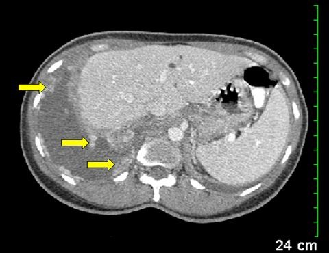
Figure 1. Transverse section of CT chest and abdomen shows enhancing pleural nodularity (yellow arrows) with a pleural effusion.


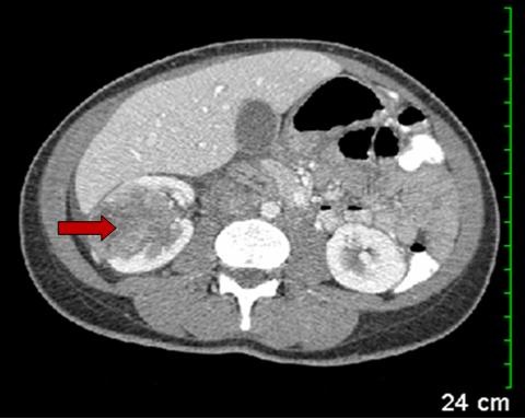
Figure 2.  Transverse section of CT abdomen shows heterogeneous enhancing mass in the right kidney (red arrow).


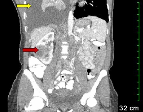
Figure 3. Coronal section of CT chest and abdomen showing a large right pleural effusion (yellow arrow) and atelectatic lung with mediastinal shift to the left. Red arrow points to the heterogeneous mass in the right kidney.


A 40-year-old woman home health nurse presented to the ED with intermittent right sided sharp chest pain and progressive dyspnea for 2 weeks. On admission she was found to be in respiratory distress. Chest x-ray revealed a massive right sided pleural effusion. Thoracic CT scan with contrast confirmed a large right pleural effusion with associated enhancing pleural nodularity also involving the diaphragmatic surface (Figure 1).  The visualized part of the abdomen revealed a mass in the midpole of right kidney (Figure 2). Subsequent CT scan of the abdomen with contrast revealed a heterogeneous enhancing mass in the right kidney suspicious for malignancy (Figure 3) and multiple paracaval lymph nodes. Thoracentesis revealed a hemorrhagic pleural effusion and during subsequent right video-assisted thoracoscopy showed disseminated tumorlets along the diaphragm and pleura. Pleural biopsy and fluid cytology was consistent with metastatic poorly differentiated collecting duct carcinoma of the kidney. The patient is currently getting outpatient chemotherapy. Collecting duct carcinoma of the kidney is an unusual variant of renal cell carcinoma and accounts for about 1% of all renal cell carcinomas (1). This variant has a poor prognosis and frequently metastasizes to the lung and liver.

Chandramohan Meenakshisundaram, MD

Nanditha Malakkla, MD

St. Francis Hospital.

Evanston, IL

Reference

  1. Wang X, Hao J, Zhou R, Zhang X, Yan T, Ding D, Shan L, Liu Z. Collecting duct carcinoma of the kidney: a clinicopathological study of five cases. Diagn Pathol. 2013;8:96. [CrossRef] [PubMed]

Reference as: Meenakshisundaram C, Malakkla N. Medical image of the week: metastatic collecting duct carcinoma. Southwest J Pulm Crit Care. 2014;9(6):348-9. doi: http://dx.doi.org/10.13175/swjpcc160-14 PDF

Wednesday
Dec242014

Medical Image of the Week: CMV Cytopathic Effect

Figure 1. Cluster of 3 large cells, most likely infected type II pneumocytes, with a single prominent red stained nuclear inclusion surrounded by a clear halo. This appearance is the “cytopathic effect” needed to definitively diagnose active CMV infection.


Figure 2. Electron microscopy (8800x) of an infected cell showing  cytomegalovirus (CMV) virions within the nuclear inclusion (small black dots encircled).


Bronchoalveolar lavage (BAL) was performed on a 45-year old man with a history of treated mycosis fungoides and Sézary syndrome, who presented with fever and pulmonary infiltrates. BAL Papanicolaou stain (Figure 1, 400x) showed single cells (lymphocytes, arrows and alveolar macrophages, stars) and a small cluster of 3 large cells, most likely infected type II pneumocytes, with a single prominent red stained nuclear inclusion surrounded by a clear halo. Nuclear chromatin was marginated on the nuclear membrane creating this “owl’s eye” appearance. In vitro, infected cells show cytomegalovirus (CMV) virions within the nuclear inclusion (Figure 2, small black dots encircled, 8,800x)

The "owl's eye" appearance (Figure 1) is the “cytopathic effect” needed to definitively diagnose active CMV infection. While cells infected with adenovirus or herpesvirus may have nuclear inclusions, the cells typically are much smaller. CMV was cultured from the BAL, and no other pathogen was identified by cytology or culture. Quantitative PCR on blood for CMV was 144359 IU/ml.

Afshin Sam, MD; Felicia Goodrum, PhD; Robert Ricciotti, MD; Ken Knox, MD and Richard Sobonya, MD

Departments of Medicine, Immunobiology, and Pathology

University of Arizona Health Sciences Center

Tucson, AZ

Reference as: Sam A, Goodrum F, Ricciotti R, Knox KS, Sobonya R. Medical image of the week: CMV cytopathic effect. Southwest J Pulm Crit Care. 2014;9(6):341-2. doi: http://dx.doi.org/10.13175/swjpcc161-14 PDF