Search Journal-type in search term and press enter
Southwest Pulmonary and Critical Care Fellowships
In Memoriam

Imaging

Last 50 Imaging Postings

(Click on title to be directed to posting, most recent listed first)

May 2024 Medical Image of the Month: Hereditary Hemorrhagic
   Telangiectasia in a Patient on Veno-Arterial Extra-Corporeal Membrane
   Oxygenation
May 2024 Imaging Case of the Month: Nothing Is Guaranteed
April 2024 Medical Image of the Month: Wind Instruments Player Exhibiting
   Exceptional Pulmonary Function
March 2024 Medical Image of the Month: Sputum Cytology in Patients with
   Suspected Lung Malignancy Presenting with Acute Hypoxic Respiratory
   Failure
February 2024 Medical Image of the Month: Pulmonary Alveolar Proteinosis
   in Myelodysplastic Syndrome
February 2024 Imaging Case of the Month: Connecting Some Unusual Dots
January 2024 Medical Image of the Month: Polyangiitis Overlap Syndrome
   (POS) Mimicking Fungal Pneumonia 
December 2023 Medical Image of the Month: Metastatic Pulmonary
   Calcifications in End-Stage Renal Disease 
November 2023 Medical Image of the Month: Obstructive Uropathy
   Extremis
November 2023 Imaging Case of the Month: A Crazy Association
October 2023 Medical Image of the Month: Swyer-James-MacLeod
   Syndrome
September 2023 Medical Image of the Month: Aspergillus Presenting as a
   Pulmonary Nodule in an Immunocompetent Patient
August 2023 Medical Image of the Month: Cannonball Metastases from
   Metastatic Melanoma
August 2023 Imaging Case of the Month: Chew Your Food Carefully
July 2023 Medical Image of the Month: Primary Tracheal Lymphoma
June 2023 Medical Image of the Month: Solitary Fibrous Tumor of the Pleura
May 2023 Medical Image of the Month: Methamphetamine Inhalation
   Leading to Cavitary Pneumonia and Pleural Complications
April 2023 Medical Image of the Month: Atrial Myxoma in the setting of
   Raynaud’s Phenomenon: Early Echocardiography and Management of
   Thrombotic Disease
April 2023 Imaging Case of the Month: Large Impact from a Small Lesion
March 2023 Medical Image of the Month: Spontaneous Pneumomediastinum
   as a Complication of Marijuana Smoking Due to Müller's Maneuvers
February 2023 Medical Image of the Month: Reversed Halo Sign in the
   Setting of a Neutropenic Patient with Angioinvasive Pulmonary
   Zygomycosis
January 2023 Medical Image of the Month: Abnormal Sleep Study and PFT
   with Supine Challenge Related to Idiopathic Hemidiaphragmatic Paralysis
December 2022 Medical Image of the Month: Bronchoesophageal Fistula in
   the Setting of Pulmonary Actinomycosis
November 2022 Medical Image of the Month: COVID-19 Infection
   Presenting as Spontaneous Subcapsular Hematoma of the Kidney
November 2022 Imaging Case of the Month: Out of Place in the Thorax
October 2022 Medical Image of the Month: Infected Dasatinib Induced
   Chylothorax-The First Reported Case 
September 2022 Medical Image of the Month: Epiglottic Calcification
Medical Image of the Month: An Unexpected Cause of Chronic Cough
August 2022 Imaging Case of the Month: It’s All About Location
July 2022 Medical Image of the Month: Pulmonary Nodule in the
   Setting of Pyoderma Gangrenosum (PG) 
June 2022 Medical Image of the Month: A Hard Image to Swallow
May 2022 Medical Image of the Month: Pectus Excavatum
May 2022 Imaging Case of the Month: Asymmetric Apical Opacity–
   Diagnostic Considerations
April 2022 Medical Image of the Month: COVID Pericarditis
March 2022 Medical Image of the Month: Pulmonary Nodules in the
   Setting of Diffuse Idiopathic Pulmonary NeuroEndocrine Cell Hyperplasia
   (DIPNECH) 
February 2022 Medical Image of the Month: Multifocal Micronodular
   Pneumocyte Hyperplasia in the Setting of Tuberous Sclerosis
February 2022 Imaging Case of the Month: Between A Rock and a
   Hard Place
January 2022 Medical Image of the Month: Bronchial Obstruction
   Due to Pledget in Airway Following Foregut Cyst Resection
December 2021 Medical Image of the Month: Aspirated Dental Implant
Medical Image of the Month: Cavitating Pseudomonas
   aeruginosa Pneumonia
November 2021 Imaging Case of the Month: Let’s Not Dance
   the Twist
Medical Image of the Month: COVID-19-Associated Pulmonary
   Aspergillosis in a Post-Liver Transplant Patient
Medical Image of the Month: Stercoral Colitis
Medical Image of the Month: Bleomycin-Induced Pulmonary Fibrosis
   in a Patient with Lymphoma
August 2021 Imaging Case of the Month: Unilateral Peripheral Lung
   Opacity
Medical Image of the Month: Hepatic Abscess Secondary to Diverticulitis
   Resulting in Sepsis
Medical Image of the Month: Metastatic Spindle Cell Carcinoma of the
   Breast
Medical Image of the Month: Perforated Gangrenous Cholecystitis
May 2021 Imaging Case of the Month: A Growing Indeterminate Solitary
   Nodule
Medical Image of the Month: Severe Acute Respiratory Distress
Syndrome and Embolic Strokes from Polymethylmethacrylate
   (PMMA) Embolization
Medical Image of the Month: Pulmonary Aspergillus Overlap Syndrome
   Presenting with ABPA, Multiple Bilateral Aspergillomas
Medical Image of the Month: Diffuse White Matter Microhemorrhages
   Secondary to SARS-CoV-2 (COVID-19) Infection
February 2021 Imaging Case of the Month: An Indeterminate Solitary
   Nodule
Medical Image of the Month: Mucinous Adenocarcinoma of the Lung
   Mimicking Pneumonia
Medical Image of the Month: Superior Vena Cava Syndrome
Medical Image of the Month: Buffalo Chest Identified at the Time of
   Lung Nodule Biopsy
November 2020 Imaging Case of the Month: Cause and Effect?


For complete imaging listings click here

Those who care for patients with pulmonary, critical care or sleep disorders rely heavily on chest radiology and pathology to determine diagnoses. The Southwest Journal of Pulmonary and Critical Care publishes case-based articles with characteristic chest imaging and related pathology. The editor of this section will oversee and coordinate the publication of a core of the most important chest imaging topics. In doing so, they encourage the submission of unsolicited manuscripts. It cannot be overemphasized that both radiologic and pathologic images must be of excellent quality. As a rule, 600 DPI is sufficient for radiographic and pathologic images. Taking pictures of plain chest radiographs and CT scans with a digital camera is strongly discouraged. The figures should be cited in the text and numbered consecutively. The stain used for pathology specimens and magnification should be mentioned in the figure legend. Those who care for patients with pulmonary, critical care or sleep disorders rely heavily on chest radiology and pathology to determine diagnoses. The Southwest Journal of Pulmonary and Critical Care publishes case-based articles with characteristic chest imaging and related pathology. The editor of this section will oversee and coordinate the publication of a core of the most important chest imaging topics. In doing so, they encourage the submission of unsolicited manuscripts. It cannot be overemphasized that both radiologic and pathologic images must be of excellent quality. As a rule, 600 DPI is sufficient for radiographic and pathologic images. Taking pictures of plain chest radiographs and CT scans with a digital camera is strongly discouraged. The figures should be cited in the text and numbered consecutively. The stain used for pathology specimens and magnification should be mentioned in the figure legend.

-------------------------------------------------------------------------------------------  

Wednesday
Jan072015

Medical Image of the Week: ECG in PE

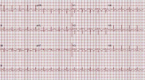
Figure 1. ECG on presentation demonstrating sinus tachycardia, anterior precordial T wave inversions and S1Q3T3, classic ECG findings of pulmonary embolism.


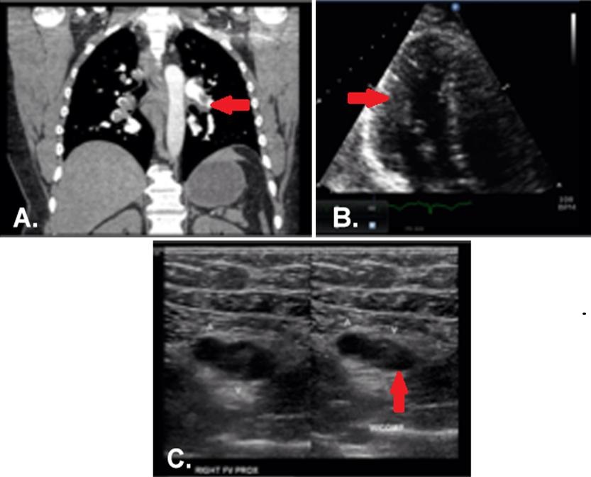
Figure 2. Panel A: CT angiogram demonstrating bilateral pulmonary embolism involving nearly every segmental and subsegmental pulmonary artery. Panel B: Echocardiogram, apical 4-chamber view, with dilated right ventricle and poor function. Panel C: Right leg ultrasound showing acute, non-occlusive thrombus; the right side of the image demonstrates incompressibility of the right femoral vein.


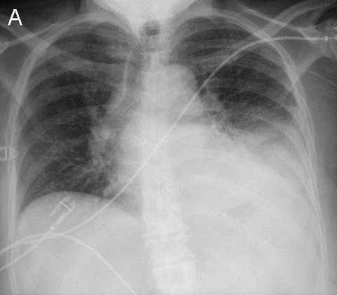
A 44-year-old male long distance truck driver with no known medical history presented with intermittent episodes of dyspnea for the past 24 hours, and an episode of exertional syncope just prior to hospitalization. The patient complained of sharp severe chest pain and reports several week history of right leg swelling. Initial Electrocardiogram (ECG, Figure 1) shows sinus tachycardia and signs of right ventricular strain with an associated troponin elevation. CT pulmonary angiography confirmed bilateral, extensive pulmonary emboli (PE) (Figure 2A, arrow at left pulmonary artery embolus). An echocardiogram showed severe right ventricular systolic dysfunction (Figure 2B, arrow indicated RV). Duplex ultrasound of the right leg showed extensive, acute, non-occlusive thrombus (Figure 2C, arrow indicates clot failing to compress). The patient received an IVC filter due to substantial clot burden. A hypercoagulability workup was negative.

The ECG is part of the typical evaluation for syncope, chest pain and shortness of breath. Multiple studies evaluating the utility of the ECG in the diagnosis of PE have been conducted (1-3). One study in patients with suspected PE undergoing diagnostic testing found that only tachycardia and incomplete right bundle branch block were significantly more prevalent in patients with PE than those without. Another study found a 39% rate of sinus tachycardia in those ultimately found to have PE compared to 24% in those who had negative studies. The S1Q3T3 phenomenon was present in 12% of those with PE vs 3% in those without. One or more traditional findings of right ventricular strain: S1Q3T3, right bundle branch block, or right axis deviation was present in only 13% of patients with PE who had RV dilation on echocardiography, however these findings were also present in 8.8% of patients with PE without evidence of RV dysfunction. Non-specific ECG findings such as sinus tachycardia and ST-T changes are the most commonly identified ECG abnormalities in patients with PE. Overall the ECG as a test for PE exhibits poor test characteristics and thus has little clinical utility for its diagnosis, despite the frequent emphasis on these findings in medical education.

Our patient’s ECG demonstrates several classic findings suggestive of PE including sinus tachycardia, S1Q3T3, and T-wave inversions in the anterior precordial leads. While certain ECG findings do correlate with the presence of PE they are frequently present in patients without PE and absent in those with the disease. ECG may have some utility in risk stratification by identifying signs of right heart strain, however echocardiography is the preferred modality.

Taylor Shekell MD2, Cameron Hypes MD MPH1,2, Yuval Raz MD1

1 Department of Medicine, Division of Pulmonary, Allergy, Critical Care, and Sleep Medicine

2 Department of Emergency Medicine

University of Arizona Medical Center

Tucson, AZ

References

  1. Rodger M, Makropoulos D, Turek M, Quevillon J, Raymond F, Rasuli P, Wells PS. Diagnostic value of the electrocardiogram in suspected pulmonary embolism. Am J Cardiol. 2000;86:807-9. [CrossRef] [PubMed]
  2. Sinha N, Yalamanchili K, Sukhija R, Aronow WS, Fleisher AG, Maguire GP, Lehrman SG. Role of the 12-lead electrocardiogram in diagnosing pulmonary embolism. Cardiol Rev. 2005;13:46-9. [PubMed]
  3. Stein P, Matta F, Sabra M, et al. Relation of electrocardiographic changes in pulmonary embolism to right ventricular enlargement. Am J Cardiol. 2013;112:1958-61. [CrossRef] [PubMed] 

Reference as: Shekell T, Hypes C, Raz Y. Medical image of the week: ECG in PE. Southwest J Pulm Crit Care. 2015;10(1):44-6. doi: http://dx.doi.org/10.13175/swjpcc162-14 PDF

Monday
Jan052015

January 2015 Imaging Case of the Month

Michael B. Gotway, MD


Department of Radiology

Mayo Clinic Arizona

Scottsdale, AZ


Clinical History: A 68-year-old woman with a history of myelodysplastic syndrome associated with transfusion-dependent anemia and thrombocytopenia presented with recent onset left chest pain and fever. The patient had a remote history of total right knee arthroplasty, hypertension, asthma, and schizoaffective disorder. Several months earlier the patient was hospitalized with methicillin-sensitive Staphylococcus aureus infection involving the right knee arthroplasty, associated with bacteremia and a septic right elbow. This infection was treated with incision and drainage of the elbow, antibiotic bead placement about the right knee arthroplasty with an antibiotic-impregnated spacer, and antibiotics (6 weeks intravenous cefazolin followed by chronic doxycycline suppression therapy, the former later switched to nafcillin and rifampin). The patient had been discharged from the hospital with only compression hose for deep venous thrombosis prophylaxis, owing to her episodes of epistaxis in the setting of transfusion-dependent anemia.

Upon presentation, the patient was hypotensive, tachycardic, and hypotensive. Laboratory data showed a white cell count of 3.9 cells x 109 / L, a platelet count of 7000 x 109 / L, and a hemoglobin level of 7 g/dL.

Frontal chest radiography (Figure 1A) was performed (a baseline chest radiograph- Figure 1B- is presented for comparison).


Figure 1. Panel A: Frontal chest radiography Panel B: Frontal chest radiograph obtained 3 months to presentation.

Which of the following statements regarding the chest radiograph is most accurate? (Click on the correct answer to proceed to the 2nd of 7 panels)

Reference as: Gotway MB. January 2015 imaging case of the month. Southwest J Pulm Crit Care. 2015;10(1):21-31. doi: http://dx.doi.org/10.13175/swjpcc003-15 PDF