Search Journal-type in search term and press enter
Southwest Pulmonary and Critical Care Fellowships
In Memoriam

Case Presentations from the Arizona Thoracic Society

(Click on title to be directed to posting or scroll down)

-------------------------------------------------------------------------------------

1. With Oxygen, More is Not Necessarily Better
2. A 32 Year Old Male With Idiopathic Hepatic Encephalopathy 
  and Necrotic Lower Extremity


The Arizona Thoracic Society meets monthly except for July and December, usually on Tuesday, from 6:30-8:00  PM at Scottsdale Healthcare Shea Hospital located at Shea and 90th Street. 2011 meetings are scheduled for January 18, February 15, March 15,  April 12, May 11 (Wednesday), June 21, August 16, September 20, October 18, and November 15. During these meetings dinner and case presentations occur. Selected case presentations are prepared for publication in the Southwest Journal of Pulmonary and Critical Care. Either the case content or the PowerPoint slides which often accompany the published cases may be used for teaching purposes. We ask only the courtesy of an acknowledgement to the Southwest Journal of Pulmonary and Critical Care and the author.

Wednesday
Jul272011

With Oxygen, More is Not Necessarily Better

Kevin Park MD

Clement U. Singarajah MD

Richard A. Robbins MD

Phoenix VA and Good Samaritan Medical Centers, Phoenix AZ

Reference as: Park K, Singarajah CU, Robbins RA. With oxygen, more is not necessarily better. Southwest J Pulm Crit Care 2011;3:15-18. (Click here for PDF version)

Abstract

We present a case of a patient with obesity-hypoventilation, sleep apnea syndrome who developed respiratory failure with administration of high doses of oxygen. The case illustrates of administering oxygen without titration can have potentially adverse effects.

Case Presentation

History of Present Illness

A 48 year old male was admitted for shortness of breath. He had been discharged 2 days earlier after a 10 day hospitalization for presumed sleep apnea, obesity-hypoventilation syndrome, hypertensive urgency and non-ST segment myocardial infarction complicated by ventricular tachycardia and hypercapnic respiratory failure.  Although he was prescribed continuous positive airway pressure (CPAP) at 10 cm H2O during sleep, this was not used since his prior admission. He had a questionable history of asthma and had been using his albuterol inhaler every 1-2 hours without relief.

Physical Examination

He was alert, obese man (weight 148 kg) who was oriented and appeared in no acute distress.  BP was elevated at 169/120. Occasional wheezes were heard on lung auscultation. He had 1+ pretibial edema.

Laboratory and Radiology

Pertinent laboratory findings include an elevated white blood cell count of 17,700 cells/microliter with a left shift. Arterial blood gases showed a PaO2 of 79, PaCO2 of 71 and a pH of 7.29 on oxygen administered at 2 l/min by nasal cannula. Brain naturetic peptide was elevated at 145 pg/ml.

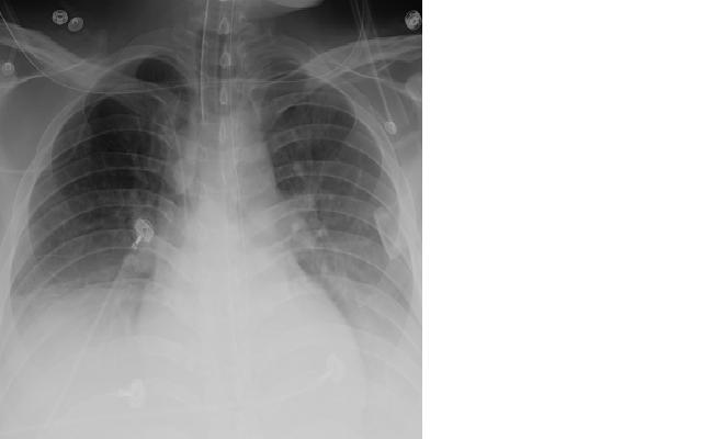
Chest x-ray was considered normal.

Hospital Course

He was treated for an asthma exacerbation with albuterol by small volume nebulizer every 4 hours and prednisone 60 mg orally. He refused CPAP therapy and because his SpO2 decreased to 87 his oxygen was increased to 4 l/min by nasal cannula. About 6 hours after admission he was lethargic, but arousable, and his oxygen was further increased to 6 l/min. He became unarousable except to painful stimuli. His arterial blood gases were repeated and showed a PaO2 of 90, PaCO2 of 92 and a pH of 7.20. He was evaluated for transfer to the intensive care unit for intubation by the pulmonary fellow, but rather than intubate the patient, the patient’s oxygen was decreased to 2 l/min by nasal cannula. The patient’s SpO2 decreased to 88 but he became awake and alert within 30 minutes. He had uneventful subsequent hospital course and was discharged 3 days later on CPAP for nighttime use.

Discussion

This case illustrates the potential dangers of inappropriate use of oxygen therapy, a problem which is being increasingly recognized. Increasing oxygen in patients with hypercapnea, particularly the hypercapnic patient with a history of respiratory depression from high oxygen, is potentially dangerous. The patient presented is typical with probable obesity-hypoventilation and/or sleep apnea, hypercapnea and a history of respiratory depression from oxygen administration. Fortunately, the respiratory depression was recognized and reduction in oxygen and administration of noninvasive positive pressure ventilation likely averted endotracheal intubation and mechanical ventilation.

High doses of oxygen can cause a number of adverse effects including absorption atelectasis and increased ventilation/perfusion mismatch, which impairs elimination of carbon dioxide potentially leading to hypercapnea and respiratory depression. Suppression of hypoxic drive is probably less important than ventilation/perfusion mismatch (1).

The British Thoracic Society (BTS) has published guidelines on the use of oxygen in adults (2). The guidelines emphasize that oxygen is a treatment for hypoxemia, not breathlessness or dyspnea. Oxygen has not been shown to have any effect on the sensation of breathlessness in non-hypoxemic patients. Therefore, increasing oxygen therapy is not only ineffective, but as illustrated by the case above, potentially dangerous.

It has become increasingly recognized that oxygen should be prescribed according to a target oxygen saturation range. Oxygen saturation can now be readily measured in most settings through the use of pulse oximetry. BTS emphasizes that those who administer oxygen therapy should monitor the patient and keep within the target saturation range. BTS suggests oxygen should be prescribed to achieve a target saturation of 94–98% for most acutely ill patients or 88–92% for those at risk of hypercapnic respiratory failure.  High-risk patients with a prior history of hypercapnic respiratory failure may usually be safely managed with an oxygen saturation in the range of 85%-88%.

In support of the concept that titration of oxygen improves outcomes, Austin et al. (3) recently compared nontitrated high flow oxygen treatment with titrated oxygen treatment in the prehospital (ambulance/paramedic) setting in patients with an exacerbation of COPD. They demonstrated that titrated oxygen titrated to an SpO2 of 88-92% had significantly reduced mortality, hypercapnia, and respiratory acidosis compared with high flow oxygen at 8-10 l/min in acute exacerbations of chronic obstructive pulmonary disease. The authors concluded that the results provide strong evidence to recommend the routine use of titrated oxygen treatment in patients with breathlessness and a history or clinical likelihood of chronic obstructive pulmonary disease in the prehospital setting.

Administration of high does of oxygen to patients without hypercapnic respiratory failure may also have adverse consequences. Kilgannon et al. (4) studied patients admitted to the intensive care unit following resuscitation from cardiac arrest. Arterial hyperoxia defined as a PaO2 of > 300 mm Hg was independently associated with increased in-hospital mortality compared with either hypoxia or normoxia. Bellomo et al. (5) also found that hyperoxia after cardiopulmonary arrest was associated with higher mortality. However, once Cox proportional hazards modeling was applied, hyperoxia defined as PaO2 > 400 mmHg was no longer predictive of hospital mortality.

The above emphasize the need to prescribe oxygen as a drug, especially in the acute setting. As with most drugs, the drug effect should be monitored, in this case by the oxygen saturation. Adjusting the oxygen to appropriate levels may well avert intubation, mechanical ventilation and even death in some patients.

References

1. Stoller JK. Clinical practice. Acute exacerbations of chronic obstructive pulmonary disease. N Engl J Med 2002;346:988-94.

2. O’Driscoll BR, Howard LS, Davison AG. BTS guideline for emergency oxygen use in adult patients. Thorax 2008;63 (Suppl VI):vi1–vi68.

3.  Austin MA, Wills KE, Blizzard L, Walters EH, Wood-Baker R.  Effect of high flow oxygen on mortality in chronic obstructive pulmonary disease patients in prehospital setting: randomised controlled trial. BMJ  2010 ;341:c5462.

4. Kilgannon JH, Jones AE, Shapiro NI, Angelos MG, Milcarek B, Hunter K, Parrillo JE, Trzeciak S; Emergency Medicine Shock Research Network (EMShockNet) Investigators. Association between arterial hyperoxia following resuscitation from cardiac arrest and in-hospital mortality. JAMA 2010;303:2165-71.

5. Bellomo R, Bailey M, Eastwood GM, Nichol A, Pilcher D, Hart GK, Reade MC, Egi M, Cooper DJ; the Study of Oxygen in Critical Care (SOCC) Group. Arterial hyperoxia and in-hospital mortality after resuscitation from cardiac arrest. Crit Care 2011 Mar 8;15:R90. [Epub ahead of print]

Presented at the Arizona Thoracic Society meeting on 11-16-2010 in Scottsdale, AZ

Friday
Nov192010

A 32 Year Old Male With Idiopathic Hepatic Encephalopathy And Necrotic Lower Extremity

Evan Denis Schmitz, MD

Robert A. Raschke, MD

Good Samaritan Regional Medical Center/Phoenix Veteran Affairs Medical Center Phoenix, AZ

Refernce as: Schmitz ED, Raschke RA. A 32 year old male with idiopathic hepatic encephalopathy and necrotic lower extremity. Southwest J Pulm Crit Care 2010;1:14-20. (Click here for PDF version)

Abstract

A 32 year old with undiagnosed alpha-1 antitrypsin deficiency had an unusual initial presentation of acute liver failure, septic shock; adult respiratory distress syndrome and what appeared to be a left lower extremity soft tissue infection. Skin biopsy revealed panniculitis. Because of the diagnosis of panniculitis, an alpha-1 antitrypsin level was ordered and demonstrated alpha-1 antitrypsin deficiency with a ZZ phenotype. The patient recovered with antibiotics and supportive therapy. This case illustrates that alpha-1 antitrypsin deficiency can present other that the more usual adult presentation of progressive emphysema. Unexplained panniculitis should prompt investigation of alpha-1 antitrypsin deficiency in the adult patient.

Case Presentation

History of Present Illness

   A 32 year old male was admitted to an outside hospital altered mental status and respiratory failure requiring intubation.  He was diagnosed with liver failure and a lower extremity soft tissue infection.  He developed E. coli bacteremia and septic shock.  He was transferred to Good Samaritan Regional Medical Center.  Two months prior to admission he developed diarrhea as well as bilateral lower extremity edema.  Odd behavior was reported by co-workers.  His skin became pale then yellow.  There was no significant past medical history.  There was no family history of liver or lung disease. He drank 3 beers a day but denied any tobacco or illicit drug use.  He was using supplements such as creatine, rip fuel and energy drinks for over a year.  He denied any anabolic steroid use.

Physical Examination

   On arrival to the ICU his vitals were as follows:  blood pressure of 88/30, heart rate of 103 and a respiratory rate of 35.  He was obtunded and intubated.  He had scleral icterus.  He was tachycardic with no murmurs, rubs or gallops.  He was tachypneic with diminished breath sounds bilaterally.  Abdomen was distended with fluid shift.  He had 4+ pitting edema and necrosis of his left leg (Figure 1).


Figure 1. Left Leg

Laboratory Findings

   Arterial blood gas revealed a pH of 7.05, PaCO2 of 37 mmHg and a PaO2 of 260 mmHg.  Hemogram showed a white blood cell count of 6.9 cells/µL, hemoglobin of 6.5 g/dL and platelet count of 289,000 cells/µL.  Serum chemistry results were sodium of 123 mEq/L, potassium of 4.2 mEq/L, chloride of 93 mEq/L, bicarbonate of 9 mEq/L, urea nitrogen of 33 mg/dL, creatinine of 2.5 mg/dL, glucose of 51 mg/dL, lactic acid level of 9.2, ammonia of 58 mg/dL, total bilirubin of 2.2 mg/dL, , albumin of 1.4 g/dL, aspartate aminotransferase of 77 U/L and an alanine aminotrasferase of 65 U/L (normal ranges10-60 and 10-50 respectively). Alkaline phosphatase was normal.  .  Coagulation study results revealed an international normalized ratio of 2.1 and a partial thromboplastin time of 44.9 seconds.  Hepatitis serologies were negative. Urinalysis was positive for bilirubin and a specific gravity of 1.020.  Blood cultures grew E. coli.

Radiography

   Chest X-ray revealed diffuse bilateral lower lobe consolidations in the lower lobes (see Figure 2). A CT scan was interpreted as not showing any significant emphysema.


Figure 2. Chest Radiography

Hospital Course

   The patient’s vitals signs stabilized with the use of pressor medications and antibiotics.  The lower extremity was debrided and skin biopsies were sent to pathology which confirmed inflammation and tissue necrosis (Figure 3, see arrows). 


Figure 3. Skin biopsy (H&E staining).

Because of the panniulitis observed on the skin biopsy, a serum α-1 antitrypsin level was obtained which revealed a level less than 30 mg/dL.  Genetic testing was also performed and the patient’s phenotype was identified as PIZZ. 

Discussion

   Manifestations of α-1 antitrypsin deficiency include emphysema, hepatic disease, panniculitis, vascular disease, inflammatory bowel disease, glomerulonephritis, and ANCA-positive vasculitis (1). Less than 50 cases of panniculitis have been reported (4). Panniculitis is not associated with any particular alpha-1 allele. There are many causes of panniculitis including Weber-Christian Disease, physical injury, multiple rheumatologic disorders, malignancies, dermatologic disorders, infections and several miscellaneous diseases including sarcoidosis in addition to alpha-1 antitrypsin deficiency.  Necrotizing panniculitis in alpha-1 antitrypsin deficiency is caused by unopposed proteolysis in the skin which leads to lobular fat necrosis of the lower reticular dermis (4).  Patients develop a hot and painful red nodule or plaque that may progress to necrosis if left untreated. Some have advocated alpha-1 replacement therapy for treatment of alpha-1 associated panniculitis (4).

   Liver disease is caused by polymerization of the variant α-1 antitrypsin protein which results in intrahepatocyte accumulation and is an unusual presentation in the adult (2,3).  PIZZ is the most common phenotype (1-3).

Conclusion

   Presentation of alpha-1 antitrypsin deficiency with panniculitis E. coli bacteremia and ARDS is unusual. Most adults present with signs and symptoms of emphysema (1).  He was successfully treated with positive pressure ventilation, antibiotics and supportive care and eventually left the hospital. It is unclear what precipitated his acute illness.

References

1. American Thoracic Society/European Respiratory Society Statement: Standards for the diagnosis and management of individuals with alpha-1 antitrypsin deficiency.  Am J Respir Crit Care Med 2003;168:818-900.

2. Teckman JH, Qu D, Perlmutter DH. Molecular pathogensis of liver disease in alpha-1 antitrypsin deficiency. Hepatology 1996;24:1504-16.

3. Perlmutter DH, Brodsky JL, Balistreri WF, Trapnell BC. Molecular pathogenesis of alpha-1 antitrypsin deficiency associated liver disease: a meeting review.  Hepatology 2007; 45:1313-23.

4. Stoller JK and Pilang M. Panniculitis in alpha-1 antitrypsin deficiency: a review. Clin Pulm Med 2008; 15:113-7.

Presented at the Arizona Thoracic Society April 20, 2010 in Scottsdale, AZ and the Great Cases Session at the American Thoracic Society International Conference, May 16, 2010 in New Orleans, LA. A link to the slides used for these presentations is below.

Click here for a PowerPoint slide presentation