Search Journal-type in search term and press enter
Southwest Pulmonary and Critical Care Fellowships
In Memoriam

Imaging

Last 50 Imaging Postings

(Click on title to be directed to posting, most recent listed first)

May 2024 Medical Image of the Month: Hereditary Hemorrhagic
   Telangiectasia in a Patient on Veno-Arterial Extra-Corporeal Membrane
   Oxygenation
May 2024 Imaging Case of the Month: Nothing Is Guaranteed
April 2024 Medical Image of the Month: Wind Instruments Player Exhibiting
   Exceptional Pulmonary Function
March 2024 Medical Image of the Month: Sputum Cytology in Patients with
   Suspected Lung Malignancy Presenting with Acute Hypoxic Respiratory
   Failure
February 2024 Medical Image of the Month: Pulmonary Alveolar Proteinosis
   in Myelodysplastic Syndrome
February 2024 Imaging Case of the Month: Connecting Some Unusual Dots
January 2024 Medical Image of the Month: Polyangiitis Overlap Syndrome
   (POS) Mimicking Fungal Pneumonia 
December 2023 Medical Image of the Month: Metastatic Pulmonary
   Calcifications in End-Stage Renal Disease 
November 2023 Medical Image of the Month: Obstructive Uropathy
   Extremis
November 2023 Imaging Case of the Month: A Crazy Association
October 2023 Medical Image of the Month: Swyer-James-MacLeod
   Syndrome
September 2023 Medical Image of the Month: Aspergillus Presenting as a
   Pulmonary Nodule in an Immunocompetent Patient
August 2023 Medical Image of the Month: Cannonball Metastases from
   Metastatic Melanoma
August 2023 Imaging Case of the Month: Chew Your Food Carefully
July 2023 Medical Image of the Month: Primary Tracheal Lymphoma
June 2023 Medical Image of the Month: Solitary Fibrous Tumor of the Pleura
May 2023 Medical Image of the Month: Methamphetamine Inhalation
   Leading to Cavitary Pneumonia and Pleural Complications
April 2023 Medical Image of the Month: Atrial Myxoma in the setting of
   Raynaud’s Phenomenon: Early Echocardiography and Management of
   Thrombotic Disease
April 2023 Imaging Case of the Month: Large Impact from a Small Lesion
March 2023 Medical Image of the Month: Spontaneous Pneumomediastinum
   as a Complication of Marijuana Smoking Due to Müller's Maneuvers
February 2023 Medical Image of the Month: Reversed Halo Sign in the
   Setting of a Neutropenic Patient with Angioinvasive Pulmonary
   Zygomycosis
January 2023 Medical Image of the Month: Abnormal Sleep Study and PFT
   with Supine Challenge Related to Idiopathic Hemidiaphragmatic Paralysis
December 2022 Medical Image of the Month: Bronchoesophageal Fistula in
   the Setting of Pulmonary Actinomycosis
November 2022 Medical Image of the Month: COVID-19 Infection
   Presenting as Spontaneous Subcapsular Hematoma of the Kidney
November 2022 Imaging Case of the Month: Out of Place in the Thorax
October 2022 Medical Image of the Month: Infected Dasatinib Induced
   Chylothorax-The First Reported Case 
September 2022 Medical Image of the Month: Epiglottic Calcification
Medical Image of the Month: An Unexpected Cause of Chronic Cough
August 2022 Imaging Case of the Month: It’s All About Location
July 2022 Medical Image of the Month: Pulmonary Nodule in the
   Setting of Pyoderma Gangrenosum (PG) 
June 2022 Medical Image of the Month: A Hard Image to Swallow
May 2022 Medical Image of the Month: Pectus Excavatum
May 2022 Imaging Case of the Month: Asymmetric Apical Opacity–
   Diagnostic Considerations
April 2022 Medical Image of the Month: COVID Pericarditis
March 2022 Medical Image of the Month: Pulmonary Nodules in the
   Setting of Diffuse Idiopathic Pulmonary NeuroEndocrine Cell Hyperplasia
   (DIPNECH) 
February 2022 Medical Image of the Month: Multifocal Micronodular
   Pneumocyte Hyperplasia in the Setting of Tuberous Sclerosis
February 2022 Imaging Case of the Month: Between A Rock and a
   Hard Place
January 2022 Medical Image of the Month: Bronchial Obstruction
   Due to Pledget in Airway Following Foregut Cyst Resection
December 2021 Medical Image of the Month: Aspirated Dental Implant
Medical Image of the Month: Cavitating Pseudomonas
   aeruginosa Pneumonia
November 2021 Imaging Case of the Month: Let’s Not Dance
   the Twist
Medical Image of the Month: COVID-19-Associated Pulmonary
   Aspergillosis in a Post-Liver Transplant Patient
Medical Image of the Month: Stercoral Colitis
Medical Image of the Month: Bleomycin-Induced Pulmonary Fibrosis
   in a Patient with Lymphoma
August 2021 Imaging Case of the Month: Unilateral Peripheral Lung
   Opacity
Medical Image of the Month: Hepatic Abscess Secondary to Diverticulitis
   Resulting in Sepsis
Medical Image of the Month: Metastatic Spindle Cell Carcinoma of the
   Breast
Medical Image of the Month: Perforated Gangrenous Cholecystitis
May 2021 Imaging Case of the Month: A Growing Indeterminate Solitary
   Nodule
Medical Image of the Month: Severe Acute Respiratory Distress
Syndrome and Embolic Strokes from Polymethylmethacrylate
   (PMMA) Embolization
Medical Image of the Month: Pulmonary Aspergillus Overlap Syndrome
   Presenting with ABPA, Multiple Bilateral Aspergillomas
Medical Image of the Month: Diffuse White Matter Microhemorrhages
   Secondary to SARS-CoV-2 (COVID-19) Infection
February 2021 Imaging Case of the Month: An Indeterminate Solitary
   Nodule
Medical Image of the Month: Mucinous Adenocarcinoma of the Lung
   Mimicking Pneumonia
Medical Image of the Month: Superior Vena Cava Syndrome
Medical Image of the Month: Buffalo Chest Identified at the Time of
   Lung Nodule Biopsy
November 2020 Imaging Case of the Month: Cause and Effect?


For complete imaging listings click here

Those who care for patients with pulmonary, critical care or sleep disorders rely heavily on chest radiology and pathology to determine diagnoses. The Southwest Journal of Pulmonary and Critical Care publishes case-based articles with characteristic chest imaging and related pathology. The editor of this section will oversee and coordinate the publication of a core of the most important chest imaging topics. In doing so, they encourage the submission of unsolicited manuscripts. It cannot be overemphasized that both radiologic and pathologic images must be of excellent quality. As a rule, 600 DPI is sufficient for radiographic and pathologic images. Taking pictures of plain chest radiographs and CT scans with a digital camera is strongly discouraged. The figures should be cited in the text and numbered consecutively. The stain used for pathology specimens and magnification should be mentioned in the figure legend. Those who care for patients with pulmonary, critical care or sleep disorders rely heavily on chest radiology and pathology to determine diagnoses. The Southwest Journal of Pulmonary and Critical Care publishes case-based articles with characteristic chest imaging and related pathology. The editor of this section will oversee and coordinate the publication of a core of the most important chest imaging topics. In doing so, they encourage the submission of unsolicited manuscripts. It cannot be overemphasized that both radiologic and pathologic images must be of excellent quality. As a rule, 600 DPI is sufficient for radiographic and pathologic images. Taking pictures of plain chest radiographs and CT scans with a digital camera is strongly discouraged. The figures should be cited in the text and numbered consecutively. The stain used for pathology specimens and magnification should be mentioned in the figure legend.

-------------------------------------------------------------------------------------------  

Wednesday
Sep202017

Medical Image of the Week: Pembrolizumab-induced Pneumonitis

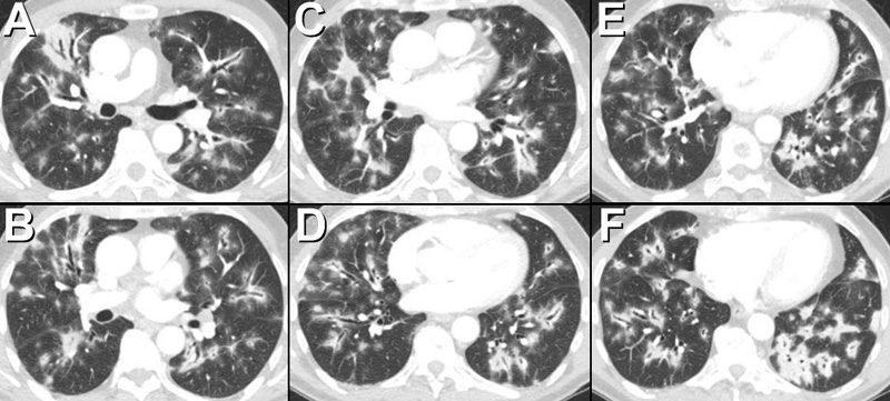
Figure 1. Thoracic CT showing multifocal, bilateral extensive lung opacities consistent with consolidation with a striking peribronchovascular distribution. Note the opacities are primarily distributed along the airways. Peripheral lung opacity, both ground-glass opacity and consolidation, is also present.


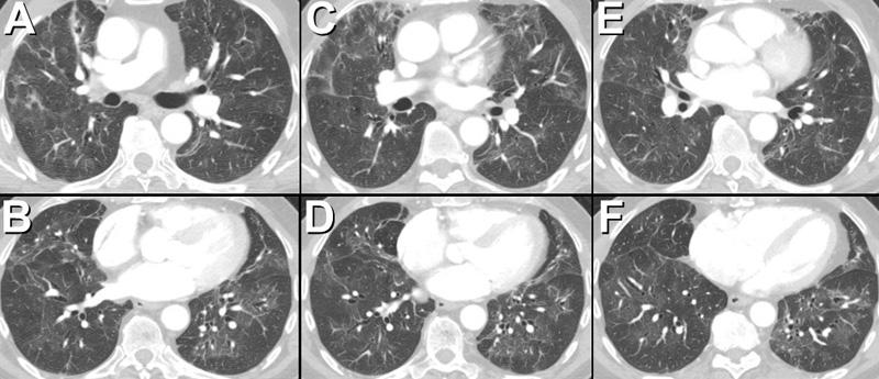
Figure 2. Axial thoracic CT performed several months after Figure 1 following discontinuation of the pembrolizumab and initiation, and subsequent tapering, of corticosteroid therapy, shows significant regression in the previously noted extensive peribronchovascular consolidation. Residual areas of consolidation and ground-glass opacity associated with architectural distortion are consistent with scarring.


A 76-year-old man with metastatic melanoma, undergoing treatment with pembrolizumab, an antibody against programmed cell death 1 (PD-1), beginning 8 months ago developed low-grade fever, non-productive cough, and shortness of breath. A thoracic CT scan showed multifocal, bilateral extensive lung opacities (Figure 1). The patient underwent bronchoscopy with bronchoalveolar lavage which showed non-specific inflammatory changes associated with foci of organizing pneumonia. Microbiologic studies, including Coccioides antibody enzyme immunoassay and Aspergillus antigen, were negative.

The patient was begun on corticosteroid therapy for presumed medication-induced pulmonary injury, manifestation as an organizing pneumonia pattern, due to pembrolizumab. Over the ensuing months, his symptoms abated and his CT scan abnormalities regressed (Figure 2).

Organizing pneumonia may occur as an idiopathic, primary pulmonary process, often referred to as “cryptogenic organizing pneumonia,” or may occur in the context of a number of systemic conditions, a situation often referred to as secondary organizing pneumonia. Among the various etiologies of secondary organizing pneumonia, medication-induced pulmonary injury is fairly common and when imaging features of organizing pneumonia are seen, careful correlation regarding the possibility of a medication-induced etiology should be undertaken. Recently, three cases of pembrolizumab-induced pneumonitis were described, two being consistent with organizing pneumonia (1).

The thoracic CT findings of organizing pneumonia include peripheral and peribronchovascular consolidation and ground-glass opacity, areas of consolidation surrounding ground-glass opacity (often referred to as the “atoll” or reverse ground-glass halo” sign- see Medical Image of the Week: The Atoll Sign in Cryptogenic Organizing Pneumonia), single and multiple nodules, and perilobular consolidation. The case illustrates a dramatically peribronchovascular distribution of pulmonary consolidation as a manifestation of medication-induced organizing pneumonia.

Michael B. Gotway, MD

Department of Radiology

Mayo Clinic Arizona

Scottsdale, AZ USA

Reference

  1. Leroy V, Templier C, Faivre JB, Scherpereel A, Fournier C, Mortier L, Wemeau-Stervinou L. Pembrolizumab-induced pneumonitis. ERJ Open Res. 2017 May 2;3(2). pii: 00081-2016. [CrossRef] [PubMed]

Cite as: Gotway MB. Medical image of the week: pembrolizumab-induced pneumonitis. Southwest J Pulm Crit Care. 2017;15(3):118-9. doi: https://doi.org/10.13175/swjpcc110-17 PDF 

Wednesday
Sep132017

Medical Image of the Week: Asbestos Related Pleural Disease

Figure 1. Chest radiograph demonstrates bilateral coarse calcification, most elongated and vertically oriented in nature (white arrows). Also note coarse calcification outlining the hemidiaphragms (dark arrows). Editor's note: the patient's only chest x-ray was two different AP views which are merged above.


Figure 2.  Holly leaf. Its shape is similar to the irregular thickened nodular edges of pleural plaques on chest radiograph, referred to as “the holly leaf sign”.


Figure 3. Thoracic CT shown in soft tissue (A: top) and lung (B: bottom) windows clearly localizes the calcifications to the parietal pleura.


Pleural plaques are strongly associated with inhalational exposure to asbestos (1). The lesions may take up to thirty years to develop. Plaques are typically bilateral, involve the parietal pleura, commonly along the sixth through ninth ribs and are usually absent at the lung apices and costophrenic sulci (Figures 1 and 3). On chest radiograph, the “holly leaf sign” refers to the shape of the calcifications with thickened rolled and nodular edges (Figure 2). The plaques per se are benign in nature. However, they can potentially impair lung function, resulting in restriction.  They are also markers of the individual’s greater risk of developing a lung cancer or mesothelioma.

Wesley Hunter MS IV1, Veronica Arteaga MD2, and Diana Palacio MD2

1College of Medicine and 2Department of Medical Imaging

University of Arizona

Tucson, AZ USA

Reference

  1. Norbet C, Joseph A, Rossi SS, Bhalla S, Gutierrez FR. Asbestos-related lung disease: a pictorial review. Curr Probl Diagn Radiol. 2015 Jul-Aug;44(4):371-82. [CrossRef] [PubMed] 

Cite as: Hunter W, Arteaga V, Palacio D. Medical image of the week: asbestos related pleural disease. Southwest J Pulm Crit Care. 2017;15(3):116-7. doi: https://doi.org/10.13175/swjpcc104-17 PDF