Search Journal-type in search term and press enter
Southwest Pulmonary and Critical Care Fellowships
In Memoriam

Imaging

Last 50 Imaging Postings

(Click on title to be directed to posting, most recent listed first)

May 2024 Medical Image of the Month: Hereditary Hemorrhagic
   Telangiectasia in a Patient on Veno-Arterial Extra-Corporeal Membrane
   Oxygenation
May 2024 Imaging Case of the Month: Nothing Is Guaranteed
April 2024 Medical Image of the Month: Wind Instruments Player Exhibiting
   Exceptional Pulmonary Function
March 2024 Medical Image of the Month: Sputum Cytology in Patients with
   Suspected Lung Malignancy Presenting with Acute Hypoxic Respiratory
   Failure
February 2024 Medical Image of the Month: Pulmonary Alveolar Proteinosis
   in Myelodysplastic Syndrome
February 2024 Imaging Case of the Month: Connecting Some Unusual Dots
January 2024 Medical Image of the Month: Polyangiitis Overlap Syndrome
   (POS) Mimicking Fungal Pneumonia 
December 2023 Medical Image of the Month: Metastatic Pulmonary
   Calcifications in End-Stage Renal Disease 
November 2023 Medical Image of the Month: Obstructive Uropathy
   Extremis
November 2023 Imaging Case of the Month: A Crazy Association
October 2023 Medical Image of the Month: Swyer-James-MacLeod
   Syndrome
September 2023 Medical Image of the Month: Aspergillus Presenting as a
   Pulmonary Nodule in an Immunocompetent Patient
August 2023 Medical Image of the Month: Cannonball Metastases from
   Metastatic Melanoma
August 2023 Imaging Case of the Month: Chew Your Food Carefully
July 2023 Medical Image of the Month: Primary Tracheal Lymphoma
June 2023 Medical Image of the Month: Solitary Fibrous Tumor of the Pleura
May 2023 Medical Image of the Month: Methamphetamine Inhalation
   Leading to Cavitary Pneumonia and Pleural Complications
April 2023 Medical Image of the Month: Atrial Myxoma in the setting of
   Raynaud’s Phenomenon: Early Echocardiography and Management of
   Thrombotic Disease
April 2023 Imaging Case of the Month: Large Impact from a Small Lesion
March 2023 Medical Image of the Month: Spontaneous Pneumomediastinum
   as a Complication of Marijuana Smoking Due to Müller's Maneuvers
February 2023 Medical Image of the Month: Reversed Halo Sign in the
   Setting of a Neutropenic Patient with Angioinvasive Pulmonary
   Zygomycosis
January 2023 Medical Image of the Month: Abnormal Sleep Study and PFT
   with Supine Challenge Related to Idiopathic Hemidiaphragmatic Paralysis
December 2022 Medical Image of the Month: Bronchoesophageal Fistula in
   the Setting of Pulmonary Actinomycosis
November 2022 Medical Image of the Month: COVID-19 Infection
   Presenting as Spontaneous Subcapsular Hematoma of the Kidney
November 2022 Imaging Case of the Month: Out of Place in the Thorax
October 2022 Medical Image of the Month: Infected Dasatinib Induced
   Chylothorax-The First Reported Case 
September 2022 Medical Image of the Month: Epiglottic Calcification
Medical Image of the Month: An Unexpected Cause of Chronic Cough
August 2022 Imaging Case of the Month: It’s All About Location
July 2022 Medical Image of the Month: Pulmonary Nodule in the
   Setting of Pyoderma Gangrenosum (PG) 
June 2022 Medical Image of the Month: A Hard Image to Swallow
May 2022 Medical Image of the Month: Pectus Excavatum
May 2022 Imaging Case of the Month: Asymmetric Apical Opacity–
   Diagnostic Considerations
April 2022 Medical Image of the Month: COVID Pericarditis
March 2022 Medical Image of the Month: Pulmonary Nodules in the
   Setting of Diffuse Idiopathic Pulmonary NeuroEndocrine Cell Hyperplasia
   (DIPNECH) 
February 2022 Medical Image of the Month: Multifocal Micronodular
   Pneumocyte Hyperplasia in the Setting of Tuberous Sclerosis
February 2022 Imaging Case of the Month: Between A Rock and a
   Hard Place
January 2022 Medical Image of the Month: Bronchial Obstruction
   Due to Pledget in Airway Following Foregut Cyst Resection
December 2021 Medical Image of the Month: Aspirated Dental Implant
Medical Image of the Month: Cavitating Pseudomonas
   aeruginosa Pneumonia
November 2021 Imaging Case of the Month: Let’s Not Dance
   the Twist
Medical Image of the Month: COVID-19-Associated Pulmonary
   Aspergillosis in a Post-Liver Transplant Patient
Medical Image of the Month: Stercoral Colitis
Medical Image of the Month: Bleomycin-Induced Pulmonary Fibrosis
   in a Patient with Lymphoma
August 2021 Imaging Case of the Month: Unilateral Peripheral Lung
   Opacity
Medical Image of the Month: Hepatic Abscess Secondary to Diverticulitis
   Resulting in Sepsis
Medical Image of the Month: Metastatic Spindle Cell Carcinoma of the
   Breast
Medical Image of the Month: Perforated Gangrenous Cholecystitis
May 2021 Imaging Case of the Month: A Growing Indeterminate Solitary
   Nodule
Medical Image of the Month: Severe Acute Respiratory Distress
Syndrome and Embolic Strokes from Polymethylmethacrylate
   (PMMA) Embolization
Medical Image of the Month: Pulmonary Aspergillus Overlap Syndrome
   Presenting with ABPA, Multiple Bilateral Aspergillomas
Medical Image of the Month: Diffuse White Matter Microhemorrhages
   Secondary to SARS-CoV-2 (COVID-19) Infection
February 2021 Imaging Case of the Month: An Indeterminate Solitary
   Nodule
Medical Image of the Month: Mucinous Adenocarcinoma of the Lung
   Mimicking Pneumonia
Medical Image of the Month: Superior Vena Cava Syndrome
Medical Image of the Month: Buffalo Chest Identified at the Time of
   Lung Nodule Biopsy
November 2020 Imaging Case of the Month: Cause and Effect?


For complete imaging listings click here

Those who care for patients with pulmonary, critical care or sleep disorders rely heavily on chest radiology and pathology to determine diagnoses. The Southwest Journal of Pulmonary and Critical Care publishes case-based articles with characteristic chest imaging and related pathology. The editor of this section will oversee and coordinate the publication of a core of the most important chest imaging topics. In doing so, they encourage the submission of unsolicited manuscripts. It cannot be overemphasized that both radiologic and pathologic images must be of excellent quality. As a rule, 600 DPI is sufficient for radiographic and pathologic images. Taking pictures of plain chest radiographs and CT scans with a digital camera is strongly discouraged. The figures should be cited in the text and numbered consecutively. The stain used for pathology specimens and magnification should be mentioned in the figure legend. Those who care for patients with pulmonary, critical care or sleep disorders rely heavily on chest radiology and pathology to determine diagnoses. The Southwest Journal of Pulmonary and Critical Care publishes case-based articles with characteristic chest imaging and related pathology. The editor of this section will oversee and coordinate the publication of a core of the most important chest imaging topics. In doing so, they encourage the submission of unsolicited manuscripts. It cannot be overemphasized that both radiologic and pathologic images must be of excellent quality. As a rule, 600 DPI is sufficient for radiographic and pathologic images. Taking pictures of plain chest radiographs and CT scans with a digital camera is strongly discouraged. The figures should be cited in the text and numbered consecutively. The stain used for pathology specimens and magnification should be mentioned in the figure legend.

-------------------------------------------------------------------------------------------  

Wednesday
Aug302017

Medical Image of the Week: The Atoll Sign in Cryptogenic Organizing Pneumonia

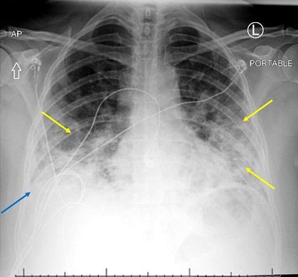
Figure 1. Portable chest X-ray shows bilateral airspace opacities (yellow arrows) and possible trace pleural effusion (blue arrow).


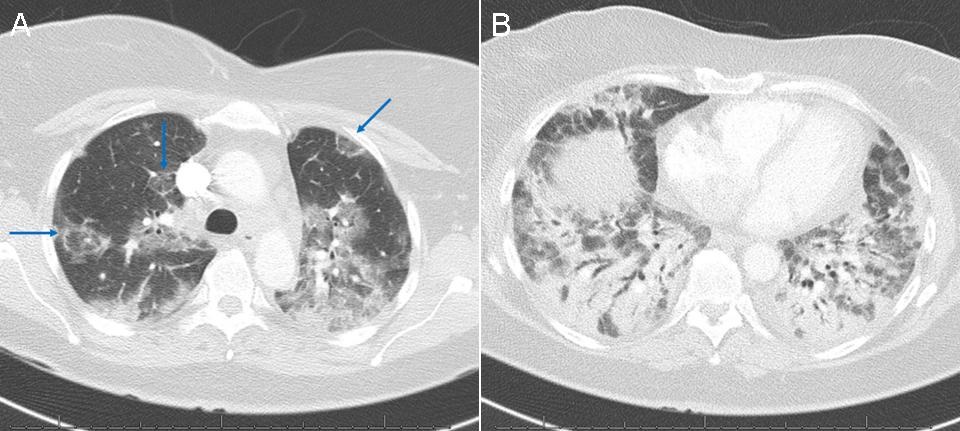
Figure 2. Computed tomography of the chest showing (A) patchy ground glass opacity in the upper lungs with additional scattered circular areas of opacity in a reverse halo configuration (blue arrows, atoll sign) and (B) extensive bibasilar consolidation with air bronchograms.


A 54-year-old woman presented to the emergency department with cough and worsening shortness of breath. Her cough began approximately 1 month prior to presentation, at which time she was diagnosed with pneumonia by her primary care physician based on a chest X-ray at an outside institution. She tried and failed courses of azithromycin, doxycycline, and levofloxacin.

The patient had an oxygen saturation of 55% and hyperpyrexia to 101.7 F in the emergency department. An initial chest X-ray was suggestive of moderate multifocal pneumonia with pleural effusion (Figure 1). Subsequent chest computed tomography (CT; Figure 2) revealed findings consistent with cryptogenic organizing pneumonia (COP) including multiple upper lobe atoll signs. Infectious and autoimmune workups were negative and the patient experienced a rapid recovery with pulse steroids, providing further evidence for the diagnosis of COP.

CT is the best imaging modality for evaluation of potential COP. Features include consolidations and nodules, bronchial wall thickening or dilatation, and ground glass opacities (1). The atoll sign, consisting of a central ground glass opacity and surrounding consolidation which may also be called a reverse halo sign, is highly specific but not sensitive for organizing pneumonia (2). Definitive diagnosis requires lung biopsy, although the disease is often managed based on a presumptive diagnosis (3).

Joseph Frankl, BS1 and Veronica A. Arteaga, MD2

1University of Arizona College of Medicine and 2Department of Medical Imaging

Banner University Medical Center Tucson

Tucson, AZ USA

References

  1. Lee JW, Lee KS, Lee HY, Chung MP, Yi CA, Kim TS, Chung MJ. Cryptogenic organizing pneumonia: serial high-resolution CT findings in 22 patients. AJR Am J Roentgenol. 2010 Oct;195(4):916-22. [CrossRef] [PubMed]
  2. Davidsen JR, Madsen HD, Laursen CB. Reversed halo sign in cryptogenic organising pneumonia. BMJ Case Rep. 2016 Feb 8;2016. pii: bcr2015213779. [CrossRef] [PubMed]
  3. Bradley B, Branley HM, Egan JJ, et al. Interstitial lung disease guideline: the British Thoracic Society in collaboration with the Thoracic Society of Australia and New Zealand and the Irish Thoracic Society. Thorax. 2008 Sep;63 Suppl 5:v1-58. [CrossRef] [PubMed]

Cite as: Frankl J, Artega VA. Medical image of the week: the atoll sign in cryptogenic organizing pneumonia. Southwest J Pulm Crit Care. 2017;15(2):92-3. doi: https://doi.org/10.13175/swjpcc100-17 PDF

Wednesday
Aug232017

Medical Image of the Week: Cannon V Waves

Figure 1. Video showing jugular venous distention to earlobes with cannon V waves.

A 66-year-old man experienced recurrent ascites of unknown etiology over six months. He had previously undergone a renal transplant secondary to complications of diabetes and hypertension and had known severe coronary artery disease. His most recent paracentesis revealed an albumin 1.6 g/dL (serum albumin 2.1) and a total protein of 3.8 g/dL. His adenosine deaminase was 11.6 U/L (normal <7.6 U/L), but repeated bacterial and mycobacterial ascites cultures were negative, as were a carcinoembryonic antigen assay and ascites cytology. Computerized tomography of the abdomen showed findings consistent with cirrhosis, but an extensive workup for common causes of cirrhosis was negative.

Physical exam showed jugular venous distention with prominent V waves and a holosystolic murmur at the left lower sternal border (Figure 1). Echocardiography showed a dilated right ventricle, moderate pulmonary and tricuspid regurgitation and an estimated right ventricular systolic pressure of 87 mm Hg. Cardiac catherization confirmed the presence of an elevated right ventricular pressure of 72/10 (22) mm Hg, an elevated pulmonary artery pressure of 75/27 (45) mm Hg and a left ventricular ejection fraction of 20-25%. The right atrial pressure was 20 and the pulmonary artery occlusion pressure was 22 mmHg.  A diagnosis of pulmonary hypertension secondary to left ventricular heart disease (type 2 pulmonary hypertension) with congestive hepatopathy and cardiac ascites was made.

The patient’s physical examination provided an important clue to the etiology of the ascites – cardiac ascites is thought to be due to chronic venous congestion of the liver due to transmission of high central venous pressures. Tricuspid regurgitation can be associated with severe hepatic congestion because of retrograde transmission of right ventricular pressure directly into the hepatic veins. In some patients (although not in this patient), careful examination will reveal that the liver in such patients is palpably pulsatile.

Cardiac ascites is typically characterized by a serum albumin gradient (SAAG) >1.1 g/dL (indicative of portal hypertension) and ascites protein level of >2.5 g/dL (1). We cannot fully explain why this patient’s SAAG was low. A complete workup for infectious and oncological etiologies of low SAAG ascites was negative. It has been noted that in patients with known cirrhosis (as in this patient), the finding of a low SAAG has a low specificity for infectious and oncological etiologies of ascites (2). Serositis which can sometimes manifest as ascites can also be a complication of tacrolimus which the patient was receiving s/p renal transplant. It’s possible that tacrolimus might have changed the nature of the ascites fluid in this patient but this is conjectural. 

Robert A. Raschke, MD

College of Medicine-Phoenix

Phoenix, AZ USA

References

  1. Sam AH, James THT. Rapid Medicine. Wiley-Blackwell; 2009: ISBN 1-4051-8323-3.
  2. Khandwalla HE, Fasakin Y, El-Serag HB. The utility of evaluating low serum albumin gradient ascites in patients with cirrhosis. Am J Gastroenterol. 2009 Jun;104(6):1401-5. [CrossRef] [PubMed] 

Cite as: Raschke RA. Medical image of the week: cannon V waves. Southwest J Pulm Crit Care. 2017;15(2):90-1. doi: https://doi.org/10.13175/swjpcc095-17 PDF