Search Journal-type in search term and press enter
Southwest Pulmonary and Critical Care Fellowships
In Memoriam

Imaging

Last 50 Imaging Postings

(Click on title to be directed to posting, most recent listed first)

May 2024 Medical Image of the Month: Hereditary Hemorrhagic
   Telangiectasia in a Patient on Veno-Arterial Extra-Corporeal Membrane
   Oxygenation
May 2024 Imaging Case of the Month: Nothing Is Guaranteed
April 2024 Medical Image of the Month: Wind Instruments Player Exhibiting
   Exceptional Pulmonary Function
March 2024 Medical Image of the Month: Sputum Cytology in Patients with
   Suspected Lung Malignancy Presenting with Acute Hypoxic Respiratory
   Failure
February 2024 Medical Image of the Month: Pulmonary Alveolar Proteinosis
   in Myelodysplastic Syndrome
February 2024 Imaging Case of the Month: Connecting Some Unusual Dots
January 2024 Medical Image of the Month: Polyangiitis Overlap Syndrome
   (POS) Mimicking Fungal Pneumonia 
December 2023 Medical Image of the Month: Metastatic Pulmonary
   Calcifications in End-Stage Renal Disease 
November 2023 Medical Image of the Month: Obstructive Uropathy
   Extremis
November 2023 Imaging Case of the Month: A Crazy Association
October 2023 Medical Image of the Month: Swyer-James-MacLeod
   Syndrome
September 2023 Medical Image of the Month: Aspergillus Presenting as a
   Pulmonary Nodule in an Immunocompetent Patient
August 2023 Medical Image of the Month: Cannonball Metastases from
   Metastatic Melanoma
August 2023 Imaging Case of the Month: Chew Your Food Carefully
July 2023 Medical Image of the Month: Primary Tracheal Lymphoma
June 2023 Medical Image of the Month: Solitary Fibrous Tumor of the Pleura
May 2023 Medical Image of the Month: Methamphetamine Inhalation
   Leading to Cavitary Pneumonia and Pleural Complications
April 2023 Medical Image of the Month: Atrial Myxoma in the setting of
   Raynaud’s Phenomenon: Early Echocardiography and Management of
   Thrombotic Disease
April 2023 Imaging Case of the Month: Large Impact from a Small Lesion
March 2023 Medical Image of the Month: Spontaneous Pneumomediastinum
   as a Complication of Marijuana Smoking Due to Müller's Maneuvers
February 2023 Medical Image of the Month: Reversed Halo Sign in the
   Setting of a Neutropenic Patient with Angioinvasive Pulmonary
   Zygomycosis
January 2023 Medical Image of the Month: Abnormal Sleep Study and PFT
   with Supine Challenge Related to Idiopathic Hemidiaphragmatic Paralysis
December 2022 Medical Image of the Month: Bronchoesophageal Fistula in
   the Setting of Pulmonary Actinomycosis
November 2022 Medical Image of the Month: COVID-19 Infection
   Presenting as Spontaneous Subcapsular Hematoma of the Kidney
November 2022 Imaging Case of the Month: Out of Place in the Thorax
October 2022 Medical Image of the Month: Infected Dasatinib Induced
   Chylothorax-The First Reported Case 
September 2022 Medical Image of the Month: Epiglottic Calcification
Medical Image of the Month: An Unexpected Cause of Chronic Cough
August 2022 Imaging Case of the Month: It’s All About Location
July 2022 Medical Image of the Month: Pulmonary Nodule in the
   Setting of Pyoderma Gangrenosum (PG) 
June 2022 Medical Image of the Month: A Hard Image to Swallow
May 2022 Medical Image of the Month: Pectus Excavatum
May 2022 Imaging Case of the Month: Asymmetric Apical Opacity–
   Diagnostic Considerations
April 2022 Medical Image of the Month: COVID Pericarditis
March 2022 Medical Image of the Month: Pulmonary Nodules in the
   Setting of Diffuse Idiopathic Pulmonary NeuroEndocrine Cell Hyperplasia
   (DIPNECH) 
February 2022 Medical Image of the Month: Multifocal Micronodular
   Pneumocyte Hyperplasia in the Setting of Tuberous Sclerosis
February 2022 Imaging Case of the Month: Between A Rock and a
   Hard Place
January 2022 Medical Image of the Month: Bronchial Obstruction
   Due to Pledget in Airway Following Foregut Cyst Resection
December 2021 Medical Image of the Month: Aspirated Dental Implant
Medical Image of the Month: Cavitating Pseudomonas
   aeruginosa Pneumonia
November 2021 Imaging Case of the Month: Let’s Not Dance
   the Twist
Medical Image of the Month: COVID-19-Associated Pulmonary
   Aspergillosis in a Post-Liver Transplant Patient
Medical Image of the Month: Stercoral Colitis
Medical Image of the Month: Bleomycin-Induced Pulmonary Fibrosis
   in a Patient with Lymphoma
August 2021 Imaging Case of the Month: Unilateral Peripheral Lung
   Opacity
Medical Image of the Month: Hepatic Abscess Secondary to Diverticulitis
   Resulting in Sepsis
Medical Image of the Month: Metastatic Spindle Cell Carcinoma of the
   Breast
Medical Image of the Month: Perforated Gangrenous Cholecystitis
May 2021 Imaging Case of the Month: A Growing Indeterminate Solitary
   Nodule
Medical Image of the Month: Severe Acute Respiratory Distress
Syndrome and Embolic Strokes from Polymethylmethacrylate
   (PMMA) Embolization
Medical Image of the Month: Pulmonary Aspergillus Overlap Syndrome
   Presenting with ABPA, Multiple Bilateral Aspergillomas
Medical Image of the Month: Diffuse White Matter Microhemorrhages
   Secondary to SARS-CoV-2 (COVID-19) Infection
February 2021 Imaging Case of the Month: An Indeterminate Solitary
   Nodule
Medical Image of the Month: Mucinous Adenocarcinoma of the Lung
   Mimicking Pneumonia
Medical Image of the Month: Superior Vena Cava Syndrome
Medical Image of the Month: Buffalo Chest Identified at the Time of
   Lung Nodule Biopsy
November 2020 Imaging Case of the Month: Cause and Effect?


For complete imaging listings click here

Those who care for patients with pulmonary, critical care or sleep disorders rely heavily on chest radiology and pathology to determine diagnoses. The Southwest Journal of Pulmonary and Critical Care publishes case-based articles with characteristic chest imaging and related pathology. The editor of this section will oversee and coordinate the publication of a core of the most important chest imaging topics. In doing so, they encourage the submission of unsolicited manuscripts. It cannot be overemphasized that both radiologic and pathologic images must be of excellent quality. As a rule, 600 DPI is sufficient for radiographic and pathologic images. Taking pictures of plain chest radiographs and CT scans with a digital camera is strongly discouraged. The figures should be cited in the text and numbered consecutively. The stain used for pathology specimens and magnification should be mentioned in the figure legend. Those who care for patients with pulmonary, critical care or sleep disorders rely heavily on chest radiology and pathology to determine diagnoses. The Southwest Journal of Pulmonary and Critical Care publishes case-based articles with characteristic chest imaging and related pathology. The editor of this section will oversee and coordinate the publication of a core of the most important chest imaging topics. In doing so, they encourage the submission of unsolicited manuscripts. It cannot be overemphasized that both radiologic and pathologic images must be of excellent quality. As a rule, 600 DPI is sufficient for radiographic and pathologic images. Taking pictures of plain chest radiographs and CT scans with a digital camera is strongly discouraged. The figures should be cited in the text and numbered consecutively. The stain used for pathology specimens and magnification should be mentioned in the figure legend.

-------------------------------------------------------------------------------------------  

Tuesday
Oct032017

October 2017 Imaging Case of the Month

Paul J. Conomos, MD1

Michael B. Gotway, MD2


1Arizona Pulmonary Specialists

Phoenix, AZ USA

2Mayo Clinic Arizona

Scottsdale, AZ USA


Clinical History: An 18-year-old man with no known previous medical history presented with complaints of intermittent cough persisting several months. No hemoptysis was noted.

Physical examination was largely unremarkable and the patient’s oxygen saturation was 99% on room air. The patient’s vital signs were within normal limits.

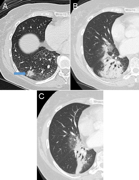
Laboratory evaluation was unremarkable.  Quantiferon testing for Mycobacterium tuberculosis was negative, and testing for coccidioidomycosis was unrevealing. Frontal and lateral chest radiography (Figure 1) was performed.

Figure 1. Figure 1. Frontal (A) and lateral (B) chest radiography.

Which of the following statements regarding the chest radiograph is most accurate? (Click on the correct answer to proceed to the second of eight pages)

  1. The chest radiograph shows asymmetric reticulation and interlobular septal thickening
  2. The chest radiograph shows bilateral reticulation associated with decreased lung volumes
  3. The chest radiograph shows focal consolidation
  4. The chest radiograph shows large lung volumes
  5. The chest radiograph shows small cavitary pulmonary nodules

Cite as: Conomos PJ, Gotway MB. October 2017 imaging case of the month. Southwest J Pulm Crit Care. 2017;15(4):138-46. doi: https://doi.org/10.13175/swjpcc119-17 PDF

Wednesday
Sep272017

Medical Image of the Week: Typical Pulmonary CT Findings Following Radiotherapy

Figure 1. Panel A: CT chest, lung windows, demonstrating a spiculated nodule, biopsy proven adenocarcinoma in the right lower lobe (arrow). Panel B: Eight months post stereotactic radiation therapy, there has been development of focal consolidation, with air bronchograms, involving the right middle and lower lobes. Notice the volumetric appearance. The primary malignancy is no longer identified as such. Panel C: Thirteen months later the consolidation has evolved into an area of volume loss, containing bronchiectasis, and sharp contours as a result of organized fibrosis.


Radiation-induced lung disease (RILD) commonly develops in patients treated with radiation for intrathoracic and chest wall malignancies.

There are two distinct radiographic patterns:

  1. Radiation pneumonitis which occurs within 4-12 weeks after completion of therapy, and is characterized by development ground-glass opacities and/or consolidation in and around the treated lesion. A somewhat nodular or patchy appearance may occur. Typically, the affected tissue conforms to the radiation ports and may cross fissures/lobes. There may be milder similar changes in the contralateral lung.
  2. A chronic phase, known as radiation fibrosis, is noticeable about 6-12 months post treatment and may progress up to 2 years, after which the findings tend to stabilize. In this stage, the areas of consolidation undergo volume loss, architectural distortion and may contain traction bronchiectasis. Linear and band scarring may also be seen. In this phase, sharper demarcation between normal and irradiated lung parenchyma is commonly seen.

Special attention to the typical radiological characteristics and timeline, in most cases allows to distinguish RILD from potential superimposed infection, subacute inflammatory diseases, locally recurrent neoplasm and radiation-induced neoplasms.

Andrew Erickson MS IV1, Berndt Schmidt MD2, Veronica Arteaga MD2, Diana Palacio MD2

1Midwestern University – Arizona College of Osteopathic Medicine

2Division of Thoracic Radiology, Department of Medical Imaging. University of Arizona, Tucson (AZ)

Reference

  1. Choi YW, Munden RF, Erasmus JJ, Joo Park K, Chung WK, Jeon SC, Park CK. Effects of radiation therapy on the lung: radiologic appearances and differential diagnosis. Radiographics. 2004 Jul;24(4):985-97. [CrossRef] [PubMed]

Cite as: Erickson A, Schmidt B, Arteaga V, Palacio D. Medical image of the week: typical pulmonary CT findings following radiotherapy. Southwest J Pulm Crit Care. 2017;15(3):120-1. doi: https://doi.org/10.13175/swjpcc112-17 PDF