Search Journal-type in search term and press enter
Southwest Pulmonary and Critical Care Fellowships
In Memoriam

Imaging

Last 50 Imaging Postings

(Click on title to be directed to posting, most recent listed first)

May 2024 Medical Image of the Month: Hereditary Hemorrhagic
   Telangiectasia in a Patient on Veno-Arterial Extra-Corporeal Membrane
   Oxygenation
May 2024 Imaging Case of the Month: Nothing Is Guaranteed
April 2024 Medical Image of the Month: Wind Instruments Player Exhibiting
   Exceptional Pulmonary Function
March 2024 Medical Image of the Month: Sputum Cytology in Patients with
   Suspected Lung Malignancy Presenting with Acute Hypoxic Respiratory
   Failure
February 2024 Medical Image of the Month: Pulmonary Alveolar Proteinosis
   in Myelodysplastic Syndrome
February 2024 Imaging Case of the Month: Connecting Some Unusual Dots
January 2024 Medical Image of the Month: Polyangiitis Overlap Syndrome
   (POS) Mimicking Fungal Pneumonia 
December 2023 Medical Image of the Month: Metastatic Pulmonary
   Calcifications in End-Stage Renal Disease 
November 2023 Medical Image of the Month: Obstructive Uropathy
   Extremis
November 2023 Imaging Case of the Month: A Crazy Association
October 2023 Medical Image of the Month: Swyer-James-MacLeod
   Syndrome
September 2023 Medical Image of the Month: Aspergillus Presenting as a
   Pulmonary Nodule in an Immunocompetent Patient
August 2023 Medical Image of the Month: Cannonball Metastases from
   Metastatic Melanoma
August 2023 Imaging Case of the Month: Chew Your Food Carefully
July 2023 Medical Image of the Month: Primary Tracheal Lymphoma
June 2023 Medical Image of the Month: Solitary Fibrous Tumor of the Pleura
May 2023 Medical Image of the Month: Methamphetamine Inhalation
   Leading to Cavitary Pneumonia and Pleural Complications
April 2023 Medical Image of the Month: Atrial Myxoma in the setting of
   Raynaud’s Phenomenon: Early Echocardiography and Management of
   Thrombotic Disease
April 2023 Imaging Case of the Month: Large Impact from a Small Lesion
March 2023 Medical Image of the Month: Spontaneous Pneumomediastinum
   as a Complication of Marijuana Smoking Due to Müller's Maneuvers
February 2023 Medical Image of the Month: Reversed Halo Sign in the
   Setting of a Neutropenic Patient with Angioinvasive Pulmonary
   Zygomycosis
January 2023 Medical Image of the Month: Abnormal Sleep Study and PFT
   with Supine Challenge Related to Idiopathic Hemidiaphragmatic Paralysis
December 2022 Medical Image of the Month: Bronchoesophageal Fistula in
   the Setting of Pulmonary Actinomycosis
November 2022 Medical Image of the Month: COVID-19 Infection
   Presenting as Spontaneous Subcapsular Hematoma of the Kidney
November 2022 Imaging Case of the Month: Out of Place in the Thorax
October 2022 Medical Image of the Month: Infected Dasatinib Induced
   Chylothorax-The First Reported Case 
September 2022 Medical Image of the Month: Epiglottic Calcification
Medical Image of the Month: An Unexpected Cause of Chronic Cough
August 2022 Imaging Case of the Month: It’s All About Location
July 2022 Medical Image of the Month: Pulmonary Nodule in the
   Setting of Pyoderma Gangrenosum (PG) 
June 2022 Medical Image of the Month: A Hard Image to Swallow
May 2022 Medical Image of the Month: Pectus Excavatum
May 2022 Imaging Case of the Month: Asymmetric Apical Opacity–
   Diagnostic Considerations
April 2022 Medical Image of the Month: COVID Pericarditis
March 2022 Medical Image of the Month: Pulmonary Nodules in the
   Setting of Diffuse Idiopathic Pulmonary NeuroEndocrine Cell Hyperplasia
   (DIPNECH) 
February 2022 Medical Image of the Month: Multifocal Micronodular
   Pneumocyte Hyperplasia in the Setting of Tuberous Sclerosis
February 2022 Imaging Case of the Month: Between A Rock and a
   Hard Place
January 2022 Medical Image of the Month: Bronchial Obstruction
   Due to Pledget in Airway Following Foregut Cyst Resection
December 2021 Medical Image of the Month: Aspirated Dental Implant
Medical Image of the Month: Cavitating Pseudomonas
   aeruginosa Pneumonia
November 2021 Imaging Case of the Month: Let’s Not Dance
   the Twist
Medical Image of the Month: COVID-19-Associated Pulmonary
   Aspergillosis in a Post-Liver Transplant Patient
Medical Image of the Month: Stercoral Colitis
Medical Image of the Month: Bleomycin-Induced Pulmonary Fibrosis
   in a Patient with Lymphoma
August 2021 Imaging Case of the Month: Unilateral Peripheral Lung
   Opacity
Medical Image of the Month: Hepatic Abscess Secondary to Diverticulitis
   Resulting in Sepsis
Medical Image of the Month: Metastatic Spindle Cell Carcinoma of the
   Breast
Medical Image of the Month: Perforated Gangrenous Cholecystitis
May 2021 Imaging Case of the Month: A Growing Indeterminate Solitary
   Nodule
Medical Image of the Month: Severe Acute Respiratory Distress
Syndrome and Embolic Strokes from Polymethylmethacrylate
   (PMMA) Embolization
Medical Image of the Month: Pulmonary Aspergillus Overlap Syndrome
   Presenting with ABPA, Multiple Bilateral Aspergillomas
Medical Image of the Month: Diffuse White Matter Microhemorrhages
   Secondary to SARS-CoV-2 (COVID-19) Infection
February 2021 Imaging Case of the Month: An Indeterminate Solitary
   Nodule
Medical Image of the Month: Mucinous Adenocarcinoma of the Lung
   Mimicking Pneumonia
Medical Image of the Month: Superior Vena Cava Syndrome
Medical Image of the Month: Buffalo Chest Identified at the Time of
   Lung Nodule Biopsy
November 2020 Imaging Case of the Month: Cause and Effect?


For complete imaging listings click here

Those who care for patients with pulmonary, critical care or sleep disorders rely heavily on chest radiology and pathology to determine diagnoses. The Southwest Journal of Pulmonary and Critical Care publishes case-based articles with characteristic chest imaging and related pathology. The editor of this section will oversee and coordinate the publication of a core of the most important chest imaging topics. In doing so, they encourage the submission of unsolicited manuscripts. It cannot be overemphasized that both radiologic and pathologic images must be of excellent quality. As a rule, 600 DPI is sufficient for radiographic and pathologic images. Taking pictures of plain chest radiographs and CT scans with a digital camera is strongly discouraged. The figures should be cited in the text and numbered consecutively. The stain used for pathology specimens and magnification should be mentioned in the figure legend. Those who care for patients with pulmonary, critical care or sleep disorders rely heavily on chest radiology and pathology to determine diagnoses. The Southwest Journal of Pulmonary and Critical Care publishes case-based articles with characteristic chest imaging and related pathology. The editor of this section will oversee and coordinate the publication of a core of the most important chest imaging topics. In doing so, they encourage the submission of unsolicited manuscripts. It cannot be overemphasized that both radiologic and pathologic images must be of excellent quality. As a rule, 600 DPI is sufficient for radiographic and pathologic images. Taking pictures of plain chest radiographs and CT scans with a digital camera is strongly discouraged. The figures should be cited in the text and numbered consecutively. The stain used for pathology specimens and magnification should be mentioned in the figure legend.

-------------------------------------------------------------------------------------------  

Wednesday
Jul052017

Medical Image of the Week: Zenker’s Diverticulum

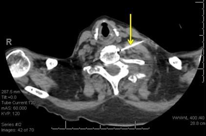
Figure 1. Panel A: PA view chest x-ray shows possible cavitation with air-fluid level in the right upper lobe (arrow). Panel B: lateral view chest x-ray shows possible cavitation with air-fluid level in the right upper lobe (arrow).


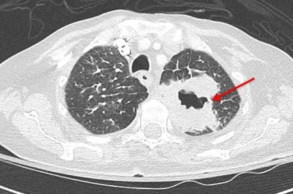
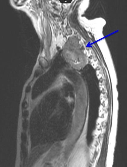
Figure 2. Coronal section of the thoracic CT scan shows focal dilation of the upper thoracic esophagus which contains fluid (arrow).


Figure 3.  Endoscopic view of the upper esophagus showing the diverticulum with impacted food bolus.


A 71-year-old man with history of recurrent aspiration pneumonia and previous esophageal surgery presented to the Emergency Department with acute hypoxia and leukocytosis. Imaging, above, showed a consolidation in the RUL and on lateral view an air fluid level. This was suspicious for infection or malignancy. For the ongoing concern for possible esophageal pathology given previous surgery, GI was consulted and upper endoscopy performed.  He was found to have an esophageal dilation at repair site of a previous Zenker’s diverticulum filled with food.

Zenker’s Diverticulum is a defect in the muscular wall of the hypopharynx in an area known as Killian's triangle. This condition is male predominant mainly occurring in the 3rd to 4th decade and/or the 7th to 8th decade of life.  The out pouching created will accumulate food and eventually lead to high incidences of aspiration pneumonia. Treatment is usually surgical in nature and can cause vocal cord damage and even recurrence of the outpouching (1).  

Chandra Stockdall MD and Roberto Swazo MD

Department of Internal Medicine

Banner University Medical Center South Campus

Tucson, AZ USA

Reference

  1. Mulder C, Van Delft F. Zenker’s diverticulum. UpToDate. May, 2017. Available at: http://www.uptodate.com/contents/zenkers-diverticulum (requires subscription, accessed 6/30/17).

Cite as: Stockdall C, Swazo R. Medical image of the week: Zenker's diverticulum. Southwest J Pulm Crit Care. 2017;15(1):15-6. doi: https://doi.org/10.13175/swjpcc075-17 PDF

Wednesday
Jun282017

Medical Image of the Week: Superior Sulcus Tumor with Neural Invasion

Figure 1. Computed tomography of the chest (axial image) shows a large left upper lobe cavitary mass (red arrow), consistent with known squamous cell carcinoma.


Figure 2. MRI of cervical and thoracic spine (sagittal image) reveals the mass abuts the spinal column with tumor invasion through the neural foramen at C7-T1 and T1-T2 (blue arrow).


Figure 3. A 22-guage needle is advanced with its tip anterior to the longus coli muscle at the level of C6 (yellow arrow). Ethanol solution was injected into this space.


A 78-year-old woman with left upper lobe squamous cell carcinoma presented with severe left arm and upper posterior chest pain. The pain was described as a severe burning sensation with “pins and needles”, and there was loss of motor function in the arm. This neuropathic pain was refractory to escalating doses of opioids and gabapentin. She was receiving chemotherapy with paclitaxel and carboplatin and completed five radiation treatments. On physical examination, there was atrophy of the left forearm and hand muscles. No evidence of Horner’s syndrome was noted.

A CT of the chest with contrast (Figure 1) showed a 5.8 cm apical segment left upper lobe cavitary mass consistent with a superior sulcus tumor and concomitant pulmonary embolism. An MRI of the cervical and thoracic spine (Figure 2) showed a large apical necrotic tumor abutting the upper thoracic spine with invasion of the neural foramina at C7-T1, T1-T2, and T2-T3, consistent with invasion into the brachial plexus.

In an attempt to improve her symptoms, the interventional radiologist performed a left stellate ganglion block with 1% lidocaine and 0.25% bupivacaine (Figure 3). There was minimal initial improvement so a repeat block was done three days later with notable reduction in arm pain. For a permanent block, a stellate ganglion block was performed with 2% lidocaine and 98% ethanol. The patient had significant palliation of the neuropathic pain in her left arm and shoulder.

Sue Cassidy ANP-BC ACHPN, Tina Skrepnik MD, Bree Johnston MD MPH, and Linda Snyder MD

University of Arizona College of Medicine

Departments of Internal Medicine and Radiation Oncology

Tucson, AZ USA

References

  1. Kratz JR, Woodard G, Jablons DM. Management of lung cancer invading the superior sulcus. Thorac Surg Clin. 2017 May;27(2):149-157. [CrossRef] [PubMed]
  2. De Leon-Casasola OA. Critical evaluation of chemical neurolysis of the sympathetic axis for cancer pain. Cancer Control. 2000 Mar-Apr;7(2):142-8. [PubMed]

Cite as: Cassidy S, Skrepnik T, Johnston B, Snyder L. Medical image of the week: superior sulcus tumor with neural invasion. Southwest J Pulm Crit Care. 2017;14(6):320-1. doi: https://doi.org/10.13175/swjpcc071-17 PDF