Search Journal-type in search term and press enter
Southwest Pulmonary and Critical Care Fellowships
In Memoriam

Imaging

Last 50 Imaging Postings

(Click on title to be directed to posting, most recent listed first)

May 2024 Medical Image of the Month: Hereditary Hemorrhagic
   Telangiectasia in a Patient on Veno-Arterial Extra-Corporeal Membrane
   Oxygenation
May 2024 Imaging Case of the Month: Nothing Is Guaranteed
April 2024 Medical Image of the Month: Wind Instruments Player Exhibiting
   Exceptional Pulmonary Function
March 2024 Medical Image of the Month: Sputum Cytology in Patients with
   Suspected Lung Malignancy Presenting with Acute Hypoxic Respiratory
   Failure
February 2024 Medical Image of the Month: Pulmonary Alveolar Proteinosis
   in Myelodysplastic Syndrome
February 2024 Imaging Case of the Month: Connecting Some Unusual Dots
January 2024 Medical Image of the Month: Polyangiitis Overlap Syndrome
   (POS) Mimicking Fungal Pneumonia 
December 2023 Medical Image of the Month: Metastatic Pulmonary
   Calcifications in End-Stage Renal Disease 
November 2023 Medical Image of the Month: Obstructive Uropathy
   Extremis
November 2023 Imaging Case of the Month: A Crazy Association
October 2023 Medical Image of the Month: Swyer-James-MacLeod
   Syndrome
September 2023 Medical Image of the Month: Aspergillus Presenting as a
   Pulmonary Nodule in an Immunocompetent Patient
August 2023 Medical Image of the Month: Cannonball Metastases from
   Metastatic Melanoma
August 2023 Imaging Case of the Month: Chew Your Food Carefully
July 2023 Medical Image of the Month: Primary Tracheal Lymphoma
June 2023 Medical Image of the Month: Solitary Fibrous Tumor of the Pleura
May 2023 Medical Image of the Month: Methamphetamine Inhalation
   Leading to Cavitary Pneumonia and Pleural Complications
April 2023 Medical Image of the Month: Atrial Myxoma in the setting of
   Raynaud’s Phenomenon: Early Echocardiography and Management of
   Thrombotic Disease
April 2023 Imaging Case of the Month: Large Impact from a Small Lesion
March 2023 Medical Image of the Month: Spontaneous Pneumomediastinum
   as a Complication of Marijuana Smoking Due to Müller's Maneuvers
February 2023 Medical Image of the Month: Reversed Halo Sign in the
   Setting of a Neutropenic Patient with Angioinvasive Pulmonary
   Zygomycosis
January 2023 Medical Image of the Month: Abnormal Sleep Study and PFT
   with Supine Challenge Related to Idiopathic Hemidiaphragmatic Paralysis
December 2022 Medical Image of the Month: Bronchoesophageal Fistula in
   the Setting of Pulmonary Actinomycosis
November 2022 Medical Image of the Month: COVID-19 Infection
   Presenting as Spontaneous Subcapsular Hematoma of the Kidney
November 2022 Imaging Case of the Month: Out of Place in the Thorax
October 2022 Medical Image of the Month: Infected Dasatinib Induced
   Chylothorax-The First Reported Case 
September 2022 Medical Image of the Month: Epiglottic Calcification
Medical Image of the Month: An Unexpected Cause of Chronic Cough
August 2022 Imaging Case of the Month: It’s All About Location
July 2022 Medical Image of the Month: Pulmonary Nodule in the
   Setting of Pyoderma Gangrenosum (PG) 
June 2022 Medical Image of the Month: A Hard Image to Swallow
May 2022 Medical Image of the Month: Pectus Excavatum
May 2022 Imaging Case of the Month: Asymmetric Apical Opacity–
   Diagnostic Considerations
April 2022 Medical Image of the Month: COVID Pericarditis
March 2022 Medical Image of the Month: Pulmonary Nodules in the
   Setting of Diffuse Idiopathic Pulmonary NeuroEndocrine Cell Hyperplasia
   (DIPNECH) 
February 2022 Medical Image of the Month: Multifocal Micronodular
   Pneumocyte Hyperplasia in the Setting of Tuberous Sclerosis
February 2022 Imaging Case of the Month: Between A Rock and a
   Hard Place
January 2022 Medical Image of the Month: Bronchial Obstruction
   Due to Pledget in Airway Following Foregut Cyst Resection
December 2021 Medical Image of the Month: Aspirated Dental Implant
Medical Image of the Month: Cavitating Pseudomonas
   aeruginosa Pneumonia
November 2021 Imaging Case of the Month: Let’s Not Dance
   the Twist
Medical Image of the Month: COVID-19-Associated Pulmonary
   Aspergillosis in a Post-Liver Transplant Patient
Medical Image of the Month: Stercoral Colitis
Medical Image of the Month: Bleomycin-Induced Pulmonary Fibrosis
   in a Patient with Lymphoma
August 2021 Imaging Case of the Month: Unilateral Peripheral Lung
   Opacity
Medical Image of the Month: Hepatic Abscess Secondary to Diverticulitis
   Resulting in Sepsis
Medical Image of the Month: Metastatic Spindle Cell Carcinoma of the
   Breast
Medical Image of the Month: Perforated Gangrenous Cholecystitis
May 2021 Imaging Case of the Month: A Growing Indeterminate Solitary
   Nodule
Medical Image of the Month: Severe Acute Respiratory Distress
Syndrome and Embolic Strokes from Polymethylmethacrylate
   (PMMA) Embolization
Medical Image of the Month: Pulmonary Aspergillus Overlap Syndrome
   Presenting with ABPA, Multiple Bilateral Aspergillomas
Medical Image of the Month: Diffuse White Matter Microhemorrhages
   Secondary to SARS-CoV-2 (COVID-19) Infection
February 2021 Imaging Case of the Month: An Indeterminate Solitary
   Nodule
Medical Image of the Month: Mucinous Adenocarcinoma of the Lung
   Mimicking Pneumonia
Medical Image of the Month: Superior Vena Cava Syndrome
Medical Image of the Month: Buffalo Chest Identified at the Time of
   Lung Nodule Biopsy
November 2020 Imaging Case of the Month: Cause and Effect?


For complete imaging listings click here

Those who care for patients with pulmonary, critical care or sleep disorders rely heavily on chest radiology and pathology to determine diagnoses. The Southwest Journal of Pulmonary and Critical Care publishes case-based articles with characteristic chest imaging and related pathology. The editor of this section will oversee and coordinate the publication of a core of the most important chest imaging topics. In doing so, they encourage the submission of unsolicited manuscripts. It cannot be overemphasized that both radiologic and pathologic images must be of excellent quality. As a rule, 600 DPI is sufficient for radiographic and pathologic images. Taking pictures of plain chest radiographs and CT scans with a digital camera is strongly discouraged. The figures should be cited in the text and numbered consecutively. The stain used for pathology specimens and magnification should be mentioned in the figure legend. Those who care for patients with pulmonary, critical care or sleep disorders rely heavily on chest radiology and pathology to determine diagnoses. The Southwest Journal of Pulmonary and Critical Care publishes case-based articles with characteristic chest imaging and related pathology. The editor of this section will oversee and coordinate the publication of a core of the most important chest imaging topics. In doing so, they encourage the submission of unsolicited manuscripts. It cannot be overemphasized that both radiologic and pathologic images must be of excellent quality. As a rule, 600 DPI is sufficient for radiographic and pathologic images. Taking pictures of plain chest radiographs and CT scans with a digital camera is strongly discouraged. The figures should be cited in the text and numbered consecutively. The stain used for pathology specimens and magnification should be mentioned in the figure legend.

-------------------------------------------------------------------------------------------  

Wednesday
Jul262017

Medical Image of the Week: Coral Reef Aorta

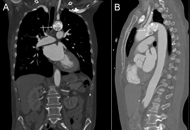
Figure 1. Coronal (A) and lateral (B) thoracic CT in soft tissue windows showing the coral reef calcification (arrows).


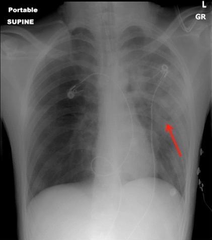
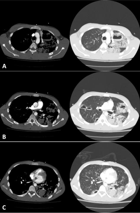
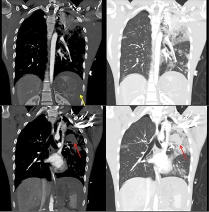
A 52-year-old woman with no past medical history presented to the emergency department with signs and symptoms concerning for pneumonia. Chest x-ray showed incidental findings of a calcified aortic mass. Subsequently, a follow up computed tomography scan (CT) was obtained which showed coral reef aorta (Figure 1). On physical examination, vital signs were only significant for mildly elevated blood pressure to 146/62 mmHg. She also had normal and equal pulses and pressures throughout all 4 extremities. In retrospect, patient had complaints of bilateral lower extremity claudication on strenuous exercise.

Coral reef aorta, a rare condition that was first described in 1984 by Qvarfordt et al. (1) is characterized by an eccentric, heavily calcified polypoid lesion and stenosis of the juxtarenal and suprarenal aorta. The rock-hard, irregular, gritty, whitish surface of the calcification strongly resembled a coral reef. The most common presentation is severe hypertension and intermittent claudication. Magnetic resonance angiogram (MRA) and CT have the ability to diagnose and appreciate the extent of this phenomenon (2).

Lance Eberson MS1 and Sehem Ghazala MD2

1College of Medicine and 2Department of Internal Medicine

University of Arizona

Tucson, Arizona, USA

References

  1. Qvarfordt PG, Reilly LM, Sedwitz MM, Ehrenfeld WK, Stoney RJ. "Coral reef" atherosclerosis of the suprarenal aorta: a unique clinical entity. J Vasc Surg. 1984 Nov;1(6):903-9. [CrossRef] [PubMed]
  2. Kopani K, Liao S, Shaffer K. The Coral Reef Aorta: Diagnosis and Treatment Following CT. Radiol Case Rep. 2016 Oct 4;4(1):209. eCollection 2009. [CrossRef] [PubMed] 

Cite as: Eberson L, Ghazala S. Medical image of the week: coral reef aorta. Southwest J Pulm Crit Care. 2017:15(1):49. doi: https://doi.org/10.13175/swjpcc080-17 PDF

Wednesday
Jul192017

Medical Image of the Week: Hematopneumatoceles from Pulmonary Lacerations

Figure 1. Chest x-ray showing irregular patchy regions of ill-defined consolidation in the left upper lobe and lingula, as well as suggestion of cystic changes (arrow).


Figure 2. Chest CT axial views, soft tissue and lung windows at the level of the aortic arch (A), right pulmonary artery (B) and the heart (C) showing mixed consolidative and nodular left lung opacities suggestive of pulmonary contusions, as well as contrecoup injury in the right lung, in addition to multiple cystic spaces containing air-fluid levels consistent with pulmonary lacerations.


Figure 3. Chest CT coronal views, soft tissue and lung windows showing consolidative and nodular lung opacities as well as fluid layering in cystic spaces (red arrows). A shattered spleen (yellow arrow) is also seen.


A 17-year-old man was brought to the emergency room after a fall from a 50-foot bridge. He was hypoxemic on presentation, requiring endotracheal intubation. Chest computed tomography (CT) revealed bilateral airspace opacities consistent with pulmonary contusions, and multiple air-fluid levels diagnostic of pulmonary lacerations (Figures 1-3).

Pulmonary lacerations are rare complications of blunt chest trauma (1). They can be contained within the lung parenchyma or may extend through the visceral pleura causing a pneumothorax. Due to its elastic recoil, the surrounding lung tissue pulls back from the laceration resulting in a round or oval cavity that may fill with air (pneumatocele), blood (hematocele) or both (hematopneumatocele). Lacerations are often obscured on chest x-ray as they are usually surrounded by contusion, requiring a CT for detection (2). They are classified into four types according to the mechanism of injury: Type 1 (compression rupture injury, most common type, usually centrally located), Type 2 (shearing against the thoracic spine, involving the paraspinal region of the lower lobes), Type 3 (rib penetration into the lung periphery, usually associated with a pneumothorax) and Type 4 (adhesion tear, in regions of pleuropulmonary adhesions) (3). Pulmonary lacerations heal more slowly than contusions and may last up to several months, over time becoming increasingly filled with blood, before regressing (2).

Our patient underwent an exploratory laparotomy with a splenectomy. The pulmonary lacerations were managed conservatively. He was successfully extubated on day#10 and discharged home on day#14 with a plan to follow his lacerations with monthly chest radiography.

Udit Chaddha MD1, Darren Maehara MD1, Ioan Puscas DO1, Ashley Prosper MD2, and Ramyar Mahdavi MD1

1Division of Pulmonary, Critical Care and Sleep Medicine and 2Department of Radiology

Keck School of Medicine of the University of Southern California

Los Angeles, CA USA

References

  1. Nishiumi N, Maitani F, Tsurumi T, Kaga K, Iwasaki M, Inoue H. Blunt chest trauma with deep pulmonary laceration. Ann Thorac Surg. 2001;71(1):314-8. [CrossRef] [PubMed]
  2. Kaewlai R, Avery LL, Asrani AV, Novelline RA. Multidetector CT of blunt thoracic trauma. Radiographics. 2008;28(6):1555-70. [CrossRef] [PubMed]
  3. Wagner RB, Crawford WO, Schimpf PP. Classification of parenchymal injuries of the lung. Radiology. 1988;167(1):77-82. [CrossRef] [PubMed]

Cite as: Chaddha U, Maehara D, Puscas I, Prosper A, Mahdavi R. Medical image of the week: hematopneumatoceles from pulmonary lacerations. Southwest J Pulm Crit Care. 2017;15(1):46-8. doi: https://doi.org/10.13175/swjpcc078-17 PDF