Search Journal-type in search term and press enter
Southwest Pulmonary and Critical Care Fellowships
In Memoriam

Imaging

Last 50 Imaging Postings

(Click on title to be directed to posting, most recent listed first)

May 2024 Medical Image of the Month: Hereditary Hemorrhagic
   Telangiectasia in a Patient on Veno-Arterial Extra-Corporeal Membrane
   Oxygenation
May 2024 Imaging Case of the Month: Nothing Is Guaranteed
April 2024 Medical Image of the Month: Wind Instruments Player Exhibiting
   Exceptional Pulmonary Function
March 2024 Medical Image of the Month: Sputum Cytology in Patients with
   Suspected Lung Malignancy Presenting with Acute Hypoxic Respiratory
   Failure
February 2024 Medical Image of the Month: Pulmonary Alveolar Proteinosis
   in Myelodysplastic Syndrome
February 2024 Imaging Case of the Month: Connecting Some Unusual Dots
January 2024 Medical Image of the Month: Polyangiitis Overlap Syndrome
   (POS) Mimicking Fungal Pneumonia 
December 2023 Medical Image of the Month: Metastatic Pulmonary
   Calcifications in End-Stage Renal Disease 
November 2023 Medical Image of the Month: Obstructive Uropathy
   Extremis
November 2023 Imaging Case of the Month: A Crazy Association
October 2023 Medical Image of the Month: Swyer-James-MacLeod
   Syndrome
September 2023 Medical Image of the Month: Aspergillus Presenting as a
   Pulmonary Nodule in an Immunocompetent Patient
August 2023 Medical Image of the Month: Cannonball Metastases from
   Metastatic Melanoma
August 2023 Imaging Case of the Month: Chew Your Food Carefully
July 2023 Medical Image of the Month: Primary Tracheal Lymphoma
June 2023 Medical Image of the Month: Solitary Fibrous Tumor of the Pleura
May 2023 Medical Image of the Month: Methamphetamine Inhalation
   Leading to Cavitary Pneumonia and Pleural Complications
April 2023 Medical Image of the Month: Atrial Myxoma in the setting of
   Raynaud’s Phenomenon: Early Echocardiography and Management of
   Thrombotic Disease
April 2023 Imaging Case of the Month: Large Impact from a Small Lesion
March 2023 Medical Image of the Month: Spontaneous Pneumomediastinum
   as a Complication of Marijuana Smoking Due to Müller's Maneuvers
February 2023 Medical Image of the Month: Reversed Halo Sign in the
   Setting of a Neutropenic Patient with Angioinvasive Pulmonary
   Zygomycosis
January 2023 Medical Image of the Month: Abnormal Sleep Study and PFT
   with Supine Challenge Related to Idiopathic Hemidiaphragmatic Paralysis
December 2022 Medical Image of the Month: Bronchoesophageal Fistula in
   the Setting of Pulmonary Actinomycosis
November 2022 Medical Image of the Month: COVID-19 Infection
   Presenting as Spontaneous Subcapsular Hematoma of the Kidney
November 2022 Imaging Case of the Month: Out of Place in the Thorax
October 2022 Medical Image of the Month: Infected Dasatinib Induced
   Chylothorax-The First Reported Case 
September 2022 Medical Image of the Month: Epiglottic Calcification
Medical Image of the Month: An Unexpected Cause of Chronic Cough
August 2022 Imaging Case of the Month: It’s All About Location
July 2022 Medical Image of the Month: Pulmonary Nodule in the
   Setting of Pyoderma Gangrenosum (PG) 
June 2022 Medical Image of the Month: A Hard Image to Swallow
May 2022 Medical Image of the Month: Pectus Excavatum
May 2022 Imaging Case of the Month: Asymmetric Apical Opacity–
   Diagnostic Considerations
April 2022 Medical Image of the Month: COVID Pericarditis
March 2022 Medical Image of the Month: Pulmonary Nodules in the
   Setting of Diffuse Idiopathic Pulmonary NeuroEndocrine Cell Hyperplasia
   (DIPNECH) 
February 2022 Medical Image of the Month: Multifocal Micronodular
   Pneumocyte Hyperplasia in the Setting of Tuberous Sclerosis
February 2022 Imaging Case of the Month: Between A Rock and a
   Hard Place
January 2022 Medical Image of the Month: Bronchial Obstruction
   Due to Pledget in Airway Following Foregut Cyst Resection
December 2021 Medical Image of the Month: Aspirated Dental Implant
Medical Image of the Month: Cavitating Pseudomonas
   aeruginosa Pneumonia
November 2021 Imaging Case of the Month: Let’s Not Dance
   the Twist
Medical Image of the Month: COVID-19-Associated Pulmonary
   Aspergillosis in a Post-Liver Transplant Patient
Medical Image of the Month: Stercoral Colitis
Medical Image of the Month: Bleomycin-Induced Pulmonary Fibrosis
   in a Patient with Lymphoma
August 2021 Imaging Case of the Month: Unilateral Peripheral Lung
   Opacity
Medical Image of the Month: Hepatic Abscess Secondary to Diverticulitis
   Resulting in Sepsis
Medical Image of the Month: Metastatic Spindle Cell Carcinoma of the
   Breast
Medical Image of the Month: Perforated Gangrenous Cholecystitis
May 2021 Imaging Case of the Month: A Growing Indeterminate Solitary
   Nodule
Medical Image of the Month: Severe Acute Respiratory Distress
Syndrome and Embolic Strokes from Polymethylmethacrylate
   (PMMA) Embolization
Medical Image of the Month: Pulmonary Aspergillus Overlap Syndrome
   Presenting with ABPA, Multiple Bilateral Aspergillomas
Medical Image of the Month: Diffuse White Matter Microhemorrhages
   Secondary to SARS-CoV-2 (COVID-19) Infection
February 2021 Imaging Case of the Month: An Indeterminate Solitary
   Nodule
Medical Image of the Month: Mucinous Adenocarcinoma of the Lung
   Mimicking Pneumonia
Medical Image of the Month: Superior Vena Cava Syndrome
Medical Image of the Month: Buffalo Chest Identified at the Time of
   Lung Nodule Biopsy
November 2020 Imaging Case of the Month: Cause and Effect?


For complete imaging listings click here

Those who care for patients with pulmonary, critical care or sleep disorders rely heavily on chest radiology and pathology to determine diagnoses. The Southwest Journal of Pulmonary and Critical Care publishes case-based articles with characteristic chest imaging and related pathology. The editor of this section will oversee and coordinate the publication of a core of the most important chest imaging topics. In doing so, they encourage the submission of unsolicited manuscripts. It cannot be overemphasized that both radiologic and pathologic images must be of excellent quality. As a rule, 600 DPI is sufficient for radiographic and pathologic images. Taking pictures of plain chest radiographs and CT scans with a digital camera is strongly discouraged. The figures should be cited in the text and numbered consecutively. The stain used for pathology specimens and magnification should be mentioned in the figure legend. Those who care for patients with pulmonary, critical care or sleep disorders rely heavily on chest radiology and pathology to determine diagnoses. The Southwest Journal of Pulmonary and Critical Care publishes case-based articles with characteristic chest imaging and related pathology. The editor of this section will oversee and coordinate the publication of a core of the most important chest imaging topics. In doing so, they encourage the submission of unsolicited manuscripts. It cannot be overemphasized that both radiologic and pathologic images must be of excellent quality. As a rule, 600 DPI is sufficient for radiographic and pathologic images. Taking pictures of plain chest radiographs and CT scans with a digital camera is strongly discouraged. The figures should be cited in the text and numbered consecutively. The stain used for pathology specimens and magnification should be mentioned in the figure legend.

-------------------------------------------------------------------------------------------  

Wednesday
Nov262014

Medical Image of the Week: Esophageal Cancer

Chandramohan Meenakshisundaram, MD 

Nanditha Malakkla, MD 


Saint Francis Hospital

Evanston, IL


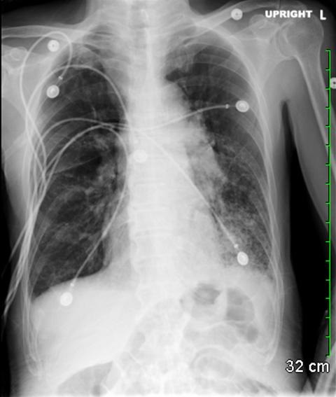
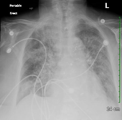
Figure 1. Admission chest x-ray showing hyper-aerated lung fields and consolidation at the left lung base.


Figure 2. Video of selected images from thoracic CT scan in soft tissue windows showing large mediastinal mass with extravasation of contrast.


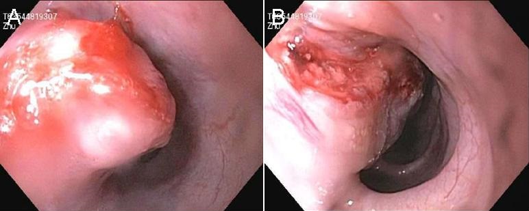
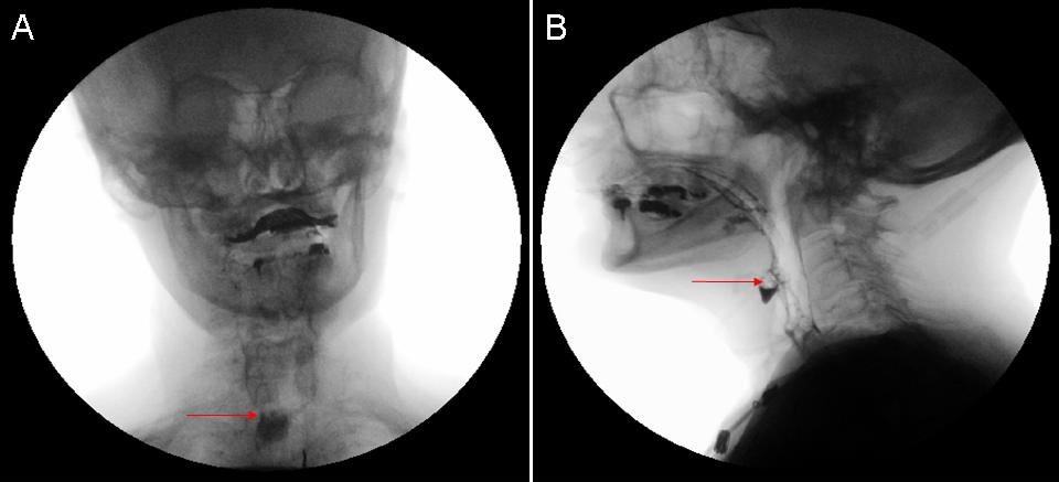
Figure 3. Views from endoscopy showing a large mass and the perforation site.

A 66-year-old Asian man with no significant past medical history was admitted with 1 week history of worsening retrosternal sharp chest pain, dyspnea on moderate exertion, and cough productive of brownish sputum. He also complained of some difficulty swallowing, decreased appetite and weight loss for the past 3 months. PPD was negative in the past. Vitals signs were significant for tachycardia and low grade fever. On physical exam he was cachectic and lung auscultation revealed bilateral scattered wheezes. Basic labs including complete blood count and comprehensive metabolic panel were unremarkable. EKG showed sinus tachycardia, serial troponin I was negative and chest x-ray revealed bilateral hyper-aerated lung fields and consolidation over the left lung base (Figure 1). He was started on antibiotics and bronchodilators. Since he continued to have chest pain and remained tachycardic, CT angiography of chest and abdomen (with IV and oral contrast) was done which revealed extravasation of contrast material into the mediastinum in the mid esophageal region representing rupture, a large mediastinal mass concerning for an abscess, and extensive infiltrates in the left lower lobe (Figure 2). During left thoracotomy, a large amount of necrotic material and phlegmon was seen in the mediastinum with adjacent area of lung necrosis which was drained and debrided. Upper GI endoscopy revealed a large mass in the mid-esophagus with perforation for which stenting was done (Figure 3). Pathology of phlegmon revealed squamous cell carcinoma with extensive necrosis. Culture of the fluid grew both viridians streptococci and anaerobes and he was started on broad spectrum antibiotics. His post-operative course was complicated and later he elected hospice care.

Reference as: Meenakshisundaram C, Malakkla N. Medical image of the week: eosphageal cancer. Southwest J Pulm Crit Care. 2014;9(5):295-6. doi: http://dx.doi.org/10.13175/swjpcc151-14 PDF

Wednesday
Nov192014

Medical Image of the Week: Killian-Jamieson Diverticulum

Figure 1. Chest x-ray showing worsening consolidation in both lungs.


Figure 2. Anterior (panel A) and lateral (panel B) fluoroscopic images showing retained contrast material in the anterior esophageal diverticulum in the hypopharynx.


An 89 year old female nursing home resident with a past medical history of hypertension and coronary artery disease was admitted with generalized weakness and vomiting for two days. Chest x-ray revealed consolidation in the left lung suggestive of pneumonia and she was started on broad spectrum antibiotics. Due to worsening consolidation in both lung fields (Figure 1) a video swallow was done for possible aspiration, which revealed contrast retained within the proximal esophagus within a diverticula in the anterior aspect (Figure 2). After excision of the diverticulum her pneumonia resolved and she was discharged back to the nursing home.

Killian-Jamieson diverticulum is a mucosal protrusion through a muscular gap in the anterolateral wall of the cervical esophagus; inferior to the cricopharyngeus and lateral to the longitudinal muscle of the esophagus just below its insertion on the posterior lamina of cricoid cartilage (gap also known as Killian-Jamieson space). This differentiates it from the Zenker’s diverticulum which arises from the muscular gap in the posterior portion of cricopharyngeus muscle (also known as Killian’s dehiscence). Killian-Jamieson diverticulum causes more non-specific symptoms than Zenker's diverticulum. Because these diverticula occur in close proximity to the recurrent laryngeal nerve, it should be carefully preserved during surgical resection.

Chandramohan Meenakshisundaram, MD and Nanditha Malakkla, MD

Medical Education

Saint Francis Hospital

Evanston, IL

References

  1. Kim DC, Hwang JJ, Lee WS, Lee SA, Kim YH, Chee HK. Surgical treatment of killian-jamieson diverticulum. Korean J Thorac Cardiovasc Surg. 2012;45(4):272-4. [CrossRef] [PubMed]
  2. Siddiq MA, Sood S, Strachan D. Pharyngeal pouch (Zenker's diverticulum). Postgrad Med J. 2001;77:506-11. [CrossRef] [PubMed]
  3. Brant WE, Helms CA. Fundamentals of Diagnostic Radiology. Lippincott Williams & Wilkins. (2007) ISBN:0781765188

Reference as: Meenakshisundaram C, Malakkla N. Medical image of the week: killian-jamieson diverticulum. Southwest J Pulm Crit Care. 2014;9(5):287-8. doi: http://dx.doi.org/10.13175/swjpcc134-14 PDF