Search Journal-type in search term and press enter
Southwest Pulmonary and Critical Care Fellowships
In Memoriam

Imaging

Last 50 Imaging Postings

(Click on title to be directed to posting, most recent listed first)

May 2024 Medical Image of the Month: Hereditary Hemorrhagic
   Telangiectasia in a Patient on Veno-Arterial Extra-Corporeal Membrane
   Oxygenation
May 2024 Imaging Case of the Month: Nothing Is Guaranteed
April 2024 Medical Image of the Month: Wind Instruments Player Exhibiting
   Exceptional Pulmonary Function
March 2024 Medical Image of the Month: Sputum Cytology in Patients with
   Suspected Lung Malignancy Presenting with Acute Hypoxic Respiratory
   Failure
February 2024 Medical Image of the Month: Pulmonary Alveolar Proteinosis
   in Myelodysplastic Syndrome
February 2024 Imaging Case of the Month: Connecting Some Unusual Dots
January 2024 Medical Image of the Month: Polyangiitis Overlap Syndrome
   (POS) Mimicking Fungal Pneumonia 
December 2023 Medical Image of the Month: Metastatic Pulmonary
   Calcifications in End-Stage Renal Disease 
November 2023 Medical Image of the Month: Obstructive Uropathy
   Extremis
November 2023 Imaging Case of the Month: A Crazy Association
October 2023 Medical Image of the Month: Swyer-James-MacLeod
   Syndrome
September 2023 Medical Image of the Month: Aspergillus Presenting as a
   Pulmonary Nodule in an Immunocompetent Patient
August 2023 Medical Image of the Month: Cannonball Metastases from
   Metastatic Melanoma
August 2023 Imaging Case of the Month: Chew Your Food Carefully
July 2023 Medical Image of the Month: Primary Tracheal Lymphoma
June 2023 Medical Image of the Month: Solitary Fibrous Tumor of the Pleura
May 2023 Medical Image of the Month: Methamphetamine Inhalation
   Leading to Cavitary Pneumonia and Pleural Complications
April 2023 Medical Image of the Month: Atrial Myxoma in the setting of
   Raynaud’s Phenomenon: Early Echocardiography and Management of
   Thrombotic Disease
April 2023 Imaging Case of the Month: Large Impact from a Small Lesion
March 2023 Medical Image of the Month: Spontaneous Pneumomediastinum
   as a Complication of Marijuana Smoking Due to Müller's Maneuvers
February 2023 Medical Image of the Month: Reversed Halo Sign in the
   Setting of a Neutropenic Patient with Angioinvasive Pulmonary
   Zygomycosis
January 2023 Medical Image of the Month: Abnormal Sleep Study and PFT
   with Supine Challenge Related to Idiopathic Hemidiaphragmatic Paralysis
December 2022 Medical Image of the Month: Bronchoesophageal Fistula in
   the Setting of Pulmonary Actinomycosis
November 2022 Medical Image of the Month: COVID-19 Infection
   Presenting as Spontaneous Subcapsular Hematoma of the Kidney
November 2022 Imaging Case of the Month: Out of Place in the Thorax
October 2022 Medical Image of the Month: Infected Dasatinib Induced
   Chylothorax-The First Reported Case 
September 2022 Medical Image of the Month: Epiglottic Calcification
Medical Image of the Month: An Unexpected Cause of Chronic Cough
August 2022 Imaging Case of the Month: It’s All About Location
July 2022 Medical Image of the Month: Pulmonary Nodule in the
   Setting of Pyoderma Gangrenosum (PG) 
June 2022 Medical Image of the Month: A Hard Image to Swallow
May 2022 Medical Image of the Month: Pectus Excavatum
May 2022 Imaging Case of the Month: Asymmetric Apical Opacity–
   Diagnostic Considerations
April 2022 Medical Image of the Month: COVID Pericarditis
March 2022 Medical Image of the Month: Pulmonary Nodules in the
   Setting of Diffuse Idiopathic Pulmonary NeuroEndocrine Cell Hyperplasia
   (DIPNECH) 
February 2022 Medical Image of the Month: Multifocal Micronodular
   Pneumocyte Hyperplasia in the Setting of Tuberous Sclerosis
February 2022 Imaging Case of the Month: Between A Rock and a
   Hard Place
January 2022 Medical Image of the Month: Bronchial Obstruction
   Due to Pledget in Airway Following Foregut Cyst Resection
December 2021 Medical Image of the Month: Aspirated Dental Implant
Medical Image of the Month: Cavitating Pseudomonas
   aeruginosa Pneumonia
November 2021 Imaging Case of the Month: Let’s Not Dance
   the Twist
Medical Image of the Month: COVID-19-Associated Pulmonary
   Aspergillosis in a Post-Liver Transplant Patient
Medical Image of the Month: Stercoral Colitis
Medical Image of the Month: Bleomycin-Induced Pulmonary Fibrosis
   in a Patient with Lymphoma
August 2021 Imaging Case of the Month: Unilateral Peripheral Lung
   Opacity
Medical Image of the Month: Hepatic Abscess Secondary to Diverticulitis
   Resulting in Sepsis
Medical Image of the Month: Metastatic Spindle Cell Carcinoma of the
   Breast
Medical Image of the Month: Perforated Gangrenous Cholecystitis
May 2021 Imaging Case of the Month: A Growing Indeterminate Solitary
   Nodule
Medical Image of the Month: Severe Acute Respiratory Distress
Syndrome and Embolic Strokes from Polymethylmethacrylate
   (PMMA) Embolization
Medical Image of the Month: Pulmonary Aspergillus Overlap Syndrome
   Presenting with ABPA, Multiple Bilateral Aspergillomas
Medical Image of the Month: Diffuse White Matter Microhemorrhages
   Secondary to SARS-CoV-2 (COVID-19) Infection
February 2021 Imaging Case of the Month: An Indeterminate Solitary
   Nodule
Medical Image of the Month: Mucinous Adenocarcinoma of the Lung
   Mimicking Pneumonia
Medical Image of the Month: Superior Vena Cava Syndrome
Medical Image of the Month: Buffalo Chest Identified at the Time of
   Lung Nodule Biopsy
November 2020 Imaging Case of the Month: Cause and Effect?


For complete imaging listings click here

Those who care for patients with pulmonary, critical care or sleep disorders rely heavily on chest radiology and pathology to determine diagnoses. The Southwest Journal of Pulmonary and Critical Care publishes case-based articles with characteristic chest imaging and related pathology. The editor of this section will oversee and coordinate the publication of a core of the most important chest imaging topics. In doing so, they encourage the submission of unsolicited manuscripts. It cannot be overemphasized that both radiologic and pathologic images must be of excellent quality. As a rule, 600 DPI is sufficient for radiographic and pathologic images. Taking pictures of plain chest radiographs and CT scans with a digital camera is strongly discouraged. The figures should be cited in the text and numbered consecutively. The stain used for pathology specimens and magnification should be mentioned in the figure legend. Those who care for patients with pulmonary, critical care or sleep disorders rely heavily on chest radiology and pathology to determine diagnoses. The Southwest Journal of Pulmonary and Critical Care publishes case-based articles with characteristic chest imaging and related pathology. The editor of this section will oversee and coordinate the publication of a core of the most important chest imaging topics. In doing so, they encourage the submission of unsolicited manuscripts. It cannot be overemphasized that both radiologic and pathologic images must be of excellent quality. As a rule, 600 DPI is sufficient for radiographic and pathologic images. Taking pictures of plain chest radiographs and CT scans with a digital camera is strongly discouraged. The figures should be cited in the text and numbered consecutively. The stain used for pathology specimens and magnification should be mentioned in the figure legend.

-------------------------------------------------------------------------------------------  

Friday
Jun032011

Ground-Glass Opacities

Reference as: Gopal V, Robbins RA. Ground-glass opacities. Southwest J Pulm Crit Care 2011;2:67-70. (Click here for PDF version)

A 54-year-old male was admitted to the medical intensive care unit complaining of abdominal pain, nausea, and vomiting for 2 days. He had a past medical history of pancreatitis in 2009, treated as outpatient, and asthma treated with albuterol inhaler as needed. His medication list included gemfibrizol, gabapentin, and amitriptyline. He drank 6-8 beers per day and smoked 1 pack-per-day for the past 40 years.

On physical examination is the patient was afebrile, his lungs are clear to auscultation, but tenderness was present in both lower quadrants. The remainder of the physical examination was normal.

Laboratory examination revealed a normal complete blood count and normal basic metabolic panel. Abnormal laboratory values included an elevated total bilirubin of 2.7 mg/dL (normal 0.2-1 mg/dL); alkaline phophatase 169 U/L (normal 10-40 U/L);  alanine aminotransferase 286 U/L (normal 10-35 U/L); amylase 468 U/L (normal 25-125 U/L), and lipase 1580 U/L (normal 8-78 U/L). Arterial blood gasmeasurements showed PaO2 = 91 mm Hg, PaCO2 = 26 mm Hg, pH = 7.52, and oxygen saturation = 98% while breathing room air.

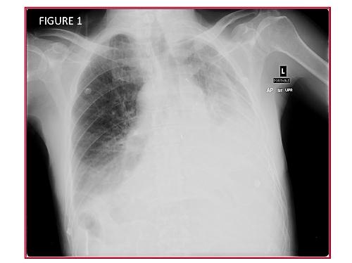
Chest radiography (Figure 1, Panel A) was interpreted as showing a “right upper lobe infiltrate which could represent an acute pneumonia”.  No distinct abnormalities were identified on abdominal radiographs (Figure 1, Panel B).


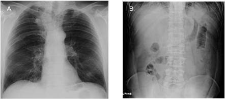
 Figure 1. Panel A. Frontal chest radiography.  Panel B. Abdominal radiography.

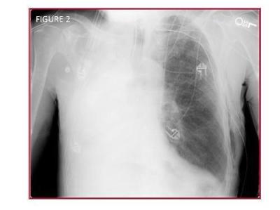
To further evaluate the possibility of a right upper lobe abnormality at chest radiography, thoracic CT was performed and as showing patchy ground-glass opacities throughout the lungs bilaterally (Figure 2).

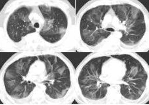
Figure 2. Representative images from thoracic CT.

Question 1. What’s the most likely diagnosis?

  1. Hypersensitivity pneumonitis
  2. Acute inhalational injury secondary to “huffing”.
  3. Drug-induced lung disease
  4. Valley Fever
  5. Ground-glass opacities associated with pancreatitis

Question 2. What would you do next?

  1. Hypersensitivity panel
  2. Bronchoscopy with bronchoalveolar lavage
  3. Begin Diflucan
  4. Broaden his antibiotic coverage
  5. Repeat the thoracic CT scan in 3-4 days.

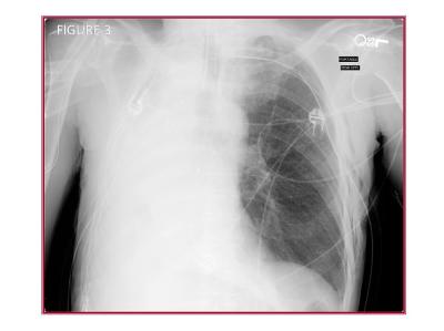
The thoracic CT was repeated four days later and the ground-glass opacities seen previously had largely resolved (Figure 3).

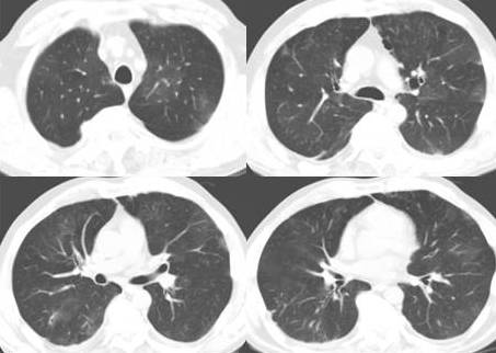
Figure 3.  Representative images from thoracic CT performed four days following the initial study Figure 2).

These ground-glass opacities likely represent subclinical non-cardiogenic pulmonary edema in the setting of acute pancreatitis. Ground-glass opacities are foci of increased lung attenuation that do not obscure underlying vessels or bronchial margins (1). Ground-glass opacities often represent parenchymal abnormalities below the spatial resolution of high-resolution CT of the lung. Although the differential diagnosis of ground-glass opacities at high-resolution CT is large, these etiologies may be broadly divided into acute or chronic causes. Table 1 lists some of the more common causes of ground-glass opacities at high-resolution CT.

Table 1: Common Etiologies for Ground-Glass Opacity at Thoracic CT

Acute

Chronic

Pulmonary edema (cardiogenic or non-cardiogenic)

Interstitial diseases (hypersensitivity pneumonitis, desquamative interstitial pneumonia, respiratory bronchiolitis-interstitial lung disease, nonspecific interstitial pneumonia, sarcoidosis, others)

Infectious pneumonitis (PJP, CMV, HSV, RSV, others)

Bronchoalveolar carcinoma

Noninfectious pneumonitis (hypersensitivity pneumonitis, acute inhalational exposures, drug-induced lung diseases)

Other causes (drug toxicity, pulmonary alveolar proteinosis, organizing pneumonia, chroic eosinophilic pneumonia, others)


Our patient had no apparent cause, other than subclinical non-cardiogenic pulmonary edema secondary to pancreatitis. Pulmonary edema is a well known complication of pancreatitis and can be severe (2). It seems likely that, as more sensitive methods for the detection of pulmonary abnormalities, such as thoracic CT, are increasingly applied to patients with pancreatitis, that subclinical pulmonary injury may be increasingly detected.

Venu Gopal, M.D.

Chief, Infectious Disease, Phoenix VA Medical Center


Richard A. Robbins, M.D.

Chief, Pulmonary and Critical Care, Phoenix VA Medical Center


Slide Set


References

  1. Miller WT Jr, Shah RM.  Isolated diffuse ground-glass opacity in thoracic CT: causes and clinical presentations.  AJR Am J Roentgenol 2005;184:613-22.
  2. Raghu MG, Wig JD, Kochhar R, Gupta D, Gupta R, Yadav TD, Agarwal R, Kudari AK, Doley RP, Javed A. Lung complications in acute pancreatitis. JOP. 2007;8:177-85.
Thursday
Dec302010

A Case of Mislabeled Identity

Reference as: Singarajah C, Park K. A case of mislabeled identity. Southwest J Pulm Crit Care 2010;1:22-27. (Click here for PDF version)

A 60-year-old man in the surgical intensive care unit for atrial fibrillation with rapid ventricular response, on his second post-operative day following colectomy, complained of worsening shortness of breath. A chest radiograph (Figure 1) was obtained. A chest radiograph performed one day previous to Figure 1 showed clear lungs, no pleural effusions, and no volume loss.

Question 1 and Figure 1: What are the abnormal findings on the chest radiograph? In particular, what technical error has occurred? 

The frontal chest radiograph shows increased opacity in the bases bilaterally, greater on the side labeled left (see “L” in the image- this is the technologist’s marker). Note the shift of the trachea towards the side labeled left. However, note also the opacity along the superior mediastinum on the right side; this opacity has the appearance of the aortic arch. Note the appearance of the stomach below the diaphragm, ipsilateral to the aortic arch. Also, the opacity at the left lung base shows a configuration resembling the heart. Taken together, these findings raise the possibility that the radiograph is mislabeled, with the “Left” marker (“L”, Figure 1) improperly placed on the patient’s right side. Prior chest radiographs not shown) confirm the patient did not have situs inversus.

There is evidence of volume loss in the right thorax (labeled incorrectly as left in this image). Note the shift of the trachea towards the side with increased lung opacity. The entire left right thorax (again, incorrectly labeled left in this image) is small, and the air column in the right mainstem bronchus abruptly terminated, suggesting endobronchial obstruction.

Furthermore, the patient had no clinical reasons for a new large pleural effusion, and recent prior films showed no pleural fluid.

Surgery was consulted and a procedure was performed. The results of this procedure are shown in Figure 2.

Question 2: What procedure was performed by the surgery team?

The surgery team improperly placed a thoracostomy tube in the left thorax because they misinterpreted Figure 1 as showing a large left pleural effusion. Figure 2 shows the tip of the thoracostomy tube in the medial superior left thorax, associated with subcutaneous emphysema. Progressive volume loss is seen on the right side, again suggesting endobronchial obstruction- note that the residual air in the right upper thorax in Figure 1 is no longer present in Figure 2. The surgery team then improperly placed a thoracostomy tube on the right side, mistaking the small, opacified right thorax for pleural effusion on that side. Figure 3 shows the new right thoracostomy tube tip located over the cranial right thorax.

The surgical team was concerned that the thoracostomy tube showed little fluid output and a second thoracotomy tube was placed on the contralateral side (Figure 3).


This tube also did not show significant output. The pulmonary / critical care medicine team was then consulted. The pulmonary / critical care medicine physician performed a procedure which partially corrected the cause of the patient’s original complaints. The chest radiograph following this procedure is shown in Figure 4. 

Question 3: What procedure (s) was performed by the pulmonary / critical care medicine team?

Bronchoscopy was performed, and showed significant mucous plugging. The mucous plugs were removed resulting in improved right lung aeration (Figure 4).

Learning Points:

  • Physical exam would have identified improperly labeled chest radiograph
  • The chest radiograph shows volume loss, suggesting endobronchial obstruction due to mucous plugging- the side of the thorax showing increased attenuation shows reduced volume. In contrast, pleural effusion would show increased opacity associated with mass effect and shift of the cardiomediastinal structures away from the side of the thorax showing increased attenuation
  • Time-outs are no substitute for clinical skills.The patient had two thoracostomy tubes, both placed for incorrect reasons, one of which was placed on the wrong side all together.
  • Therapy for atelectasis and mucous plugging includes the following:
    •  Patient mobilization, ambulation, sitting up;
    • Minimize anti-tussive meds (narcotics, etc), minimizes sedation;
    • Chest physiotherapy for the affected lung;
    • Continuous lateral rotation therapy for patients with altered mental status who cannot mobilize  (for example, a Triadyne bed  made by KCI or manual turning);
    • Bronchoscopy, and;
    • Mucolytics are of equivocal benefit


Clement Singarajah MD.  Associate Chief Pulmonary and Critical Care Fellowship, Phoenix VA Hospital and Good Samaritan Regional Medical Center, Phoenix AZ.

Kevin Park, MD, Pulmonary and Critical Care Medicine fellow, Phoenix VA Hospital and Good Samaritan Regional Medical Center, Phoenix AZ.

Email csingarajah@earthlink.net