Search Journal-type in search term and press enter
Southwest Pulmonary and Critical Care Fellowships
In Memoriam

Critical Care

Last 50 Critical Care Postings

April 2024 Critical Care Case of the Month: A 53-year-old Man Presenting
   with Fatal Acute Intracranial Hemorrhage and Cryptogenic Disseminated
   Intravascular Coagulopathy 
Delineating Gastrointestinal Dysfunction Variants in Severe Burn Injury
   Cases: A Retrospective Case Series with Literature Review
Doggonit! A Classic Case of Severe Capnocytophaga canimorsus Sepsis
January 2024 Critical Care Case of the Month: I See Tacoma
October 2023 Critical Care Case of the Month: Multi-Drug Resistant
   K. pneumoniae
May 2023 Critical Care Case of the Month: Not a Humerus Case
Essentials of Airway Management: The Best Tools and Positioning for 
   First-Attempt Intubation Success (Review)
March 2023 Critical Care Case of the Month: A Bad Egg
The Effect of Low Dose Dexamethasone on the Reduction of Hypoxaemia
   and Fat Embolism Syndrome After Long Bone Fractures
Unintended Consequence of Jesse’s Law in Arizona Critical Care Medicine
Impact of Cytomegalovirus DNAemia Below the Lower Limit of
Quantification: Impact of Multistate Model in Lung Transplant Recipients
October 2022 Critical Care Case of the Month: A Middle-Aged Couple “Not
   Acting Right”
Point-of-Care Ultrasound and Right Ventricular Strain: Utility in the
   Diagnosis of Pulmonary Embolism
Point of Care Ultrasound Utility in the Setting of Chest Pain: A Case of
   Takotsubo Cardiomyopathy
A Case of Brugada Phenocopy in Adrenal Insufficiency-Related Pericarditis
Effect Of Exogenous Melatonin on the Incidence of Delirium and Its 
   Association with Severity of Illness in Postoperative Surgical ICU Patients
Pediculosis As a Possible Contributor to Community-Acquired MRSA
Bacteremia and Native Mitral Valve Endocarditis
April 2022 Critical Care Case of the Month: Bullous Skin Lesions in
   the ICU
Leadership in Action: A Student-Run Designated Emphasis in
   Healthcare Leadership
MSSA Pericarditis in a Patient with Systemic Lupus
   Erythematosus Flare
January 2022 Critical Care Case of the Month: Ataque Isquémico
   Transitorio in Spanish 
Rapidly Fatal COVID-19-associated Acute Necrotizing
   Encephalopathy in a Previously Healthy 26-year-old Man 
Utility of Endobronchial Valves in a Patient with Bronchopleural Fistula in
   the Setting of COVID-19 Infection: A Case Report and Brief Review
October 2021 Critical Care Case of the Month: Unexpected Post-
   Operative Shock 
Impact of In Situ Education on Management of Cardiac Arrest after
   Cardiac Surgery
A Case and Brief Review of Bilious Ascites and Abdominal Compartment
   Syndrome from Pancreatitis-Induced Post-Roux-En-Y Gastric Remnant
   Leak
Methylene Blue Treatment of Pediatric Patients in the Cardiovascular
   Intensive Care Unit
July 2021 Critical Care Case of the Month: When a Chronic Disease
   Becomes Acute
Arizona Hospitals and Health Systems’ Statewide Collaboration Producing a
   Triage Protocol During the COVID-19 Pandemic
Ultrasound for Critical Care Physicians: Sometimes It’s Better to Be Lucky
   than Smart
High Volume Plasma Exchange in Acute Liver Failure: A Brief Review
April 2021 Critical Care Case of the Month: Abnormal Acid-Base Balance
   in a Post-Partum Woman
First-Attempt Endotracheal Intubation Success Rate Using A Telescoping
   Steel Bougie 
January 2021 Critical Care Case of the Month: A 35-Year-Old Man Found
   Down on the Street
A Case of Athabaskan Brainstem Dysgenesis Syndrome and RSV
   Respiratory Failure
October 2020 Critical Care Case of the Month: Unexplained
   Encephalopathy Following Elective Plastic Surgery
Acute Type A Aortic Dissection in a Young Weightlifter: A Case Study with
   an In-Depth Literature Review
July 2020 Critical Care Case of the Month: Not the Pearl You Were
   Looking For...
Choosing Among Unproven Therapies for the Treatment of Life-Threatening
   COVID-19 Infection: A Clinician’s Opinion from the Bedside
April 2020 Critical Care Case of the Month: Another Emerging Cause
   for Infiltrative Lung Abnormalities
Further COVID-19 Infection Control and Management Recommendations for
   the ICU
COVID-19 Prevention and Control Recommendations for the ICU
   Loperamide Abuse: A Case Report and Brief Review
Single-Use Telescopic Bougie: Case Series
Safety and Efficacy of Lung Recruitment Maneuvers in Pediatric Post-
   Operative Cardiac Patients
January 2020 Critical Care Case of the Month: A Code Post Lung 
   Needle Biopsy
October 2019 Critical Care Case of the Month: Running Naked in the
   Park
Severe Accidental Hypothermia in Phoenix? Active Rewarming Using 
   Thoracic Lavage
Left Ventricular Assist Devices: A Brief Overview


For complete critical care listings click here.

The Southwest Journal of Pulmonary and Critical Care publishes articles directed to those who treat patients in the ICU, CCU and SICU including chest physicians, surgeons, pediatricians, pharmacists/pharmacologists, anesthesiologists, critical care nurses, and other healthcare professionals. Manuscripts may be either basic or clinical original investigations or review articles. Potential authors of review articles are encouraged to contact the editors before submission, however, unsolicited review articles will be considered.

------------------------------------------------------------------------------------

Tuesday
Oct022012

October 2012 Critical Care Case of the Month

Henry Luedy, MD

Clement U. Singarajah, MD

Phoenix VA Medical Center

Phoenix, AZ

History of Present Illness

An 85 year old patient was admitted with hypotension and respiratory failure. He was intubated shortly after arrival and mechanical ventilation was begun. Fluids and vasopressors were begun for his hypotension.

PMH, SH, FH

His past medical history included peripheral vascular disease, abdominal aortic aneurysm repair, type 2 diabetes mellitus, hypertension, alcohol use, coronary artery disease, chronic obstructive pulmonary disease and hyperlipidemia.

Physical Examination

His vital signs were a temperature of 98.6 degrees F, heart rate 110 beats/min, respiratory rate 14 breaths per minute while intubated and receiving mechanical ventilation, and BP of 95/65 mmHg on vasopressors.

He was sedated. Lungs were clear and the heart had a regular rhythm without murmur or gallop. Abdominal examination was unremarkable and neurologic exam was limited because of sedation but without localizing signs. Plantar reflexes were down-going.

Admission Laboratory

Significant initial laboratory findings included a white blood cell count of 21,000 cells/μL, blood lactate level of 10 mmol/L and creatinine of 12 mg/dL. Urinanalysis showed pyuria and was positive for nitrates. At this time which of the following are diagnostic possibilities?

  1. Sepsis secondary to urinary tract infection (urosepsis)
  2. Community-acquired pneumonia
  3. Cardiogenic shock secondary to myocardial infarction
  4. Critical illness related corticosteroid insufficiency
  5. All of the above

Reference as: Luedy H, Singarajah CU. October 2012 critical care case of the month. Southwest J Pulm Crit Care 2012;5:179-85. PDF

Tuesday
Sep042012

September 2012 Critical Care Case of the Month

Robert A. Raschke, MD

Banner Good Samaritan Regional Medical Center

Phoenix, AZ

History of Present Illness

A 45 year old man was transferred from another medical center. He was found unresponsive, with muscle spasticity. After arrival at the outside medical center his vital signs were temperature 106.4 degrees F, heart rate 160 beats/min, respiratory rate 44 breaths per minute, and BP of 70/45 mm Hg. He was orally intubated for respiratory distress with induced by vecuronium.  His white blood cell count was 21,000 cells/μL. Chest x-ray showed bilateral consolidations and he was given fluids and gatifloxacin. His blood pressure improved to 130/94 and he was transferred.

PMH, SH, FH

He has a past medical history of quadriplegia at the C6 level with a history of severe back pain because syringomyelia.  He has a history of autonomic dysreflexia. Despite his disability he is quite functional working as a personal injury lawyer. He had been managed with a variety of medications including benzodiazepams, narcotics and baclofen. The later two were administered via an intrathecal pump which had been weaned over several weeks, and totally discontinued the day prior to admission. There is no history of smoking or alcohol abuse.

Physical Examination

His vital signs were temperature of 102.6 degrees F, heart rate 160 beats/min,  respiratory rate 14 breaths per minute, and BP of 130/50 mmHg.

He was paralyzed and mechanically ventilated. There was tenting of the skin and mottling of neck and knees. He had calloused hands and excoriated forearms. Lungs had diffuse rales and the heart rate was regular but rapid. A subcutaneous pump device was palpable in the left lower abdominal quadrant. There was a pressure sore on the coccyx.

Admission Laboratory and X-ray

His admission chest x-ray showed a diffuse 5-lobe consolidation. White blood cell count was elevated at 21,000 cells/μL.

At this time which of the following are diagnostic possibilities?

  1. Sepsis secondary to Staphylococcus aureus
  2. Pneumonia secondary to aspiration
  3. Neuroleptic malignant syndrome
  4. Benzodiazepam withdrawal
  5. All of the above

Reference as: Raschke RA. September 2012 critical care case of the month. Southwest J Pulm Crit Care 2012;5:121-5. (Click here for a PDF version)

Thursday
May102012

Correlation of Compliance with Central Line Associated Blood Stream Infection Guidelines and Outcomes: A Review of the Evidence

Jessica Hurley, MD1

Roxanne Garciaorr, MD1

Henry Luedy, MD1

Christan Jivcu, MD1

Emad Wissa, MD1

Joshua Jewell, MD1

Tonya Whiting, MD1

Richard Gerkin, MD1

Clement U. Singarajah, MD2

Richard A. Robbins, MD2

1Banner Good Samaritan Medical Center and 2Phoenix Pulmonary and Critical Care Medicine Research and Education Foundation, Phoenix, AZ

Abstract

Background 

Clinical practice guidelines are developed to assist in patient care but the evidence basis for many guidelines has been called into question.

Methods 

We conducted a literature review using PubMed and analyzed the overall quality of evidence and made strength of recommendation behind 8 Institute of Health Care (IHI) guidelines for prevention of central line associated blood stream infection (CLABSI). Quality of evidence was assessed by the American Thoracic Society (ATS) levels of evidence (levels I through III). We also examined data from our intensive care units (ICUs) for evidence of a correlation between guideline compliance and the development of VAP.

Results 

None of the guidelines was graded at level I. Two of the guidelines were graded at level II and the remaining 6 at level III. Despite the lack of evidence, 2 of the guidelines (hand hygiene, sterile gloves) were given a strong recommendation. Chlorhexidine and use of nonfemoral sites were given a moderate recommendation. In our ICUs compliance with the use of chlorhexidine correlated with a reduction in CLABSI (p<0.02) but the remainder did not.

Conclusions 

The IHI CLABSI guidelines are based on level II or III evidence. Data from our ICUs supported the use of chlorhexidine in reducing CLABSI. Until more data from well-designed controlled clinical trials become available, physicians should remain cautious when using current IHI guidelines to direct patient care decisions or as an assessment of the quality of care.

Introduction

The past three decades have seen the growth of numerous medical regulatory organizations. Many of these organizations have developed medical regulatory guidelines with over 10,000 listed under treatment/intervention in the National Guideline Clearinghouse (1). Many of these guidelines were rapidly adopted by healthcare organizations as a method to improve care. However, recent evidence suggests that many are based on opinion rather than randomized trials and most have not been shown to improve patient outcomes (2-5). We examined the IHI guidelines for prevention of CLABSI because these guidelines have been widely implemented despite what appeared to be a weak evidence basis (6). 

Methods

The study was approved by the Western Institutional Review Board.

Literature Search

In each instance PubMed was searched using central line associated blood stream infection which was cross referenced with each component of the CLABSI bundle (as modified by the Veterans Administration) using the following search terms: 1. hand hygiene; 2. cap (worn by inserter); 3. mask (worn by inserter); 4. sterile gloves (worn by inserter); 5. sterile gown (worn by inserter); 6. full body drape; 7. chlorhexidine used instead of povidone iodine (betadine); and 8. femoral sites not used. Additional studies were identified from the Related Citations in PubMed and the manuscript bibliographies. Each study was assessed for appropriateness to the guideline. Studies were required to be prospective and controlled in design. Only studies that used the incidence of CLABSI as a primary outcome measure were included in assessing the quality of evidence. 

Grading of level of evidence. The American Thoracic Society grading system was used to assess the underlying quality of evidence for the IHI CLABSI guidelines (7) (Table 1). A consensus was reached in each case. 

Table I. Levels of Evidence

Level of Evidence

     Definition


Level I (high)

Evidence from well-conducted, randomized controlled trials. 

Level II (moderate)


Evidence from well-designed, controlled trials without randomization (including cohort, patient series, and case-control studies). Level II studies also include any large case series in which systematic analysis of disease patterns was conducted, as well as reports of data on new therapies that were not collected in a randomized fashion.

Level III (low)


Evidence from case studies and expert opinion. In some instances, therapy recommendations come from antibiotic susceptibility data without clinical observations.


Strength of recommendations. Seven pulmonary and critical care fellows made strength of recommendations for each guideline. This was based not only the strength of evidence but also on clinical knowledge and judgment.

Guideline Compliance and CLABSI Incidence. We also assessed our ICUs for additional evidence of the effectiveness of the individual components of the CLABSI bundle. Data were collected monthly for a period of 50 months from January, 2007 through February, 2011. This was after the Veterans Administration requirements for CLABSI reporting and compliance was instituted. Diagnosis and compliance were assessed by a single quality assurance nurse using a standardized protocol (8). Compliance with each component of the bundle was analyzed individually and expressed as a percentage. This was correlated with the incidence of CLABSI during that month expressed in line-days. Each of the following was recorded by the quality assurance nurse: (1) line days, (2) number of CLABSI and (3) the number of audits or checklists completed during central line insertion and from those checklists (4) the number of times individual bundle practices were used including femoral location. In addition to data being kept locally, data was also entered into a centralized VA website. Entry into the website required completion of a learning session and a test correctly identifying CLABSI infections in case scenarios based on CDC definitions. The program included audits because the audit tool teaches critical bundle elements and facilitates communication about bundle adherence between team members.

Statistical analysis.  In some cases data were reanalyzed from original papers by Fisher’s exact test with a two-tailed comparison. For the data from our ICUs analyses were done using a Pearson correlation coefficient with a two-tailed test. Significance was defined as p<0.05. 

Results

Literature Review

Numbers of articles identified by PubMed search and used for grading the level of evidence and strength of recommendation are given in Table 2. Also included are the level of evidence and the strength of the recommendation.

Table 2.

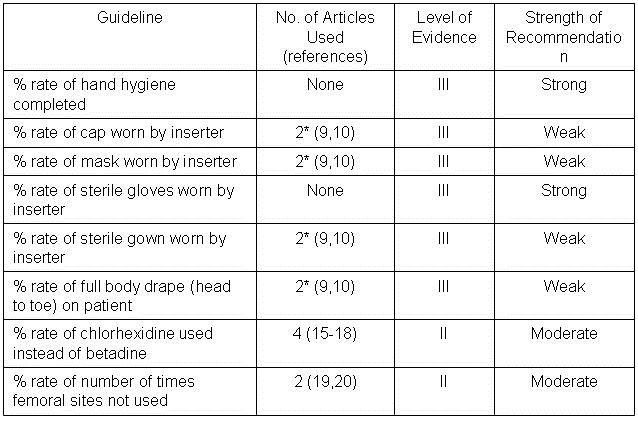
*Includes maximum barrier compared to standard barrier studies.

Barrier Protection. Five of the guidelines (cap worn by inserter; mask worn by inserter; sterile gown worn by inserter; and full body drape on patient) can be grouped under maximal barrier precautions as originally described by Raad et al. (9). This single site study compared 343 randomized patients to have nontunneled central catheters inserted under maximal sterile barrier precautions or control precautions (sterile gloves and small drape only). The catheters were for non-emergency venous access for chemotherapy and/or bone marrow transplantation. All patients were followed for 3 months and there were a total of four catheter infections in the test group and 12 in the control group (p= 0.03, chi-square test). However, examination of Table 3 of the manuscript revealed that 6 of the 12 in the control and 3 of the 4 in the test group had colonization rather than septicemia. Furthermore, of the remaining 7 patients only 2 developed septicemia within 30 days (both in the control group at 7 and 10 days). The remaining patients developed septicemia long after any expected benefit from barrier precautions during insertion (35-98 days). Development of septicemia this long after insertion and would seem more likely to represent contamination during handling of the catheter. Eliminating those subjects with colonization alone and septicemia after 30 days leaves 2 of 167 in the control and none of 176 in the maximum barrier precautions group. Recalculation reveals no statistically significant reduction by (p=0.24). Consistent with this reanalysis, a recent randomized, multicenter trial comparing maximum sterile barrier precautions vs. standard precautions reported by Ishikawa et al. (10) did not demonstrate a reduction in CLABSI. CLABSI developed in 5 of 211 patients with the use of maximum sterile barrier precautions compared to 6 of 213 patients with standard precautions (gloves and drape, p=1.00). Combining Raad’s revised data with Ishikawa’s data did not demonstrate a statistically significant difference between maximum and standard barrier precautions (p=0.42).  

Two other studies were considered. One was a prospective but observational study by Lee et al. (11). Data from this study demonstrate a lower rate of infection with maximum sterile precautions (p=0.02) but was excluded because of the nonrandomization. Another study by Rijnders et al. (12) was a randomized study comparing maximum barrier precautions and standard precautions but with arterial lines. Maximum barrier precautions did not significantly lower the infection rate (p>0.1) but the study was excluded because it dealt with arterial rather than central venous lines.

It is also important to note that CLABSI has been directly linked to the organisms growing on the skin at the insertion site. Carrer et al. (21) found maximal sterile barrier precautions, when compared to standard care, decreased skin colonization at the insertion site for the first 48 hours (39% vs. 69%). However, within 48 hours skin colonization was no different than at 5 days. Furthermore there was no statistical difference in device colonization found between the groups (p=0.10) indicating barrier precautions did not change the rate of CLABSI. Kim and colleagues (22) found the maximal barrier precautions successfully decreased the number of gram-positive infections (p=0.05) but actually increased fungal infections (p=0.04) while having no effect on gram-negative organisms. Given the natural flora of the skin is nearly all gram-positive organisms, sole reduction of gram-positive infections alone in this study reiterates the minimal overall effect maximal barrier precautions has on CLABSI in relation to the skin organisms at the central line insertion site and suggests CLABSI often occurs in the setting of future contamination post-insertion (22).

Based on the data and a lack of clear cut rationale into a mechanism of why maximum barrier precautions should reduce CLABSI, a weak recommendation was given to each of the components of maximum barrier precautions (cap, mask, gown and drape).

Hand hygiene and sterile gloves. The effects of hand hygiene and/or sterile gloves on the development of CLABSI have not been validated in randomized controlled trials. Observational studies have demonstrated a significant decrease in the incidence of nosocomial infections with improvements in hand hygiene and use of sterile gloves (13). Decreased mortality associated with the implementation of hand hygiene dates back to Semmelweis in 1847 (14). As the simplest and least expensive means of reducing CLABSI, hand hygiene and the use of sterile gloves were strongly recommended.

Chlorhexidine. One single institution study compared 492 arterial line insertions combined with 176 central venous catheters (15). Patients were randomized to povidone iodine (227 patients), alcohol (227 patients) and chlorhexidine (224 patients) for use in insertion as well as every 48 hour cleansing of the insertion site. Seven, six and one of the patients in each group developed bacteremia respectively. Chlorhexidine use resulted in a statistically significant reduction in bacteremia if compared to the combined povidone iodine and alcohol groups. However, statistical significance was lost when analyzed on a 3x2 table or comparing povidone iodine or alcohol with chlorhexidine individually (p>0.05, all comparisons).

Mimoz et al. (16) reported a single center study in both arterial and central venous line insertions. Patients were randomly assigned to either a solution composed of 0.25% chlorhexidine gluconate, 0.025% benzalkonium chloride, and 4% benzyl alcohol or 10% povidone iodine. The same solution was used for skin disinfection from the time of catheter insertion to the time of removal of each catheter. The use of the chlorhexidine containing solution was more efficacious in preventing line related sepsis compared to povidone iodine in preventing Gram + but not Gram negative infections but there was no overall reduction of CLABSI with chlorhexidine (3 CLABSI out of 170) compared to povidone-iodine (4 CLABSI out of 145).

Another multicenter prospective, randomized, controlled trial reported by Humar et al. (17) compared 0.5% tincture of chlorhexidine to 10% povidone-iodine as cutaneous antisepsis for central venous catheter in intensive care units. Four cases of documented catheter-related bacteremia out of 193 patients were found in the chlorhexidine group compared to 5 of 181 the povidone-iodine group (p>0.05).

Combining the above studies resulted in no significant reduction with the use of chlorhexidine (8 CLABSI out of 577) compared to povidone-iodine (15 CLABSI out of 553, p=0.14).

The results of a more recent 3-year, multi-institutional, interrupted time-series design (October 2006 to September 2009), with historical control data in the pediatric intensive care unit produced differing results (18). A nested, 18-month, nonrandomized, factorial design was used to evaluate chlorhexidine scrub and chlorhexidine-impregnated sponge compliance rates. Neither was associated with a reduction in CLABSI. Due to the results only being reported in CLABSI rate per 1000 line days it was not possible to combine the data with the other studies.

One of the randomized studies showed decreased CLABSI with chlorhexidine (15). Two of the three studies showed decreased colonization as well as decreased rates of line related sepsis while the third showed decreased exit site infections (all other findings of that study did not quite make significance). All three studies were in the ICU setting, two in surgical, evaluating both central venous catheters as well as arterial lines. In this setting with likely minimal cost difference, equal to better ease of use, and smaller studies of both venous and arterial lines, the strength of recommendation was judged as moderate.

Insertion Site. One randomized study compared infections using the femoral and internal jugular sites (19). In this study the rate of CLABSI did not differ (3/313 vs. 5/313). Another study compared the femoral and subclavian sites. It also did not show a reduction with a nonfemoral site (2/127 vs. 6/100) (20). Combining the two studies did not show a significantly lower infection rate with a nonfemoral site (7/440 nonfemoral vs. 9/413 femoral, p= 0.45). Two other nonrandomized studies examined femoral compared to subclavian and internal jugular sites (23,24). One did not show a difference between the sites (23). The other, larger study showed a lower rate with non-femoral sites (24). Three other studies were considered but were found to be nonrandomized (25-27). However, complications appear higher with the femoral route including thrombosis and hematomas (28,29).

The one observational study and the higher rate of non-infectious complications resulted in the group recommending a non-femoral site when possible. The strength of this recommendation was judged as moderate.

Guideline Compliance CLABSI Incidence. In our ICUs, 1133 audits representing 11470 line-days were assessed monthly (Appendix 1). An average of 1.3 CLABSI infections/1000 line-days occurred. Correlation between the monthly compliance with each component of the CLABSI bundle with the monthly CLABSI incidence revealed only chlorhexidine use was associated with reduced CLABSI (r=-0.35, p=0.01).

Discussion

This manuscript questions the validity of the CLABSI bundles as proposed by the IHI. We found that a systematic review of the literature revealed predominantly weak evidence to support these guidelines. Only one guideline (chlorhexidine) was supported by a randomized trial (15). However, data from our own ICUs showed a correlation between use of chlorhexidine and a reduction in CLABSI.

The diagnosis of CLABSI is difficult, requiring clinical judgment even in the presence of objective clinical criteria (8). The difficulty in diagnosis, along with the negative consequences for failure to follow the IHI guidelines, makes before and after comparisons of the incidence of CLABSI unreliable. Therefore, we sought evidence for the effectiveness of CLABSI prevention guidelines reasoning that the better the compliance with the guidelines, the lower the incidence of CLABSI. We were unable to show that improved CLABSI guideline compliance correlated with a reduced incidence of CLABSI with the exception of use of chlorhexidine.

Particularly disappointing is the data on maximum barrier precautions and reduction in CLABSI. The evidence presented in the first randomized trial was weak (9). Furthermore, when we carefully examined the data we found inclusion of catheter colonization and delays in diagnosis of over 30 days in the time from catheter insertion. It seems unlikely that contamination at the time of insertion would take over 30 days to present with sepsis. If the insertion technique was faulty (e.g., no barrier use) the infection should present within a matter of days not weeks. Many studies conflate catheter colonization with a true catheter related infection. The two entities are managed quite differently and thus need to be carefully separated (8). Reanalysis of the data eliminating the colonized patients and those who took over 30 days to present showed no reduction in CLABSI with maximum barrier protection. A more recent randomized, multicenter study would support the conclusion that there is no significant difference between maximum and standard barrier precautions (10).

We could find no randomized studies of hand hygiene and gloves in the context of CLABSI prevention. However, studies in the operating room and the intensive care unit have both demonstrated that hand hygiene decreases infection (13). Both have become standards of practice. Therefore, our group felt ardently that this should be a strong recommendation.

Use of povidone iodine or chlorhexidine is largely dependent on what is stocked at the time of central line insertion. We are unaware of data supporting physician preference for povidone iodine over chlorhexidine; in fact, our group almost universally prefers chlorhexidine. Although the evidence basis for chlorhexidine over povidone iodine is marginal, it seems reasonable to use chlorhexidine until the time that additional data are available, and therefore, chlorhexidine use was given a moderate recommendation.

The data using non-femoral sites showed no clear cut reduction in CLABSI. The femoral site may have advantages particularly in emergency situations including ease of placement, compressibility and being distant from the head and neck during resuscitation. However, it appears to come at a higher price of both hematomas and thrombosis (19,20). Based on this our group felt a moderate recommendation was justified in nonemergent situations.

Our study has several limitations. No literature review is totally comprehensive. It is possible that studies relevant to the IHI CLABSI guidelines, especially those written in a foreign language, were not identified. Second, the Phoenix VA data may be underpowered to show a small beneficial effect despite having over 11,000 line-days. Third, as with other healthcare facilities, the CLABSI guidelines at our institution were mandated and monitored. The threat of negative consequences may have compromised the objective assessment of the self-reported data, likely invalidating a before and after comparison. Fourth, correlation between guideline compliance and CLABSI incidence is not a substitute for a randomized trial. Unfortunately, the later is not possible given that guideline compliance is mandated.

In the above context, this report both confirms some aspects but differs in others from a recent report by the Veterans Administration (30). In this report the VA reported data from all ICUs and found a reduction in CLABSI and an increase in compliance from 2006-9. Although the database is much larger than the data in this report from a single institution, it suffers from the same weaknesses as our data. Not reported are the death or morbidity rates from CLABSI. Interestingly, CLABSI rates were much lower in smaller hospitals (level 4). Whether these hospitals had increased compliance was not reported, but these smaller hospitals are known to have higher all cause mortality, surgical mortality and surgical morbidity (31).

Guidelines have taken on the aura of law which is substituted for clinical judgment. For example, a nurse practitioner attempted to prevent a senior critical care physician in one of our facilities from inserting a central line because the physician was not wearing a cap. However, since the patient had no line access and was in extremis, the physician decided to proceed. Other examples are the decisions to place a femoral catheter during emergencies, when other venous access is unavailable, when a pneumothorax might be catastrophic, or when major bleeding is a risk (the femoral vein is compressible). Clinical judgment might weigh the risks of internal jugular or subclavian insertions compared to the femoral vein and conclude that the femoral site might be the best choice for the patient.

It is unclear why the IHI guidelines have received such wide acceptance given their weak evidence basis. Agencies involved in guideline writing should show restraint in guideline formulation based on opinion or weak or conflicting evidence. Only those interventions based on strong evidence which can make a real difference to patients should be designated as guidelines.

Acknowledgements

The authors acknowledge Janice Allen, MSN, RN who collected the CLABSI data reported from the Phoenix VA.

References

  1. http://www.guideline.gov/ Accessed 12-1-2011.
  2. Lee DH, Vielemeyer O. Analysis of overall level of evidence behind infectious diseases society of America practice guidelines. Arch Intern Med 2011;171:18-22.
  3. Kett DH, Cano E, Quartin AA, Mangino JE, Zervos MJ, Peyrani P, Cely CM, For KD, Scerpella EG, Ramirez JA. Implementation of guidelines for management of possible multidrug-resistant pneumonia in intensive care: an observational, multicentre cohort study. Lancet Infect Dis 2011;11:181-9.
  4. Padrnos L, Bui T, Pattee JJ, Whitmore EJ, Iqbal M, Lee S, Singarajah CU, Robbins RA. Analysis of overall level of evidence behind the Institute of Healthcare Improvement ventilator-associated pneumonia guidelines. Southwest J Pulm Crit Care 2011;3:40-8.
  5. Robbins RA, Gerkin R, Singarajah CU. Relationship between the Veterans Healthcare Administration Hospital Performance Measures and Outcomes. Southwest J Pulm Crit Care 2011;3:92-133.
  6. http://www.ihi.org/explore/centrallineinfection/Pages/default.aspx (accessed 12-1-11).
  7. Schünemann H, Jaeschke R, Cook DJ, et al. An Official ATS Statement: Grading the Quality of Evidence and Strength of Recommendations in ATS Guidelines and Recommendations. Am J Resp Crit Care Med 2006;174:605-14.
  8. Horan TC, Andrus M, Dudeck MA. CDC/NHSN surveillance definition of health care-associated infection and criteria for specific types of infections in the acute care setting. Am J Infect Control 2008;36:309-32.
  9. Raad II, Hohn DC, Gilbreath BJ, Suleiman N, Hill LA, Bruso PA, Marts K, Mansfield PF, Bodey GP. Prevention of central venous catheter-related infections by using maximal sterile barrier precautions during insertion. Infect Control Hosp Epidemiol 1994;15:231-8.
  10. Ishikawa Y, Kiyama T, Haga Y, Ishikawa M, Takeuchi H, Kimura O, Harihara Y, Sunouchi K, Furuya T, Kimura M. Maximal sterile barrier precautions do not reduce catheter-related bloodstream infections in general surgery units: a multi-institutional randomized controlled trial. Ann Surg 2010 ;251:620-3.
  11. Lee D-h, Jung KY, Choi Y-H. Use of maximal sterile barrier precautions and/or antimicrobial- coated catheters to reduce the risk of central venous catheter–related bloodstream infection. Infect Control Hosp Epidemiol 2008;29:947-50.
  12. Rijnders BJ, Van Wijngaerden E, Wilmer A, Peetermans WE. Use of full sterile barrier precautions during insertion of arterial catheters: a randomized trial. Clin Infect Dis 2003;36:743-8.
  13. Cork DP, Maxwell PJ 4th, Yeo CJ. Remembering Semmelweis: hand hygiene and its importance on today's clinical practice. Am Surg 2011;77:123-5.
  14. Maki DG, Ringer M, Alvarado CJ. Prospective randomised trial of povidone-iodine, alcohol, and chlorhexidine for prevention of infection associated with central venous and arterial catheters. Lancet. 1991;338:339-43.
  15. Mimoz O, Pieroni L, Lawrence C, Edouard A, Costa Y, Samii K, et al. Prospective, randomized trial of two antiseptic solutions for prevention of central venous or arterial catheter colonization and infection in intensive care unit patients. Crit Care Med 1996;24:1818-23.
  16. Humar A, Ostromecki A, Direnfeld J, Marshall JC, Lazar N, Houston PC, Boiteau P, Conly JM. Prospective randomized trial of 10% povidone-iodine versus 0.5% tincture of chlorhexidine as cutaneous antisepsis for prevention of central venous catheter infection. Clin Infect Dis. 2000;31:1001-7.
  17. Parienti JJ, Thirion M, Mégarbane B, Souweine B, Ouchikhe A, Polito A, Forel JM, Marqué S, Misset B, Airapetian N, Daurel C, Mira JP, Ramakers M, du Cheyron D, Le Coutour X, Daubin C, Charbonneau P; Members of the Cathedia Study Group. Femoral vs jugular venous catheterization and risk of nosocomial events in adults requiring acute renal replacement therapy: a randomized controlled trial. JAMA 2008;299:2413-22.
  18. Merrer J, De Jonghe B, Golliot F, Lefrant JY, Raffy B, Barre E, Rigaud JP, Casciani D, Misset B, Bosquet C, Outin H, Brun-Buisson C, Nitenberg G; French Catheter Study Group in Intensive Care. Complications of femoral and subclavian venous catheterization in critically ill patients: a randomized controlled trial. JAMA 2001;286:700-7.
  19. Carrer S, Bocchi A, Bortolotti M, Braga N, Gilli G, Candini M, Tartari S. Effect of different sterile barrier precautions and central venous catheter dressing on the skin colonization around the insertion site. Minerva Anestesiol 2005;71:197-206.
  20. Kim JS. Holtom P. Vigen C. Reduction of catheter-related bloodstream infections through the use of a central venous line bundle: epidemiologic and economic consequences. American Journal of Infection Control 2011;39:640-6.
  21. Deshpande KS, Hatem C, Ulrich HL, Currie BP, Aldrich TK, Bryan-Brown CW, et al. The incidence of infectious complications of central venous catheters at the subclavian, internal jugular, and femoral sites in an intensive care unit population. Critical Care Medicine 2005;33:13-20.
  22. Lorente L, Henry C, Martin MM, Jimenez A, Mora ML. Central venous catheter-related infection in a prospective and observational study of 2,595 catheters. Critical Care (London, England) 2005;9:R631-5.
  23. Casanegra AI, Brannan S, Dadu R, Ong P, Berner M, Spinning K, Hughes T, Marrinan G, Zinn K, Manthous C. Short-term femoral vein catheterization rarely causes thrombosis or bacteremia. J Hosp Med 2011;6:33-6.
  24. Hsu JF, Tsai MH, Huang HR, Lien R, Chu SM, Huang CB. Risk factors of catheter-related bloodstream infection with percutaneously inserted central venous catheters in very low birth weight infants: a center's experience in Taiwan. Pediatr Neonatol 2010;51:336-42.
  25. Tsai MH, Lien R, Wang JW, Huang HR, Chiang CC, Chu SM, Hsu JF, Huang YC. Complication rates with central venous catheters inserted at femoral and non-femoral sites in very low birth weight infants. Pediatr Infect Dis J 2009;28:966-70.
  26. Trottier SJ, Veremakis C, O’Brien J, Auer AI. Femoral deep vein thrombosis associated with central venous catheterization: results from a prospective, randomized trial. Crit Care Med 1995;23:52-9.
  27. Ge X, Cavallazzi R, Li C, Pan SM, Wang YW, Wang FL. Central venous access sites for the prevention of venous thrombosis, stenosis and infection. Cochrane Database Syst Rev 2012;3:CD004084.
  28. Render ML, Hasselbeck R, Freyberg RW, Hofer TP, Sales AE, Almenoff PL. Reduction of central line infections in Veterans Administration intensive care units: an observational cohort using a central infrastructure to support learning and improvement. BMJ Qual Saf 2011;20:725-732.
  29. Robbins RA, Gerkin R, Singarajah CU. Relationship between the veterans healthcare administration hospital performance measures and outcomes. Southwest J Pulm Crit Care 2011;3:92-133.

Refernce as: Hurley J, Garciaorr R, Luedy H, Jivcu C, Wissa E, Jewell J, Whiting T, Gerkin R, Singarajah CU, Robbins RA. Correlation of compliance with central line associated blood stream infection guidelines and outcomes: a review of the evidence. Southwest J Pulm Crit Care 2012;4:163-73. (Click here for a PDF version of manuscript)

Tuesday
Oct182011

Critical Care Review: the High Price of Sugar

Reference as: Robbins RA, Singarajah CU. Critical care review: the high price of sugar. Southwest J Pulm Crit Care 2011;3: 78-86. (Click here for PDF version)

Richard A. Robbins, MD

Clement U. Singarajah, MD

The Phoenix Pulmonary and Critical Care Research and Education Foundation, Phoenix, AZ

Abstract

The intensive control of blood glucose had been proposed to be important in increasing survival in the intensive care unit (ICU) despite only one positive randomized trial. The concept was supported by guidelines released by several regulatory organizations including the Joint Commission of Healthcare Organizations and the Institute of Healthcare Improvement. However, the large, randomized, multi-center NICE-SUGAR trial published in 2009 showed tight control of glucose in the ICU is actually hazardous with a 14% increase in mortality. The historical basis and data used to support intense control of glucose in the ICU are reviewed and illustrate the harm that can result when guidelines are based on weak evidence.

Intensive Control of Glucose in Diabetes

Diabetes has long been associated with vascular complications. These are divided into microvascular complications (retinopathy, nephropathy, and neuropathy) and macrovascular complications (coronary artery disease, stroke, and peripheral vascular disease). The concept that intense control of glucose results in improved vascular outcomes in diabetes dates back decades but has been plagued with controversy. The University Group Diabetes Program Study (UGDPS), which began in 1959, was designed to evaluate the relationship between blood sugar control and vascular complications in patients with newly diagnosed type II diabetes. The investigators found that control of blood sugar levels was ineffective in preventing the micro- and macrovascular complications associated with diabetes, regardless of the type of therapy (1). This prompted the American Diabetes Association (ADA) and the American Medical Association to withdrawn their support of UDGPS (2). In 1978, at a meeting of diabetes researchers, clinicians, and epidemiologists from the ADA, the National Institutes of Health (NIH), the Centers for Disease Control, and various university centers, it was concluded that there was “no definite evidence that treatment to regulate blood sugar levels is effective beyond relieving symptoms and controlling acute metabolic disturbances” (2).

This controversy prompted the NIH to organize the Diabetes Control and Complications Trial. This was a large, multi-center, randomized study which compared intensive to conventional treatment in preventing vascular complications in insulin-dependent, type I diabetics. Published in 1993, the results of this trial demonstrated that intensive therapy effectively delayed the onset and slowed the progression of diabetic retinopathy, nephropathy, and neuropathy in patients with insulin-dependent diabetes (3). However, the mortality rate, incidence of macrovascular complications, and incidence of diabetic ketoacidosis were not significantly reduced. Weight gain and episodes of hypoglycemia were significantly more common in the intensive therapy group.

Published in 1998 but started in 1977, the UK Prospective Diabetes Study (UKPDS) was designed to determine if intensive blood glucose control reduced the risk of micro- or macrovascular complications in type II diabetes (4). This study is important since over 90% of adult diabetics, including the majority of diabetics in an adult ICU, have type II diabetes. This large, multi-center, randomized study compared conventional therapy with diet alone to an intense glucose control with diet and either a sulphonylurea (chlorpropamide, glibenclamide, or glipizide) or insulin. The goals of the study were to maintain fasting blood glucose of less than 270 mg/dL (15 mmol/L) in the conventional group and less than 108 mg/dL (6 mmol/L) in the intensive control group. Consistent with the blood sugar goals of the study, the hemoglobin A1C was reduced in the intensive therapy group compared to the conventional group (7.0% vs. 7.9%, p<0.05). The results in this study of type II diabetics were similar to the Diabetes Control and Complications Trial in type I diabetics. Microvascular complications, particularly retinal complications, were significantly reduced in the intensive therapy group but macrovascular complications were not. Mortality was not reduced and hypoglycemia and weight gain were more common in the intensive therapy group.

Intensive Control of Blood Glucose in the ICU

Hyperglycemia associated with insulin resistance is common in critically ill patients, even those who have not previously had diabetes (5-7). It had been reported that pronounced hyperglycemia might lead to complications. For example, studies reported that in acute myocardial infarction therapy to maintain blood glucose below 215 mg /dL (11.9 mmol/L) improved long-term outcomes (8-10). Furthermore, high serum levels of insulin-like growth factor-binding protein 1, which reflect insulin resistance, increase the risk of death (11, 12).

Spurred by the above data and the overwhelming opinion of diabetes experts that intensive control of glucose improves outcomes in diabetes and should in the ICU, van den Berge et al. (13) compared intensive insulin therapy (maintenance of blood glucose at a level between 80 and 110 mg/dL) to conventional treatment (infusion of insulin only if the blood glucose level exceeded 215 mg/dL and maintenance of glucose at a level between 180 and 200 mg/dL) in ICU patients. The study was large with 1548 subjects but was a single center study from a surgical intensive care unit with 63% of the patients post-cardiac surgery. Reported in 2001, the results showed that intensive insulin therapy reduced mortality during intensive care from 8.0 percent with conventional treatment to 4.6 percent (p<0.04). The benefit of intensive insulin therapy was attributable to its effect on mortality among patients who remained in the intensive care unit for more than five days (20.2 percent with conventional treatment, as compared with 10.6 percent with intensive insulin therapy; p=0.005).

The results of van den Berge’s original study were supported by a nonrandomized, single center study reported by Krinsley (14) in 2004. This study from a combined 14 bed medical/surgical ICU consisted of 800 consecutive patients after initiation of a intensive control protocol compared to 800 patients admitted immediately preceding initiation, i.e., a before and after design. The protocol involved intensive monitoring and treatment to maintain plasma glucose values lower than 140 mg/dL. Hospital mortality decreased 29.3% (p=0.002), and length of stay in the ICU decreased 10.8% (p=0.01) with intensive control of glucose. Despite the before and after comparison, some considered this single center study as confirmatory evidence for the mortality benefit of intensive glucose control.

It has been pointed out that van den Berge’s study had multiple limitations (15). Van den Berge’s 2001 study was a non-blinded, single center and including predominately patients after cardiac surgery, Other limitations included the unusual practices of most patients receiving intravenous glucose on arrival at the intensive care unit (ICU) at 200 to 300 g/d (the equivalent of 2-3 L of 10% glucose per day) and initiation of total parenteral nutrition, or enteral feeding, or combined feeding for all patients within 24 hours. Also, the mortality of cardiac surgery patients in the control group was 5.1% which is unacceptably high in most centers.

Kringsley’s study also had limitations (15). This was a single-center, retrospective, unblinded study and likely reflect a powerful Hawthorne effect (intense glucose control = investigator commitment and bedside presence, more tests, more attention, more patient visits, more interventions, and overall better care). Intensive insulin therapy comes at a substantial price: a greater than 6-fold increase in the risk of hypoglycemia and a marked increase in bedside nurse workload.

When many regulatory guidelines were initiated in the mid 2000’s,  not all data about glucose control and insulin in acute illness pointed to a benefit. The Diabetes Mellitus, Insulin Glucose Infusion in Acute Myocardial Infarction (DIGAMI) 2 study with more than 1000 randomized patients with myocardial infarction to intense compared to conventional glucose control failed to show a mortality benefit (16). Similarly, the Reviparin and Metabolic Modulation in Acute Myocardial Infarction Treatment Evaluation (CREATE)-Estudios Cardiologicas Latin America Study Group (ECLA) study with over 20,000 randomized patients with myocardial infarction failed to show a benefit of a glucose, insulin and potassium infusion regimen compared to usual care (17).

Regulatory Guidelines

By 2005 the Joint Commission on Accreditation of Healthcare Organization (Joint Commission) and the Institute for Healthcare Improvement (IHI) recommended tight glucose control for the critically ill as a core quality of care measure for all U.S. hospitals (18). Furthermore, an international initiative by several professional societies, including the American Thoracic Society, promoted a care “bundle” for severe sepsis that also includes intensive glycemic control.

Concerns about Intensive Glucose Control in the ICU

The medical literature is rife with initially positive trials followed by studies with equivocal or negative trials and occasionally followed by studies with actual harm to patients (19). Intensive control of glucose is a good example of this progression in medical research.

In late 2005, editorials urged waiting on further studies before widespread implement of tight control of glucose as usual care in the ICU. Bellomo and Egi (17) recommended awaiting the results of two large multi-center, randomized trials of tight control of glucose in the ICU, the GluControl study and the NICE SUGAR study. Angus and Abraham (18) echoed the limitations of van den Berge’s study and also advocated caution in the widespread initiation of intensive glucose control in the ICU.

Van den Berge’s group that initially reported the positive results in surgical ICU patients followed their 2001 publication with a report of medical ICU patients in 2006 (20).  In this prospective, randomized study of adult patients admitted to the medical ICU, the authors were unable to reproduce the reduction of in-hospital mortality with intensive glucose control seen in their surgical ICU patients (40.0 vs. 37.3% mortality, p= 0.33). However, the authors reported a significant improvement in morbidity with a reduction in newly acquired kidney injury, accelerated weaning from mechanical ventilation, and accelerated discharge from the ICU and the hospital. However, among the 433 patients who stayed in the medical ICU for less than three days, mortality was greater among those receiving intensive insulin therapy.  Since the mean length of stay in our medical intensive care at the Phoenix VA was a little less than 3 days, many of our group became concerned that intensive control of glucose would not improve mortality and might actually prove harmful.

The GluControl study was undertaken in 2004 to test the hypothesis that intensive control of glucose (80-110 mg/dL) improves survival of patients treated in  medical/surgical intensive care units (ICU) compared to a control target of 140-180 mg/dL. Planned enrollment was 3500 subjects but the trial was stopped in 2006 after a little over 1000 subjects because interim analysis revealed numerous protocol violations resulting in hypoglycemia. The results were initially reported as an abstract at the 20th Congress of the European Society of Intensive Care in 2008 and a full length manuscript was published in 2009 (21,22). ICU, 28-day and hospital mortality were similar in both groups. ICU and hospital length of stay were identical. Hypoglycemia defined as a blood glucose below 40 mg/dL was seen in 8.7% of the intensive therapy group vs. 2.7% in the conventional group.

Further concern about the concept of intensive glucose control was raised by Weiner et al. (23) in 2008. They searched the medical literature (MEDLINE, the Cochrane Library, clinical trial registries, reference lists, and abstracts from conferences from both the American Thoracic Society and the Society of Critical Care Medicine) and identified 29 randomized controlled trials totaling 8432 patients.  A meta-analysis did not reveal a significant difference between intensive glucose control and usual care overall (21.6% vs. 23.3%) but did reveal an increased risk of hypoglycemia (glucose ≤40 mg/dL, 13.7% vs. 2.5%). In fact, the only study that showed a mortality advantage was van den Berge’s original study in 2001.

The NICE SUGAR Study

The landmark NICE SUGAR study (24) was published in the spring of 2009. This large study randomized 6104 patients to either intensive glucose control, with a target blood glucose range of 81 to 108 mg/dL, or conventional glucose control, with a target of <180 mg/dL. The main finding of the study was that intensive glucose control resulted in a 14% increase in morality. Furthermore, the adverse treatment effect on mortality did not differ significantly between surgical patients and medical patients. As in previous trials, severe hypoglycemia (blood glucose level ≤40 mg /dL) was significantly more common in the intensive-control group (6.8%) compared to the conventional-control group (0.5%, p<0.001). There was no significant difference between the two treatment groups in the median number of days in the ICU or hospital, the median number of days of mechanical ventilation or days of  renal-replacement therapy (p>0.05, all comparisons).

Follow up data was presented by Egi et al. (25) in patients admitted to 2 ICUs. The authors analyzed all those who had a blood glucose of <81 mg/dL to determine if there was an independent association between hypoglycemia and outcome. Of the 4946 patients admitted to the ICUs, 1109 had at least 1 episode of hypoglycemia. Mortality was higher in these patients (36.6%) compared with 19.7% in the nonhypoglycemic control patients (p<0.001). Mortality increased significantly with increasing severity of hypoglycemia (p<0.001). In fact, a minimum glucose of <36 mg/dL was associated with over a four-fold increase in ICU mortality compared to a minimum blood sugar of 72-81 mg/dL. After adjustment for insulin therapy, hypoglycemia was independently associated with increased risk of death, cardiovascular death, and death due to infectious disease.

Regulatory Agency Guidelines Following the NICE SUGAR Study

Following publication of the NICE SUGAR study most regulatory agencies dropped their recommendations for intensive glucose control in the ICU. However, remnants of the concept persist. IHI continues to promote “…effective glucose control in the intensive care unit (ICU) [which] has been shown to decrease morbidity across a large range of conditions and also to decrease mortality” (26). In another posting entitled “Establish a Glycemic Control Policy in Your ICU” (27) IHI states, “Typically, clinicians’ fear of inducing hypoglycemia is the first obstacle to overcome in launching an improvement effort. Doctors remain wary of inducing hypoglycemia and may not have confidence in selecting appropriate doses. Nurses fear hypoglycemia and remain concerned about protocolized adjustments to intravenous insulin rates of administration. The balance of evidence suggests, however, that once these barriers are addressed, ICU patients receive better care with appropriate glycemic control.”  Since hypoglycemia is associated with increased mortality in the ICU (22), this doctor and nurse fear of hypoglycemia seems well founded.

Hyperglycemia

Even though hypoglycemia is associated with excess mortality, hyperglycemia is also undesirable. As Falciglia et al. (28) point out, mortality increases with increasing admission glucose in the ICU. Although this is not the same as saying correcting the hyperglycemia improves mortality, it does suggest that hyperglycemia is undesirable. Furthermore, it has long been known that mortality is increased in patients with myocardial infarction and hyperglycemia (29). However, this increase in mortality with hyperglycemia does not apply to all disease states. For example, hyperglycemia in COPD or liver failure is not associated with increased mortality (28). This may have implications if the patients in a particular ICU population have predominately cardiac, respiratory or liver disease. However, even in this study an increase in mortality was noted with an admission blood sugar of <70 mg/dL to the ICU compared to a blood sugar of 70-100 mg/dL and approximates the mortality seen with an admission glucose of >300 mg/dL.

Conclusions and Recommendations

Based on the available evidence, we would suggest maintaining blood glucose levels of less than 180-200 mg/dL while avoiding blood sugars less than 80 mg/dL in the ICU. Intensive control of glucose is not evidence based, harmful, and should be discouraged. One might be somewhat more aggressive to maintain the blood sugar below 150 mg/dL in patients who are post-operative cardiac patients or receiving large infusions of glucose such as in van den Berge’s original study (13). However, avoidance of hypoglycemia is probably more important than maintaining a blood sugar below a certain level.

The rush to publish guidelines creating a standard of care of intensive regulatory control of glucose in the ICU seems irrational in retrospect and demonstrates a potentially continued threat to patient safety. In addition, these guidelines increased the workload of both nurses and clinicians. Although often thought to be revenue neutral, these mandates come at the price of increasing personnel costs both in implementation and monitoring of a guideline. Since personnel costs account for about 60-70% of the total costs in most health care systems, such mandates may be quite costly, or as the mandate for intensive glucose regulation illustrate, may actually be harmful. If the increase in mortality of 14% with intense glucose control is true as in the NICE SUGAR trial, this would calculate to one excess death for every 84 patients treated with this protocol (24,30). It seems unlikely that any ICU guidelines mandated in the future could compensate for the excess deaths caused by the mandated implementation of intense control of glucose. Fortunately, it is doubtful that implementation was 100%.

In an editorial entitled “Intensive insulin therapy in critical illness: when is the evidence enough?” Angus and Abraham (18) addressed the issue of when there is sufficient evidence for a concept to be widely applied as a guideline. Comparing the evaluation of intensive control of glucose in the ICU to evaluation of novel pharmacologic therapies, they point out that promising phase II studies are insufficient for regulatory approval. Instead, one, and usually two, large multicenter phase III trials are necessary to confirm reliability. The same principle is echoed in evidence-based medicine, where grade A recommendations are based on two or more large, positive, randomized, and multicenter trials. This seems a reasonable suggestion. Strong recommendations of this clinical importance should only be made when two or more large randomized controlled trials concur. However, it also seems unlikely that a mere review article such as this or the multiple recommendations from clinicians such as occurred with intensive control of glucose in the ICU will attenuate the exuberance of regulatory agents to mandate physicians and nurses to conform to their guidelines. Perhaps what is needed is an independent Federal or private agency to review and approve guidelines, and as Angus and Abraham suggest require at least two randomized, multicenter trials before implementation. As long as regulatory agencies accept no responsibility for harmful recommendations, it seems likely that in the absence of regulation, mistakes similar to the mandate to intensively regulate glucose in the ICU are likely to reoccur.

References

  1. University Group Diabetes Program: A study of the effects of 9. hypoglycemic agents on vascular complications in patients with adult-onset diabetes (parts I and II). Diabetes 1970;19:747-830.
  2. Kilo C. Value of glucose control in preventing complications of diabetes. Am J Med 1985;79:33-7.
  3. The diabetes control and complications trial research group. The effect of intensive treatment of diabetes on the development and progression of long-term complications in insulin-dependent diabetes mellitus. N Engl J Med 1993;329:977-86.
  4. UK Prospective Diabetes Study (UKPDS) Group. Intensive blood-glucose control with sulphonylureas or insulin compared with conventional treatment and risk of complications in patients with type 2 diabetes (UKPDS 33). Lancet 1998;352:837-853. 
  5. Wolfe RR, Allsop JR, Burke JF. Glucose metabolism in man: responses to intravenous glucose infusion. Metabolism 1979;28:210-20.
  6. Wolfe RR, Herndon DN, Jahoor F, Miyoshi H, Wolfe M. Effect of severe burn injury on substrate cycling by glucose and fatty acids. N Engl J Med 1987;317:403-8.
  7. Shangraw RE, Jahoor F, Miyoshi H, et al. Differentiation between septic and postburn insulin resistance. Metabolism 1989;38:983-9.
  8. Malmberg K, Norhammar A, 8. Wedel H, Ryden L. Glycometabolic state at admission: important risk marker of mortality in conventionally treated patients with diabetes mellitus and acute myocardial infarction: long-term results from the Diabetes and Insulin-Glucose Infusion in Acute Myocardial Infarction (DIGAMI) study. Circulation 1999;99:2626-32.
  9. Malmberg K. Prospective randomised study of intensive insulin treatment on long term survival after acute myocardial infarction in patients with diabetes mellitus. BMJ 1997;314:1512-5.
  10. Malmberg K, Ryden L, Efendic S, et al. A randomized trial of insulin glucose infusion followed by subcutaneous insulin treatment in diabetic patients with acute myocardial infarction (DIGAMI study): effects of mortality at 1 year. J Am Coll Cardiol 1995;26:57-65.
  11. Van den Berghe G, Wouters P, Weekers F, et al. Reactivation of pituitary hormone release and metabolic improvement by infusion of growth hormone-releasing peptide and thyrotropin-releasing hormone in patients with protracted critical illness. J Clin Endocrinol Metab 1999;84:1311-23.
  12. Van den Berghe G, Baxter RC, Weekers F, Wouters P, Bowers CY, Veldhuis JD. A paradoxical gender dissociation within the growth hormone/ insulin-like growth factor I axis during protracted critical illness. J Clin Endocrinol Metab 2000;85:183-92.
  13. Van den Berghe G, Wouters P, Weekers F, Verwaest C, Bruyninckx F, Schetz M, Vlasselaers D, Ferdinande P, Lauwers P, Bouillon R. Intensive insulin therapy in the critically ill patients. N Engl J Med 2001;345:1359-67.
  14. Krinsley JS. Effect of an intensive glucose management protocol on the mortality of critically ill adult patients. Mayo Clin Proc 2004;79:992-1000.
  15. Bellomo R, Egi M. Glycemic Control in the Intensive Care Unit: Why We Should Wait for NICE-SUGAR. Mayo Clin Proc 2005;80:1546-8.
  16. Malmberg K, Ryden L, Wedel H, et al., DIGAMI 2 Investigators. Intense metabolic control by means of insulin in patients with diabetes mellitus and acute myocardial infarction (DIGAMI 2): effects on mortality and morbidity. Eur Heart J 2005;26:650-661. 
  17. Mehta SR, Yusuf S, Diaz R, et al. CREATE-ELCA Trial Group Investigators. ST-segment elevation myocardial infarction: the REATE-ECLA randomized controlled trial. JAMA 2005;293:437-446.
  18. Angus DC, Abraham E. Intensive insulin therapy in critical illness: when is the evidence enough? Am J Resp Crit Care 2005;172:1358-9.
  19. McGauran N, Wieseler B, Kreis J, Schüler YB, Kölsch H, Kaiser T. Reporting bias in medical research - a narrative review. Trials 2010;11:37.
  20. Van den Berghe G, Wilmer A, Hermans G, Meersseman W, Wouters PJ, Milants I, Van Wijngaerden E, Bobbaers H, Bouillon R. Intensive insulin therapy in the medical ICU. New Engl J Med 2006;354:449-61.
  21. Devos P, Preiser JC, Melot C. Impact of tight glucose control by intensive insulin therapy on ICU mortality and the rate of hypoglycaemia: final results of the Glucontrol study. Intensive Care Med 2007;33:S189 [abstract].
  22. Preiser JC, Devos P, Ruiz-Santana S, Mélot C, Annane D, Groeneveld J, Iapichino G, Leverve X, Nitenberg G, Singer P, Wernerman J, Joannidis M, Stecher A, Chioléro R. A prospective randomised multi-centre controlled trial on tight glucose control by intensive insulin therapy in adult intensive care units: the Glucontrol study. Intensive Care Med 2009;35:1738-48.
  23. Wiener RS, Wiener DC, Larson RJ. Benefits and risks of tight glucose control in critically ill adults: a meta-analysis. JAMA 2008;300:933-44.
  24. NICE-SUGAR Study Investigators. Intensive versus conventional insulin therapy in critically ill patients. N Engl J Med 2009;360:1283-97.
  25. Egi M, Bellomo R, Stachowski E, et al. Hypoglycemia and outcome in critically ill patients. Mayo Clin Proc 2010;85:217-224.
  26. http://www.ihi.org/knowledge/Pages/Changes/ImplementEffectiveGlucoseControl.aspx (accessed 9-15-11)
  27. http://www.ihi.org/knowledge/Pages/Changes/EstablishaGlycemicControlPolicyinYourICU.aspx (accessed 9-9-11).
  28. Falciglia M,Freyberg RW, Almenoff PL, D'Alessio DA, Render ML. Hyperglycemia-related mortality in critically ill patients varies with admission diagnosis. Crit Care Med 2009;37:3001-9.
  29. Capes SE, Hunt D, Malmberg K, Gerstein HC. Stress hyperglycaemia and increased risk of death after myocardial infarction in patients with and without diabetes: a systematic review. Lancet 2000:355:773-8.
  30. Robbins RA. Changes in medicine: the decline of physician autonomy. Southwest J Pulm Crit Care 2011;3:49-51.
Monday
Aug222011

Analysis of Overall Level of Evidence Behind The Institute of Healthcare Improvement Ventilator-Associated Pneumonia Guidelines 

Reference as: Padrnos L, Bui T, Pattee JJ, Whitmore EJ, Iqbal M, Lee S, Singarajah CU, Robbins RA. Analysis of overall level of evidence behind the Institute of Healthcare Improvement ventilator-associated pneumonia guidelines. Southwest J Pulm Crit Care 2011;3:40-8. (Click here for PDF version of manuscript)

Leslie Padrnos1,4(lpadrnos@email.arizona.edu)

Tony Bui1,4 (tony.bui@cox.net)

Justin J. Pattee2,4 (backageyard@gmail.com)

Elsa J. Whitmore2,4 (elsa_whitmore@hotmail.com)

 Maaz Iqbal1,4 (maaziqbal@gmail.com)

Steven Lee3,4 (timmah2k@gmail.com)

Clement U. Singarajah2,4 (clement.singarajah@va.gov)

 Richard A. Robbins1,4,5 (rickrobbins@cox.net)


1University of Arizona College of Medicine

2Midwestern University-Arizona College of Osteopathic Medicine

3Kirksville College of Osteopathic Medicine

4Phoenix VA Medical Center

5Phoenix Pulmonary and Critical Care Research and Education Foundation


None of the authors report any significant conflicts of interest.


Abstract

Background 

Clinical practice guidelines are developed to assist in patient care but the evidence basis for many guidelines has recently been called into question.

Methods 

We conducted a literature review using PubMed and analyzed the overall quality of evidence and made strength of recommendation behind 6 Institute of Health Care (IHI) guidelines for prevention of ventilator associated pneumonia (VAP). Quality of evidence was assessed by the American Thoracic Society levels of evidence (levels I through III) with addition of level IV when evidence existed that the guideline increased VAP. We also examined our own intensive care units (ICUs) for evidence of a correlation between guideline compliance and the development of VAP.

Results 

None of the guidelines could be given more than a moderate recommendation. Only one of the guidelines (head of bed elevation) was graded at level II and could be given a moderate recommendation. One was graded at level IV (stress ulcer disease prophylaxis). The remainder were graded level III and given weak recommendations. In our ICUs compliance with the guidelines did not correlate with a reduction in VAP (p<0.05).

Conclusions 

Most of the IHI guidelines are based on level III evidence. Data from our ICUs did not support guideline compliance as a method of reducing VAP. Until more data from well-designed controlled clinical trials become available, physicians should remain cautious when using current IHI VAP guidelines to direct patient care decisions or as an assessment of the quality of care.


Introduction

The growth of guideline publications addressing nearly every aspect of patient care has been remarkable. Over the past 30 years numerous medical regulatory organizations have been founded to improve the quality of care. Many of these organizations have developed medical regulatory guidelines with 6870 listed in the National Guideline Clearinghouse (1). Many of these guidelines were rapidly adopted by healthcare organizations as a method to improve care.

Interest has grown in critically appraising not only individual clinical practice guidelines but also entire guideline sets of different medical (sub)specialties based on their rapid proliferation and in many instances an overall lack of efficacy in improving care (2,3). We assessed the quality of evidence underlying recommendations from one medical regulatory organization, the Institute for Healthcare Improvement (IHI), regarding one set of guidelines, the ventilator associated pneumonia (VAP) guidelines or VAP bundles (4). This was done by senior medical students during a month long rotation in the Phoenix Veterans Administration ICU. 


Methods

The study was approved by the Western Institutional Review Board.

Literature Search

In each instance PubMed was searched using VAP which was cross referenced with each component of the VAP bundle (as modified by the Veterans Administration) using the following MESH terms: 1. Elevation of the head of the bed; 2. Daily sedation vacation; 3. Daily readiness to wean or extubate; 4. Daily spontaneous breathing trial; 5. Peptic ulcer disease prophylaxis; and 6. Deep venous thrombosis prophylaxis. In addition, each individual component of the term was cross referenced with VAP. We also reviewed “Related citations” as listed on PubMed. Additional studies were identified using the “Related citations” in Pubmed from studies listed as supporting evidence on the IHI website and from the references of these studies.

Each study was assessed for appropriateness to the guideline. Studies were required to be prospective and controlled in design. Only studies demonstrating a reduction in VAP were considered, i.e., surrogate outcomes such as reduction in duration of mechanical ventilation were not considered. 

The American Thoracic Society grading system was used to assess the underlying quality of evidence for the IHI VAP guidelines (5) (Table 1). Only evidence supporting a reduction in VAP was considered. We added category IV when there was literature evidence of potentially increasing VAP with the use of the recommendation. A consensus was reached in each case. 

Table I. Levels of Evidence

Level of Evidence

Definition

Level I (high)


Evidence from well-conducted, randomized controlled trials.


Level II (moderate)


Evidence from well-designed, controlled trials without randomization (including cohort, patient series, and case-control

Studies). Level II studies also include any large case series in which systematic analysis of disease patterns was conducted, as well as reports of data on new therapies that were not collected in a randomized fashion.

Level III (low)


Evidence from case studies and expert opinion. In some instances, therapy recommendations come from antibiotic susceptibility data without clinical observations.

Level IV

No evidence of improvement with some evidence of an increase in a negative outcome.


Guideline Compliance and VAP Incidence

We also assessed our ICUs for additional evidence of the effectiveness of the VAP bundle. Data was collected for a period of 50 months from January, 2007 through February, 2011. This was after the Veterans Administration requirements for VAP reporting and IHI compliance was instituted. Diagnosis and compliance were assessed by a single quality assurance nurse using a standardized protocol (6). Statistical analysis was done using a Pearson correlation coefficient with a two-tailed test. Significance was defined as p<0.05.


Results

Literature Review

Numbers of articles identified by PubMed search and used for grading the level of evidence and strength of recommendation are given in Table 2. Also included are the level of evidence and the strength of the recommendation.

Table 2.


Guideline

Total Articles

No. of Articles Used (references)

Level of Evidence

Strength of Recommendation


Elevation of the head of the bed

31

8 (7-14)

II

Moderate

Daily sedation vacation

66

4 (15-18)

III

Weak

Daily readiness to wean or extubate

47

3 (19-21)

III

Weak

Daily spontaneous breathing trial

29

1 (22)

III

Weak

Peptic ulcer disease prophylaxis

52

9 (23-29)

IV

Weak

Deep venous thrombosis prophylaxis.

14

2 (30-31)

III

Weak

Head of Bed Elevation

A literature search identified 31 articles of which 8 were used in evaluating this guideline (7-14). However, only 2 specifically studied head of bed elevation with one supporting and another not supporting the intervention (7,8). Consequently it was graded as level II and the strength of recommendation was graded as moderate.

Daily Spontaneous Breathing Trial, Daily Readiness to Wean, and Daily Sedation Vacation

From 1-4 studies were identified for each of these interventions, however, none demonstrated a reduction in VAP. Consequently, it was judged that the evidence basis was level III and the strength of recommendation was graded as weak.

Stress Ulcer Disease Prophylaxis

We found no evidence that stress ulcer disease prophylaxis decreased VAP (23-29). There was some evidence that acid suppressive therapy increased pneumonia and VAP. Consequently, it was judged to be a level IV (possibly increasing VAP). 

Deep Venous Thrombosis Prophylaxis

We could find no evidence that deep venous thrombosis prophylaxis decreased VAP (30,31).

Guideline Compliance and VAP Incidence

Beginning in the first quarter of fiscal year 2007 there was a significant decrease in the incidence of VAP in our hospital (33). This coincided with the requirement for the monitoring of VAP, compliance with the VAP bundles and our adoption of endotracheal aspiration with nonquantitative culture of the aspirate as opposed to bronchoalveolar lavage which had been out standard practice. We changed practices because bronchoalveolar lavage with quantitative cultures appeared to offer no improvement in clinical outcomes to endotracheal aspiration (34). In our medical and surgical ICUs, 5097 audits representing 5800 ventilator-days were assessed. Nineteen cases of VAP were identified with an average of 2.1 VAP infections/1000 ventilator-days.  We assessed our surgical and medical ICUs, combined and separately, for a correlation between total bundle compliance and each component of the VAP bundle with VAP incidence (Appendices 1-3). There was no significant correlation between compliance with the bundles and VAP (p<0.05).


Discussion

This manuscript questions the validity of the VAP bundles as proposed by the IHI. We found that a systematic review of the literature revealed predominately weak evidence to support these guidelines. Only one guideline (head of bed elevation) was supported by a randomized trial (7), but an additional, larger trial showed no decrease in VAP (8). Furthermore, data from our own ICUs showed no evidence of IHI VAP guideline compliance with a reduction in VAP.

Head of bed elevation is a relatively simple and easy to perform intervention which may reduce VAP. Studies examining aspiration have shown a reduction in critical care patients with the head of bed elevation but it is unclear whether this translates into a reduction in VAP (36,37). Drakulovic et al. (7) reported a randomized controlled trial in 86 mechanically ventilated patients assigned to semi-recumbent or supine body position.  The trial demonstrated that suspected cases of ventilator-associated pneumonia had an incidence of 34 percent while in the semi-recumbent position suspected cases had an incidence of 8 percent (p=0.003).  However, another study in 221 subjects demonstrated that the target head elevation of 45 degrees was not achieved for 85% of the study time, and these patients more frequently changed position than supine-positioned patients (8). The achieved difference in treatment position (28 degrees vs. 10 degrees) did not prevent the development of ventilator-associated pneumonia. The other 5 articles identified either did not identify head of bed elevation directly or as part of a bundle. Most were a before and after design and not randomized. Therefore, it is difficult to draw any meaningful conclusions.

The IHI groups daily "sedation vacations" and assessing the patient’s “readiness to extubate.” The logic is that more rapid extubation leads to a reduction in VAP. Kress et al. (15) conducted a randomized controlled trial in 128 adult patients on mechanical ventilation, randomized to either daily interruption of sedation irrespective of clinical state or interruption at the clinician’s discretion. Daily interruption resulted in a reduction in the duration of mechanical ventilation from 7.3 days to 4.9 days (p=0.004). However, in a retrospective review of the data, the authors were unable to show a significant reduction in VAP (16).

Stress ulcer prophylaxis and deep venous thrombosis prophylaxis are routine in most ICUs. However, stress ulcer prophylaxis with enteral feeding is probably as effective as acid suppressive therapy and acid suppressive therapy may increase the incidence of VAP (38). Deep venous thrombosis prophylaxis has been shown to decrease the incidence of pulmonary emboli but not improve mortality (32). Although we use these interventions in our ICU, we would suggest that these would be more appropriate for recommendations rather than guidelines.

The diagnosis of VAP is difficult, requiring clinical judgment even in the presence of objective clinical criteria (6). The difficulty in diagnosis, along with the negative consequences for failure to follow the IHI guidelines, makes before and after comparisons of the incidence of VAP unreliable. Therefore, we sought evidence for the effectiveness of VAP prevention guidelines reasoning that the better the compliance with the guidelines, the lower the incidence of VAP. We were unable to show that improved VAP guideline compliance correlated with a reduced incidence of VAP.

The IHI guidelines would not meet the criteria outlined earlier in an editorial in the Southwest Journal of Pulmonary and Critical Care for a good guideline:

Our study has several limitations. No literature review is totally comprehensive. It is possible that studies relevant to the IHI VAP guidelines, especially those written in a foreign language, were not identified. Second, the Phoenix VA data may be underpowered to show a small beneficial effect despite having over 5000 patient audits. Third, as with other healthcare facilities, the VAP guidelines at our institution were mandated and monitored. The threat of negative consequences may have compromised the objective assessment of the data, likely invalidating a before and after comparison. Fourth, correlation between guideline compliance and VAP incidence is not a substitute for a randomized trial. Unfortunately, the later is not possible given that guideline compliance is mandated.

It is unclear why the IHI guidelines have received such wide acceptance given their weak evidence basis. Agencies involved in guideline writing should show restraint in guideline formulation based on opinion or weak or conflicting evidence. Only those interventions based on strong evidence which can make a real difference to patients should be designated as guidelines.


Acknowledgements

The authors acknowledge Janice Allen, MSN, RN who collected the VAP data reported from the Phoenix VA.

References

  1. http://www.guideline.gov/ Accessed 3-16-2011.
  2. Lee DH, Vielemeyer O. Analysis of overall level of evidence behind infectious diseases society of America practice guidelines. Arch Intern Med 2011;171:18-22.
  3. Kett DH, Cano E, Quartin AA, Mangino JE, Zervos MJ, Peyrani P, Cely CM, For KD, Scerpella EG, Ramirez JA. Implementation of guidelines for management of possible multidrug-resistant pneumonia in intensive care: an observational, multicentre cohort study.  Lancet Infect Dis 2011 Jan 19. [Epub ahead of print].
  4. http://www.ihi.org/IHI/Topics/CriticalCare/IntensiveCare/Changes/ImplementtheVentilatorBundle.htm. Accessed 3-16-2011.
  5. Schünemann H, Jaeschke R, Cook DJ, et al. An Official ATS Statement: Grading the Quality of Evidence and Strength of Recommendations in ATS Guidelines and Recommendations. Am J Resp Crit Care Med 2006;174:605-14.
  6. Horan TC, Andrus M, Dudeck MA. CDC/NHSN surveillance definition of health care-associated infection and criteria for specific types of infections in the acute care setting. Am J Infect Control 2008;36:309-32.
  7. Drakulovic MB, Torres A, Bauer TT, Nicolas JM, Nogue S, Ferrer M. Supine body position as a risk factor for nosocomial pneumonia in mechanically ventilated patients: A randomised trial. Lancet 1999;354:1851-8.
  8. van Nieuwenhoven CA, Vandenbroucke-Grauls C, van Tiel FH, Joore HC, van Schijndel RJ, van der Tweel I, Ramsay G, Bonten MJ. Feasibility and effects of the semirecumbent position to prevent ventilator-associated pneumonia: a randomized study. Crit Care Med 2006;34:396-402.
  9. Baxter AD, Allan J, Bedard J, Malone-Tucker S, Slivar S, Langill M, Perreault M, Jansen O. Adherence to simple and effective measures reduces the incidence of ventilator-associated pneumonia. Can J Anaesth 2005;52:535-41.
  10. Muscedere J, Dodek P, Keenan S, Fowler R, Cook D, Heyland D; VAP Guidelines Committee and the Canadian Critical Care Trials Group. Comprehensive evidence-based clinical practice guidelines for ventilator-associated pneumonia: prevention. J Crit Care 2008;23:126-37.
  11. Wip C, Napolitano L. Bundles to prevent ventilator-associated pneumonia: how valuable are they? Curr Opin Infect Dis. 2009;22:159-66.
  12. Laux L, Dysert K, Kiely S, Weimerskirch J. Trauma VAP SWAT team: a rapid response to infection prevention. Crit Care Nurs Q 2010;33:126-31.
  13. Bird D, Zambuto A, O'Donnell C, Silva J, Korn C, Burke R, Burke P, Agarwal S. Adherence to ventilator-associated pneumonia bundle and incidence of ventilator-associated pneumonia in the surgical intensive care unit.  Arch Surg 2010;145:465-70.
  14. Torres A, Serra-Batlles J, Ros E, Piera C, Puig de la Bellacasa J, Cobos A, Lomeña F, Rodríguez-Roisin R. Pulmonary aspiration of gastric contents in patients receiving mechanical ventilation: the effect of body position. Ann Intern Med 1992;116:540-3.
  15. Kress JP, Pohlman, AS, O'Connor, MF, Hall,JB. Daily interruption of sedative infusions in critically ill patients undergoing mechanical ventilation. N Engl J Med 2000; 342:1471-1477.
  16. Schweickert WD, Gehlbach BK, Pohlman AS, Hall JB,  Kress JP. Daily interruption of sedative infusions and complications of critical illness in mechanically ventilated patients. Crit Care Med 2004; 32:1272–6.
  17. Mehta, S. A randomized trial of daily awakening in critically ill patients managed with a sedation protocol: a pilot trial. Critical Care Medicine 2008; 36:2092-9.
  18. Girard TD, Kress JP, Fuchs BD, et al. Efficacy and safety of a paired sedation and ventilator weaning protocol for mechanically ventilated patients in intensive care (Awakening and Breathing Controlled Trial): A randomized contolled trial.  Lancet 2008;371:126-34.
  19. Marelich GP, Murin S, Battistella F, Inciardi J, Vierra T, Roby M. Protocol weaning of mechanical ventilation in medical and surgical patients by respiratory care practitioners and nurses: Effect on weaning time and incidence of ventilator-associated pneumonia. Chest 2000;118:459-67.
  20. Jain M, Miller L, Belt D, King D, Berwick DM.  Decline in ICU adverse events, nosocomial infections and cost through a quality improvement initiative focusing on teamwork and culture change.  Qual Saf Health Care 2006; 15: 235–239.
  21. Resar RK. Making noncatastrophic health care processes reliable: learning to walk before running in creating high-reliability organizations. Health Serv Res 2006; 41: 1677–89.
  22. Liang JF, Tian R, Feng L. Clinical experience of spontaneous breathing trial in weaning mechanical ventilation.  Zhongguo Wei Zhong Bing Ji Jiu Yi Xue 2009;21:617-20.
  23. Yildizdas D, Yapicioglu H, Yilmaz HL. Occurrence of ventilator-associated pneumonia in mechanically ventilated pediatric intensive care patients during stress ulcer prophylaxis with sucralfate, ranitidine, and omeprazole. J Crit Care 2002;17:240-5.
  24. Lopriore E, Markhorst DG,  Gemke RJ. Ventilator-associated pneumonia and upper airway colonization with Gram negative bacilli: the role of stress ulcer prophylaxis in : the role of stress ulcer prophylaxis in children. Intensive Care Med 2002;28:763–767.
  25. Dellinger RP, Carlet JM, Masur H, et al. Surviving Sepsis Campaign guidelines for management of severe sepsis and septic shock. Crit Care Med 2004;32:858-873.
  26. Guidelines for the management of adults with hospital-acquired, ventilator-associated, and healthcare-associated pneumonia. Am J Respir Crit Care Med 2005;171:388-416.
  27. Laheij RJ, Sturkenboom MC, Hassing RJ, Dieleman J, Stricker BH, Jansen JB. Risk of community-acquired pneumonia and use of gastric acid-suppressive drugs. JAMA 2004;292:1955-60.
  28. Cook DJ, Laine LA, Guyatt GH, Raffin TA. Nosocomial pneumonia and the role of gastric pH: A meta-analysis. Chest 1991;100:7-13.
  29. Chua LC, Mehta M, MD, Bhutani S, Schorr C, Milcarek B, Gerber D.  Early ventilator-associated pneumonia in patients on outpatient acid-suppressive therapy.  Chest  2010;138:730A [Abstract].
  30. Wahl WL, Talsma A, Dawson C, Dickinson S, Pennington K, Wilson D, Arbabi S, Taheri PA. Use of computerized ICU documentation to capture ICU core measures Surgery 2006; 140:684-9.
  31. Pronovost PJ, Berenholtz SM, Ngo K, McDowell M, Holzmueller C, Haraden C, Resar R, Rainey T, Nolan T, Dorman T. Developing & pilot testing quality indicators in the intensive care unit. Journal of Critical Care 2003;18:145-55.
  32. Dentali F, Douketis JD, Gianni M, Lim W, Crowther MA. Meta-analysis: anticoagulant prophylaxis to prevent symptomatic venous thromboembolism in hospitalized medical patients. Ann Intern Med 2007;146:278-88.
  33. Benneyan JC, Lloyd RC, Plsek PE. Statistical process control as a tool for research and healthcare improvement. Qual Safe Health Care 2003 ;12:458-64.
  34. Canadian Critical Care Trials Group. A randomized trial of diagnostic techniques for ventilator-associated pneumonia. N Engl J Med 2006;355:2619-30.
  35. Robbins RA, Thomas AT Raschke RA. Guidelines, recommendations and improvement in healthcare. Southwest J Pulm Crit Care 2011;2;34-37.
  36. Torres A, Serra-Batlles J, Ros E, Piera C, Puig de la Bellacasa J, Cobos A, Lomeña F, Rodríguez-Roisin R. Pulmonary aspiration of gastric contents in patients receiving mechanical ventilation: the effect of body position. Ann Intern Med 1992;116:540-3.
  37. Orozco-Levi M, Torres A, Ferrer M, Piera C, el-Ebiary M, de la Bellacasa JP, Rodriguez-Roisin R. Semirecumbent position protects from pulmonary aspiration but not completely from gastroesophageal reflux in mechanically ventilated patients. Am J Respir Crit Care Med 1995;152:1387-90.
  38. Marik PE, Vasu T, Hirani A, Pachinburavan M. Stress ulcer prophylaxis in the new millennium: A systematic review and meta-analysis. Critical Care Medicine 2010:38;2222-8.

Appendices

Click here for Appendix 1. VAP rate in all ICUs

Click here for Appendix 2. VAP in medical ICU

Click here for Appendix 3. VAP in surgical ICU