Search Journal-type in search term and press enter
Southwest Pulmonary and Critical Care Fellowships
In Memoriam

Critical Care

Last 50 Critical Care Postings

April 2024 Critical Care Case of the Month: A 53-year-old Man Presenting
   with Fatal Acute Intracranial Hemorrhage and Cryptogenic Disseminated
   Intravascular Coagulopathy 
Delineating Gastrointestinal Dysfunction Variants in Severe Burn Injury
   Cases: A Retrospective Case Series with Literature Review
Doggonit! A Classic Case of Severe Capnocytophaga canimorsus Sepsis
January 2024 Critical Care Case of the Month: I See Tacoma
October 2023 Critical Care Case of the Month: Multi-Drug Resistant
   K. pneumoniae
May 2023 Critical Care Case of the Month: Not a Humerus Case
Essentials of Airway Management: The Best Tools and Positioning for 
   First-Attempt Intubation Success (Review)
March 2023 Critical Care Case of the Month: A Bad Egg
The Effect of Low Dose Dexamethasone on the Reduction of Hypoxaemia
   and Fat Embolism Syndrome After Long Bone Fractures
Unintended Consequence of Jesse’s Law in Arizona Critical Care Medicine
Impact of Cytomegalovirus DNAemia Below the Lower Limit of
Quantification: Impact of Multistate Model in Lung Transplant Recipients
October 2022 Critical Care Case of the Month: A Middle-Aged Couple “Not
   Acting Right”
Point-of-Care Ultrasound and Right Ventricular Strain: Utility in the
   Diagnosis of Pulmonary Embolism
Point of Care Ultrasound Utility in the Setting of Chest Pain: A Case of
   Takotsubo Cardiomyopathy
A Case of Brugada Phenocopy in Adrenal Insufficiency-Related Pericarditis
Effect Of Exogenous Melatonin on the Incidence of Delirium and Its 
   Association with Severity of Illness in Postoperative Surgical ICU Patients
Pediculosis As a Possible Contributor to Community-Acquired MRSA
Bacteremia and Native Mitral Valve Endocarditis
April 2022 Critical Care Case of the Month: Bullous Skin Lesions in
   the ICU
Leadership in Action: A Student-Run Designated Emphasis in
   Healthcare Leadership
MSSA Pericarditis in a Patient with Systemic Lupus
   Erythematosus Flare
January 2022 Critical Care Case of the Month: Ataque Isquémico
   Transitorio in Spanish 
Rapidly Fatal COVID-19-associated Acute Necrotizing
   Encephalopathy in a Previously Healthy 26-year-old Man 
Utility of Endobronchial Valves in a Patient with Bronchopleural Fistula in
   the Setting of COVID-19 Infection: A Case Report and Brief Review
October 2021 Critical Care Case of the Month: Unexpected Post-
   Operative Shock 
Impact of In Situ Education on Management of Cardiac Arrest after
   Cardiac Surgery
A Case and Brief Review of Bilious Ascites and Abdominal Compartment
   Syndrome from Pancreatitis-Induced Post-Roux-En-Y Gastric Remnant
   Leak
Methylene Blue Treatment of Pediatric Patients in the Cardiovascular
   Intensive Care Unit
July 2021 Critical Care Case of the Month: When a Chronic Disease
   Becomes Acute
Arizona Hospitals and Health Systems’ Statewide Collaboration Producing a
   Triage Protocol During the COVID-19 Pandemic
Ultrasound for Critical Care Physicians: Sometimes It’s Better to Be Lucky
   than Smart
High Volume Plasma Exchange in Acute Liver Failure: A Brief Review
April 2021 Critical Care Case of the Month: Abnormal Acid-Base Balance
   in a Post-Partum Woman
First-Attempt Endotracheal Intubation Success Rate Using A Telescoping
   Steel Bougie 
January 2021 Critical Care Case of the Month: A 35-Year-Old Man Found
   Down on the Street
A Case of Athabaskan Brainstem Dysgenesis Syndrome and RSV
   Respiratory Failure
October 2020 Critical Care Case of the Month: Unexplained
   Encephalopathy Following Elective Plastic Surgery
Acute Type A Aortic Dissection in a Young Weightlifter: A Case Study with
   an In-Depth Literature Review
July 2020 Critical Care Case of the Month: Not the Pearl You Were
   Looking For...
Choosing Among Unproven Therapies for the Treatment of Life-Threatening
   COVID-19 Infection: A Clinician’s Opinion from the Bedside
April 2020 Critical Care Case of the Month: Another Emerging Cause
   for Infiltrative Lung Abnormalities
Further COVID-19 Infection Control and Management Recommendations for
   the ICU
COVID-19 Prevention and Control Recommendations for the ICU
   Loperamide Abuse: A Case Report and Brief Review
Single-Use Telescopic Bougie: Case Series
Safety and Efficacy of Lung Recruitment Maneuvers in Pediatric Post-
   Operative Cardiac Patients
January 2020 Critical Care Case of the Month: A Code Post Lung 
   Needle Biopsy
October 2019 Critical Care Case of the Month: Running Naked in the
   Park
Severe Accidental Hypothermia in Phoenix? Active Rewarming Using 
   Thoracic Lavage
Left Ventricular Assist Devices: A Brief Overview


For complete critical care listings click here.

The Southwest Journal of Pulmonary and Critical Care publishes articles directed to those who treat patients in the ICU, CCU and SICU including chest physicians, surgeons, pediatricians, pharmacists/pharmacologists, anesthesiologists, critical care nurses, and other healthcare professionals. Manuscripts may be either basic or clinical original investigations or review articles. Potential authors of review articles are encouraged to contact the editors before submission, however, unsolicited review articles will be considered.

------------------------------------------------------------------------------------

Saturday
Feb022013

February 2013 Critical Care Case of the Month: Thoracentesis Through the Looking Glass

Clement U. Singarajah MD

Jay E. Blum

Allen R. Thomas MD

Henry Luedy MD

Elijah Poulos MD

Tonya Whiting DO


Phoenix VA Medical Center

Phoenix, AZ


History of Present Illness

A 62 year old man was recently diagnosed with Stage 4 squamous cell left lung cancer with metastases to the pleura, brain and mediastinum. He also had known chronic obstructive pulmonary disease (COPD) with a FEV1 = 1.96 L and a known left side pleural effusion (see Figure 1).

Figure 1. Baseline chest radiograph showing left pleural effusion (red arrow).

He was seen as an outpatient for symptomatic shortness of breath and underwent real time ultrasound guided left sided thoracentesis removing 500 ml of straw-colored fluid. The procedure was uneventful except that near the end, the patient started to cough.  He denied any symptoms post procedure apart from some minor puncture site pain. A routine post procedure chest x-ray was performed (Figure 2).

Figure 2. Post-thoracentesis x-ray (Panel A) and its negative image (Panel B).

What new abnormality is identified on the post-procedure chest x-ray?

  1. Left pneumothorax
  2. Right pneumothorax
  3. Lung “sliding” on the left
  4. New pneumonia in the left upper lobe
  5. Left hilar retraction

Reference as: Singarajah CU, Blum JE, Thomas AR, Luedy H, Poulos E, Whiting T. February 2013 critical care case of the month: thoracentesis through the looking glass. Southwest J Pulm Crit Care. 2013;6(2):63-74. PDF


Wednesday
Jan022013

January 2013 Critical Care Case of the Month: Different Name, Same Disease...or Is It?

Nathaniel Reyes MD (NReyes@deptofmed.arizona.edu)

Jarrod Mosier MD (JMosier@aemrc.arizona.edu)


University of Arizona- AHSC/Pulmonary

1501 N Campbell Ave.

Tucson, AZ 85724-5030


History of Present Illness

A 50-year-old female who presented with 2-weeks of worsening cough and shortness of breath.  She presented to another hospital 2-weeks prior to presentation complaining of cough productive of yellow sputum and was diagnosed with bronchitis and discharged home with a normal chest x-ray.  Her symptoms persisted and one day prior to admission she experienced one episode of hemoptysis which prompted her presentation to our emergency department.  She denied fever, chills, night sweats, and complained only of dyspnea on exertion.

PMH/SH

Granulomatous polyangitis (GPA) was diagnosed by renal biopsy in 2004. She subsequent developed end-stage renal disease and has been receiving peritoneal dialysis.  She has never required immunosuppresive therapy. There is no history of tobacco use.  She has lived in Arizona for many years.  She is retired but previously worked as an information technology manager. 

Physical Exam

Vital signs were normal except for an O2 saturation of 91% on room air.  Physical exam was significant only for pale sclerae and bilateral dry crackles.

Laboratory Data

Hemoglobin: 5.4 g/dL; Hematocrit: 17%.  BUN: 43 mg/dL; creatinine: 10.7 mg/dL.   

ABG: PaO2 75; PaCO2 39; pH 7.43 on 2L O2.

P-ANCA: Positive

Myeloperoxidase antibody titer: 83 U/mL

C-ANCA/proteinase 3 antibody titer/Anti-GBM antibodies: negative.

Imaging

Chest X-ray showed diffuse areas of consolidation (Figure 1).

Figure 1. PA Chest X-ray

Which of the following is not appropriate in her management?

  1. Coccidioidomycosis serology

The authors report no conflict of interest.

Reference as: Reyes N, Mosier J. Critical care case of the month: different name, same disease...or is it? Southwest J Pulm Crit Care 2013;6(1):5-11. PDF


Saturday
Dec012012

December 2012 Critical Care Case of the Month: Sepsis-like Syndrome in a Returning Traveler

Eric Chase, MD

Eric Ong, MD

John Bloom, MD


University of Arizona

Division of Pulmonary and Critical Care Medicine

Tucson, AZ


History of Present Illness

The patient is a 56 year old male with a past medical history that is significant only for well controlled hypertension presenting with acute onset of fever, hematuria, jaundice and fatigue. He had been hospitalized in Mexico for the last 5 days. When he failed to improve his friends chartered an airplane and brought him to the U.S. Prior to his hospitalization in Mexico he had traveled to Sierra Leone related to his work as a geologist.

PMH, SH, FH

Past Medical History: Hypertension, gastroesophageal reflux disease

Past Surgical History: Vasectomy

Medications:  Omeprazole, Lisinopril

Social History:  Works as a geologist with recent travel to Sierra Leone, no history of alcohol abuse, intravenous drug abuse, or HIV

Physical Examination

Vital signs: Temperature 97.5° F, Pulse 87 beats/min, Respiratory Rate 18 breaths/min, Blood Pressure 111/84 mm Hg, and SaO2 89% on room air. 

The patient was initially alert, oriented and appropriate.

His pulmonary examination revealed faint bibasilar rales. 

His abdomen was obese, soft, non-tender and non-distended. 

His skin had obvious jaundice and his sclerae were icteric. 

He later decompensated, became altered and developed significant tachypnea.

Admission Laboratory Studies

Significant initial laboratory studies are as follows:  Hemoglobin 11.5 g/dl, Hematocrit 35%, Platelet Count 25,000/uL, Chloride 115 mMol/L, CO2 17 mMol/L, BUN 35mg/dL, Creatinine 1.6 mg/dL, Albumin 1.5 g/dL, Total Bilirubin 13.2 mg/dL, ALT 38 IU/L, AST 97 IU/L, INR 1.7, Fibrinogen 270 mg/dL, D-Dimer 8.37 ug/ml, Venous Lactate 3.9 mMol/L, Urinalysis: Small Blood, 2 RBCs/HPF, Moderate Bilirubin, Urobilinogen 2.0 mg/dL.                    

As part of the workup for possible hemolysis a peripheral blood smear was obtained (Figure 1).

Figure 1. Peripheral smear of the patient’s blood.

Which of the following is the diagnosis?

  1. Malaria
  2. Babesosis
  3. Ehrlichiosis
  4. Relapsing fever
  5. American trypanosomiasis (Chagas disease)

Reference as: Chase E, Ong E, Bloom J. December 2012 critical care case of the month: sepsis-like syndrome in a returning traveler. Southwest J Pulm Crit Care 2012;5:279-85. PDF


Thursday
Nov082012

Fatal Dynamic Hyperinflation Secondary to a Blood Clot Acting As a One-Way Valve at the Internal Orifice of a Tracheostomy Tube

Robert A Raschke MD MS

robert.raschke@bannerhealth.com 


Professor of Clinical Medicine

Banner Good Samaritan Regional Medical Center

Phoenix, Arizona


Abstract

We report the case of a patient who suffered fatal cardiopulmonary effects of a mobile blood clot adherent to the internal orifice of her tracheostomy tube. We believe the clot acted as a one-way valve, leading to dynamic hyperinflation and elevated intrinsic positive end expiratory pressure (iPEEP). This complication of a tracheostomy tube was suggested by clinical findings of expiratory wheezing, hypotension, increasing peak inspiratory pressure, and unusual but distinctive radiographic findings. Confirmation of one-way tracheostomy tube obstruction was difficult, even with a bronchoscopic examination. When this diagnosis is suspected, tracheostomy tube exchange should be rapidly performed.

Case Report 

The patient was a 59-year old woman who had undergone elective colostomy for symptomatic colonic atony. The patient developed a post-operative anastomotic leak, and septic shock. Despite surgical intervention and broad-spectrum antibiotics, acute respiratory distress syndrome ensued, necessitating prolonged mechanical ventilation. On the 29th day of admission, an 8.0 DCT Shiley tracheostomy tube was placed in an open procedure.

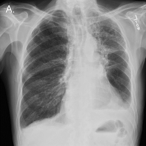
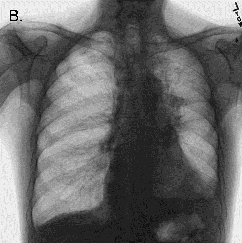
On day 33, a chest radiograph demonstrated persistent diffuse pulmonary infiltrates that had not significantly improved over the preceding 3 weeks (Figure 1).

Figure 1.  Portable chest x-ray the morning before the code arrest.

Minor bleeding was noted from the tracheostomy tube. Shortly thereafter, peak inspiratory pressures suddenly rose to the point that adequate tidal volumes could not be delivered by a mechanical ventilator. The inner cannula of the tracheostomy tube was removed. A suction catheter passed easily though the external cannula lumen, and a small amount of blood was suctioned out. However, attempts to bag-ventilate the patient became progressively more difficult. The patient's head and neck became cyanotic and mottled, and a pulse could no longer be detected. Advanced cardiac life support was initiated. Examination was significant for pan-expiratory wheezes throughout the thorax interrupted only by strenuous attempts to at bag-mask inspiration. The trachea was midline, and there was no subcutaneous crepitus. The abdomen was soft.  A bronchoscope passed through the tracheostomy tube easily, revealing a widely patent trachea and major airways. Bag ventilation transiently improved, cyanosis resolved, and a blood pressure of 150/85 was briefly obtained.  Inhaled albuterol and intravenous corticosteroids were administered.  A chest x-ray was performed (Figure 2). 

Figure 2. CXR performed during the code arrest, showing flattening of the diaphragms, and acute narrowing of the cardiac silhouette/vascular pedicle, and acute clearing of pulmonary infiltrates, consistent with hyperinflation.

Bag-ventilation became progressively more difficult, and the patient once more became hypotensive and cyanotic. The bronchoscope again passed easily through the tracheostomy and revealed the same findings as before. Needle thoracostomy was considered to treat possible pneumothorax, but the chest x-ray returned to the bedside demonstrated no evidence of barotrauma. The radiograph demonstrated striking improvement in pulmonary edema, a reduction in the size of the cardiac silhouette and vascular pedicle, and flattening of the diaphragms (see Figure 2 - note: the large radio-opacity overlying the mid-portion of the left lung is the shadow of an adherent transcutaneous pacing pad, not a pneumothorax). Further resuscitative efforts were unsuccessful.

The possibility of tracheostomy dysfunction was re-considered at some length in a postmortem debriefing. We concluded that the most likely explanation for the patient's clinical and radiological findings was dynamic hyperventilation and hemodynamic consequences of severe iPEEP induced by a dysfunction of the tracheostomy tube. 

Autopsy Findings

The tracheostomy tube was left in place, and the pathologist carefully dissected the trachea open from the carina in a caudal direction to expose the internal tip of the tracheostomy tube in-situ. A blood clot was found that nearly completely occluded the internal orifice of the tube (Figure 3, Panel A). The clot swung out of the way of some IV tubing passed inward through the external orifice of the tracheostomy tube, but swung shut again when the IV tube was removed, like a trap door (Figure 3, Panel B).

Figure 3.  Longitudinal view of the open tracheal lumen at autopsy.  Orientation: the left side of the figure is rostral. In panel A, the distal orifice of the tracheostomy tube can be seen to be nearly completely obstructed by a thrombus (black arrow). In panel B, the thrombus (black arrow) can be seen to be pushed aside by the passage of a plastic catheter (white arrow),

This clot appeared to function as a one-way valve, allowing inward passage of air, suction catheters, and a bronchoscope, but severely obstructing exhalation. We reasoned that such an obstruction could lead to wheezing and dynamic hyperinflation, and could explain the clinical and radiographic findings. Ultimately, severe iPEEP compromised cardiac preload, leading to pulselessness and death. 

No other cause for the patient's clinical syndrome was found - specifically, the patient had no antecedent history of asthma, had received no new medications on the day of the arrest, nor had any dermatological findings suggestive of anaphylaxis. The autopsy failed to reveal pulmonary embolism, mucous plugging, pneumothorax, or any histological evidence of asthma. 

Discussion

We are not the first to report dynamic hyperinflation as a complication of uni-directional tracheostomy tube obstruction (1). Several experienced clinicians at our institution recall dealing with this entity before, therefore, we suspect that it is not as rare as the paucity of clinical reports suggests. We felt that the clinical, radiological and postmortem findings in our case are sufficiently interesting, and the danger of missing this diagnosis sufficiently great, to warrant a brief review. 

Other types of tracheostomy tube dysfunction can cause high airway pressure and hypotension. Bi-directional tube obstruction from blood, dried secretions, or balloon hyperinflation is the most common (2,3). Barotrauma related to tracheostomy tubes may occur when they become displaced into the soft tissues of the neck, or into the pleural space, or when the cutaneous tracheostomy wound is sutured in an overly constrictive manner (4). 

We learned three important lessons from this unfortunate case:

  1. Clinical and radiographic findings can suggest the diagnosis of expiratory tracheostomy obstruction in a patient ventilated through a tracheostomy tube. The key clinical findings are: expiratory wheezing, hypotension, increasing iPEEP, and increasing peak inspiratory pressure.  Unexpected radiographic improvement in pulmonary edema may suggest the presence of occult iPEEP if it is not directly measured. 
  2. The diagnosis of unidirectional obstruction of a tracheostomy tube can be difficult to confirm.  The easy passage of suction catheters, or a bronchoscope, does not rule it out.  If bronchoscopy is performed emergently, the internal lumen and internal orifice of the tracheostomy tube should be examined with extreme deliberation.  This can be difficult during cardiopulmonary resuscitation.  If visualized, the potential detriment of small mobile clots should not be under-estimated.
  3. Alternative airway access should be immediately pursued in patients with tracheostomy tubes who are difficult to ventilate.  In dire clinical situations, the best diagnostic test might be to simply see if the patient improves with a new airway.  If the tracheostomy tract is likely to be mature (> 5 days old), the tracheostomy tube can simply be exchanged.  If the tract is immature, or if tube displacement is suspected, oral laryngoscopic intubation should be performed immediately.  The tracheostomy tube may need to be pulled out in order to accommodate the endotracheal tube in the trachea.  Either of these actions would likely have saved our patient's life.

References

  1. Timmus HH.  Tracheostomy: An Overview of implications, management, and morbidity.  Advances in Surgery 1973;7:199-233.
  2. Saini S, Taxak S, Singh MR.  Tracheostomy tube obstruction caused by an overinflated cuff.  Otolaryngol Head Neck Surg 2000;122:768-9.
  3. Rowe BH, Rampton J, Bota GW.  Life-threatening luminal obstruction due to mucous plugging in chronic tracheostomies: three case reports and a review of the literature.  J Emerg Med 1996;14:565-7.
  4. Tayal VS. Tracheostomies. Emerg Med Clin North Am 1994;12:707-27.

The author reports no financial support and no conflict of interest for this publication.

Reference as: Raschke RA. Fatal dynamic hyperinflation secondary to a blood clot acting as a one-way valve at the internal orifice of a tracheostomy tube. Southwest J Pulm Crit Care 2012;5:256-61. PDF

Friday
Nov022012

November 2012 Critical Care Case of the Month: I Just Can’t Do It Captain! I Can’t Get the Sats Up!

Bridgett Ronan, MD

Department of Pulmonary Medicine

Mayo Clinic Arizona

Scottsdale, AZ

History of Present Illness

A 61 year old man was seen in consultation after undergoing a laparoscopic repeat Nissen fundoplication with mesh reinforcement.  He developed worsening hypoxia postoperatively. He was initially extubated without difficulty to nasal cannula. However, he had progressive hypoxemia requiring a nonrebreathing mask, followed by BiPAP and eventually reintubation. Discussion with the surgeons revealed he had gastric contents present on intraoperative esophagogastroduodenoscopy (EGD). There was a small perforation of the fundus, with possible contamination of the peritoneum.

PMH, FH, SH

He has a long history of a paraesophageal hernia and reflux esophagitis and had previously undergone a Nissen fundoplication. There was also a history of atrial flutter and a 4.8 cm thoracic aortic aneurysm. A pre-operative echocardiogram was othewise normal. There was no remarkable family history. He was a non-drinker and non-smoker.

Physical Examination

Vital signs: Heart rate 79 beats/min, BP 95/67 mm Hg, Temperature 99.4°F, SpO2 78% on 100% FiO2.

His lungs were clear interiorly.

No murmurs or gallops were heard on cardiac auscultation.

His abdomen was post-surgical and distended but soft and nontender.

Which of the following is true regarding hypoxemia?

  1. Most hypoxia is secondary to alveolar-capillary block
  2. A normal pCO2 excludes hypoventilation as a cause of hypoxemia
  3. Low inspired FiO2 is a common cause of hypoxia in the ICU because of attaching air to the oxygen line on the ventilator.
  4. A normal chest x-ray excludes ventilation-perfusion mismatch as a cause of hypoxemia
  5. The patient’s age of 61 excludes a congenital heart lesion

Reference as: Ronan B. November 2012 critical care case of the month: I just can’t do it captain! I can’t get the sats up! Southwest J Pulm Crit Care 2012;5:235-41. PDF