
April 2022 Critical Care Case of the Month: Bullous Skin Lesions in the ICU
Margaret Wat MD PhD, Jawad Bilal MD, Martin Chacon MD, Stephen Klotz MD, and Janet Campion MD
University of Arizona College of Medicine-Tucson
Tucson, AZ USA
History of Present Illness: A 29-year-old woman with past medical history of mixed connective tissue disease [lupus predominant], prior pulmonary embolism complained of a 2-week history of nonproductive cough. The cough began after her son was diagnosed with respiratory syncytial virus (RSV). Symptoms progressively worsened and now she is admitted from the emergency department (ED) with generalized weakness and progressive shortness of breath. Earlier in the day at an outside hospital, she tested positive for RSV, negative for COVID-19 and had normal O2 saturations and was discharged home. She has not received COVID-19 vaccine. Symptoms progressed, 911 called and in the ED, she was found to have temperature = 104°F, SpO2 = 64% on room air, and fasting blood sugar in the 40s. She was lethargic with visible respiratory distress and unable to answer questions.
Past Medical History:
- Mixed connective tissue disease [features of systemic lupus erythematosus, rheumatoid arthritis, polymyositis, scleroderma]
- Membranous lupus nephritis [class V]
- History of pulmonary embolus
- Posterior intracranial artery infarct with venous sinus thrombosis in February 2020
- Hypertension
- Recent septic shock due to pneumococcal bacteremia 2 months prior to admission
- Post-op C section
Medications:
- Atovaquone 750 mg BID
- Eliquis 5 mg BID
- Fluconazole 150 mg Q 72h
- Hydroxychloroquine 200 mg daily
- Nifedipine 30 mg daily
- Pantoprazole 40 mg BID
- Prednisone 5 mg daily
- Vitamin D3 2000 IU daily
- Albuterol PRN SOB
- Ferrous sulfate 325 mg daily
- Losartan 25 mg daily
Social History and Family History
- Married, nonsmoker, rare social ethanol use, no recreational drug use
- Father with hypertension, mother with autoimmune disease
Physical Examination
- T = 40°C, heart rate = 130 beats/min, respiratory rate = 28 breaths/min, BP = 100/61 mm Hg, SpO2 = 95% on 100% nonrebreathing mask, BMI = 24
- General: Lethargic well-nourished young woman unable to answer questions, accessory respiratory muscle use
- HEENT: Dry mucosa, no scleral icterus, injected conjunctiva
- Pulmonary: No audible wheeze, crackles, rhonchi
- CV: Tachycardic, regular, no murmur
- Abd: Tender bilateral upper quadrants, nondistended, no HSM
- Neurological: Moving extremities but unable to follow commands, CN grossly intact
- Psychiatric: Unable to assess, mentation/mood normal earlier in day per her husband
- Extremities: Warm with mottled UE and LE digits, scattered areas of purpura (Figure 1)
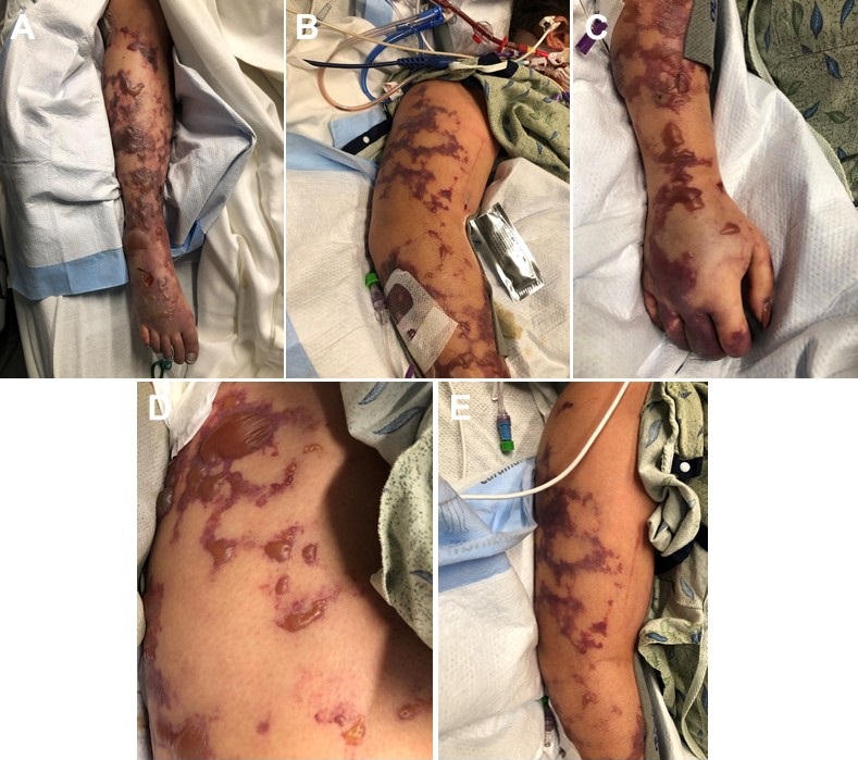
Figure 1. Photographs of extremities taken during day 1 and 2 in the ICU.
With this patient's presentation, what is the most likely cause of the purpura? (Click on the correct answer to be directed to the second of six pages)
- Angioinvasive fungal infection
- Thrombotic related to cryoglobulinemia
- Septic emboli
- Thrombosis from disseminated intravascular coagulation (purpura fulminans)
- Depositional vessel disease from calciphylaxis
 Post a Comment
Reader Comments