
Correct!
5. All of the above
The differential diagnosis of shock is large but falls into three major categories (1):
These can usually be separated by common clinical ICU measurements (Table 1).
Table 1. Characterization of different types of shock by clinical ICU measurements.
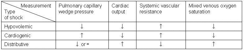
In some cases clinical substitutes for pulmonary capillary wedge pressure such as stroke volume variation and central venous pressure (CVP).
In our patient’s case initial fluid resuscitation was guided by goal directed therapy (preload as monitored by stroke volume variation and CVP, ScVO2 and falling lactate). Cardiac evaluation showed no significant rise in troponins and bedside echocardiography was not consistent with a cardiogenic cause of his shock. Hydrocortisone was started for possible critical illness related corticosteroid insufficiency. E. coli was cultured from the urine and was sensitive to the antibiotic started on day 1 within 4 hours of admission – ceftriaxone. His clinical picture was most consistent with distributive shock secondary to sepsis.
By 24 hours, lactate levels had fallen from 10 to 6 mmol/L but stabilized at around 6 despite treatment despite continued treatment. The patient's acute kidney injury had improved marginally from a creatinine of 12 to 9 mg/dL with the resuscitation.
An abdominal x-ray was taken for taken for feeding tube placement (Figure 1).
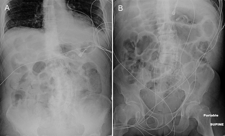
Figure 1. Panel A. Abdominal film taken at admission. Panel B. Abdominal film taken during prior admission for comparison.
What abnormalities are seen on the abdominal film?