Search Journal-type in search term and press enter
Southwest Pulmonary and Critical Care Fellowships
In Memoriam

Sleep

(Click on title to be directed to posting , most recent listed first)

July 2023 Sleep Case of the Month: Fighting for a Good Night’s Sleep
Associations Between Insomnia and Obstructive Sleep Apnea with
   Nutritional Intake After Involuntary Job Loss
January 2023 Sleep Case of the Month: An Unexpected EEG Abnormality
July 2022 Sleep Case of the Month: A Sleepy Scout
Assessing Depression and Suicidality Among Recently Unemployed
   Persons with Obstructive Sleep Apnea and Socioeconomic
   Inequality
Impact of Recent Job Loss on Sleep, Energy Consumption and Diet
Long-term All-Cause Mortality Risk in Obstructive Sleep Apnea Using
   Hypopneas Defined by a ≥3 Percent Oxygen Desaturation or Arousal
The Association Between Obstructive Sleep Apnea Defined by 3 Percent
   Oxygen Desaturation or Arousal Definition and Self-Reported
Cardiovascular Disease in the Sleep Heart Health Study
Informe de políticas: Fatiga, sueño y salud del personal de enfermería, y 
   cómo garantizar la seguridad de los pacientes y el público
Sleep Tips for Shift Workers in the Time of Pandemic
Tips for Circadian Sleep Health While Working from Home
Impacto del Sueño y la Modalidad de Diálisis sobre la Calidad de Vida en
   una Población
The Effect of CPAP on HRQOL as Measured by the Quality of Well-Being
   Self-Administered Questionnaire (QWB-SA)
Declaración de posición: Reducir la fatiga asociada con la deficiencia de 
   sueño y las horas de trabajo en enfermeras
Impact of Sleep and Dialysis Mode on Quality of Life in a Mexican Population
Out of Center Sleep Testing in Ostensibly Healthy Middle Aged to Older
   Adults
Sleep Related Breathing Disorders and Neurally Mediated Syncope (SRBD
   and NMS)
Sleep Board Review Question: Restless Legs
Impact of Sleep Duration and Weekend Oversleep on Body Weight
   and Blood Pressure in Adolescents
Role of Spousal Involvement in Continuous Positive Airway Pressure
   (CPAP) Adherence in Patients with Obstructive Sleep Apnea (OSA)
The Impact of an Online Prematriculation Sleep Course (Sleep 101) on
   Sleep Knowledge and Behaviors in College Freshmen: A Pilot Study
Obstructive Sleep Apnea and Quality of Life: Comparison of the SAQLI,
   FOSQ, and SF-36 Questionnaires
Gender Differences in Real-Home Sleep of Young and Older Couples
Brief Review: Sleep Health and Safety for Transportation Workers
Lack of Impact of Mild Obstructive Sleep Apnea on Sleepiness, Mood and
   Quality of Life
Alpha Intrusion on Overnight Polysomnogram
Sleep Board Review Question: Insomnia in Obstructive Sleep Apnea
Long-Term Neurophysiologic Impact of Childhood Sleep Disordered 
   Breathing on Neurocognitive Performance
Sleep Board Review Question: Hyperarousal in Insomnia
Sleep Board Review Question: Epilepsy or Parasomnia?
Sleep Board Review Question: Nocturnal Hypoxemia in COPD
Sleep Board Review Questions: Medications and Their
   Adverse Effects
Sleep Board Review Questions: The Restless Sleeper
Obstructive Sleep Apnea and Cardiovascular Disease:
Back and Forward in Time Over the Last 25 Years
Sleep Board Review Questions: The Late Riser
Sleep Board Review Questions: CPAP Adherence in OSA
Sleep Board Review Questions: Sleep Disordered Breathing 
That Improves in REM
The Impact Of Sleep-Disordered Breathing On Body
   Mass Index (BMI): The Sleep Heart Health Study (SHHS)
Incidence and Remission of Parasomnias among Adolescent Children in the 
   Tucson Children’s Assessment of Sleep Apnea (TuCASA) Study 
A 45-Year Old Man with Excessive Daytime Somnolence, 
   and Witnessed Apnea at Altitude


The Southwest Journal of Pulmonary and Critical Care and Sleep publishes articles related to those who treat sleep disorders in sleep medicine from a variety of primary backgrounds, including pulmonology, neurology, psychiatry, psychology, otolaryngology, and dentistry. Manuscripts may be either basic or clinical original investigations or review articles. Potential authors of review articles are encouraged to contact the editors before submission, however, unsolicited review articles will be considered.

-------------------------------------------------------------------------------------

Wednesday
Sep052012

Sleep Board Review Questions: CPAP Adherence in OSA

Carmen Luraschi-Monjagatta, MD1

Rohit Budhiraja, MD1,2

1 Department of Medicine, Section of Pulmonary, Allergy, Critical Care and Sleep Medicine, University of Arizona, Tucson, AZ, 85724, USA. mdelcarmen@deptofmed.arizona.edu

2 Department of Medicine, Southern Arizona Veterans Affairs Health Care System (SAVAHCS), Tucson, AZ 85723, USA. rohit.budhiraja@va.gov

Which of the following has been shown to be associated with a better adherence to positive airway pressure (PAP) therapy in adults with obstructive sleep apnea (OSA)?

Reference as: Luraschi-Monjagatta C, Budhiraja R. Sleep board review questions: CPAP adherence in OSA. Southwest J Pulm Crit Care 2012;5:135-7. (Click here for a PDF version)

Friday
Aug312012

Sleep Board Review Questions: Sleep Disordered Breathing That Improves in REM

Rohit Budhiraja, MD

Pulmonary, Allergy, Critical Care and Sleep Medicine

University of Arizona


Which of the following breathing disorders is usually less severe in rapid eye movement (REM) sleep compared to non-rapid eye movement (NREM) sleep?

  1. Sleep-related hypoxemia in COPD
  2. Obstructive Sleep Apnea
  3. Cheyne Stokes Breathing
  4. Hypoxemia in Pulmonary Hypertension

Reference as: Budhiraja R. Sleep board review questions: sleep disordered breathing that improves in REM. Southwest J Pulm Crit Care;2012:106-7. (Click here for a PDF version)

Thursday
Dec082011

The Impact of Sleep-Disordered Breathing on Body Mass Index (BMI): The Sleep Heart Health Study (SHHS)

Mark A. Brown, M.D. 1

James L. Goodwin, Ph.D.2

Graciela E. Silva, Ph.D, MPH.3

Ajay Behari, M.D.4

Anne B. Newman, M.D., M.P.H5,6

Naresh M. Punjabi, M.D., Ph.D.7

Helaine E. Resnick, Ph.D., M.P.H.8

John A. Robbins, M.D., M.S.H.9

Stuart F. Quan, M.D.2,10


1Department of Psychiatry, Kaiser Permanente, Portland, OR (markbrownmd@gmail.com); 

2Sleep and Arizona Respiratory Centers, University of Arizona College of Medicine, Tucson, AZ(jamieg@arc.arizona.edu);

3College of Nursing & Health Innovation, Arizona State University, Tempe, AZ (Graciela.Silva@asu.edu); 

4Pulmonary and Critical Care Associates of Baltimore, Baltimore, MD (ajaybehari@yahoo.com);

5Graduate School of Public Health, Department of Epidemiology, University of Pittsburgh, Pittsburgh, PA

6Division of Geriatric Medicine, University of Pittsburgh School of Medicine, Pittsburgh, PA (NewmanA@edc.pitt.edu);

7Department of Medicine, Johns Hopkins University School of Medicine, Baltimore, MD (npunjabi@jhmi.edu);

8American Association of Homes and Services for the Aging, Washington, DC (heresnick@gmail.com); 

9Center for HealthCare Policy and Research, University of California, Davis, Sacramento, CA (jarobbins@ucdavis.edu);

10Division of Sleep Medicine, Harvard Medical School, Boston, MA (squan@arc.arizona.edu)


Address for correspondence and reprint requests: Stuart F. Quan, M.D., Division of Sleep Medicine, Harvard Medical School, 401 Park Dr., 2nd Floor East, Boston, MA 02215, Tel (617) 998-8842, Fax (617) 998-8823, Email: squan@arc.arizona.edu

Conflict of Interest Statement: None of the authors have conflicts of interest pertinent to the subject matter of this manuscript. 

Reference as: Brown MA, Goodwin JL, Silva GE, Behari A, Newman AB, Punjabi NM, Resnick HE, Robbins JA, Quan SF. The impact of sleep-disordered breathing on body mass index (BMI): the sleep heart health study (SHHS). Southwest J Pulm Crit Care 2011;3:159-68. (Click here for PDF version of the manuscript)

Abstract

Introduction: It is well known that obesity is a risk factor for sleep-disordered breathing (SDB). However, whether SDB predicts increase in BMI is not well defined. Data from the Sleep Heart Health Study (SHHS) were analyzed to determine whether SDB predicts longitudinal increase in BMI, adjusted for confounding factors.

Methods: A full-montage unattended home polysomnogram (PSG) and body anthropometric measurements were obtained approximately five years apart in 3001 participants. Apnea-hypopnea index (AHI) was categorized using clinical thresholds: < 5 (normal), ≥ 5 to <15 (mild sleep apnea), and ³ 15 (moderate to severe sleep apnea). Linear regression was used to examine the association between the three AHI groups and increased BMI. The model included age, gender, race, baseline BMI, and change in AHI as covariates.

Results: Mean (SD) age was 62.2 years (10.14), 55.2% were female and 76.1% were Caucasian. Five-year increase in BMI was modest with a mean (SD) change of 0.53 (2.62) kg/m2 (p=0.071). A multivariate regression model showed that subjects with a baseline AHI between 5-15 had a mean increase in BMI of 0.22 kg/m2 (p=0.055) and those with baseline AHI ≥ 15 had a BMI increase of 0.51 kg/m2 (p<0.001) compared to those with baseline AHI of <5.

Conclusion: Our findings suggest that there is a positive association between severity of SDB and subsequent increased BMI over approximately 5 years. This observation may help explain why persons with SDB have difficulty losing weight.

Key Words: Sleep Apnea, Weight Gain, Obesity

Abbreviation List: PSG-polysomnogram, SDB-sleep disordered breathing, AHI-apnea hypopnea index, SHHS-Sleep Heart Health Study, BMI-body mass index, SD-standard deviation, SEM-standard error of the mean, ANOVA-analysis of variance

Introduction

There is overwhelming epidemiological and clinical data indicating that obesity is a risk factor for sleep disordered breathing (SDB).1-8 The association between obesity and SDB is substantial, with high body mass index (BMI) contributing to moderate to severe SDB in 58% of affected persons.9 The effect of obesity is greater in men than women1,10-12 although it decreases with increasing age.6,7 In addition, weight loss has been demonstrated to decrease the severity of SDB.10,13,14 Longitudinal data from population studies including the Sleep Heart Health Study (SHHS),10 the Wisconsin Sleep Cohort,15 and the Cleveland Family Study6 have initially focused on the impact of increased weight on SDB severity. However, examination of the opposite causal pathway has yet to be prospectively addressed.

Anecdotally, patients with SDB appear to have more difficulty losing weight than obese patients without SDB. They also report marked weight gain prior to confirmation of their diagnosis. Two small studies support these empiric observations.5,16  Given this limited information on the impact of SDB on BMI, data from SHHS was analyzed to examine the impact of SDB on BMI after controlling for change in AHI and severity of SDB.

Methods

Study Design and Population. The SHHS is a multi-center, community-based prospective cohort study of the natural history and cardiovascular consequences of SDB. Details of the study design, sampling, and procedures have been reported.17 Briefly, between November 1995 and January 1998 participants were recruited from several ongoing prospective cohort studies--the Framingham Offspring and Omni Studies, the Atherosclerosis Risk in Communities Study, the Cardiovascular Health Study, the Strong Heart Study, and the cohort studies of respiratory disease in Tucson and of hypertension in New York. Participants were eligible if they were ≥ 40 years of age and were not being treated for sleep apnea with positive pressure therapy, an oral appliance, oxygen, or a tracheostomy. Habitual snorers < 65 years were over sampled to increase the prevalence of obstructive sleep apnea. Subjects were required to provide written consent and the protocol was approved by the institutional review boards of each of the eight investigative sites. 

Data Collection. A total of 6,441 subjects completed the baseline polysomnogram (PSG), and 4,586 consented to have a second evaluation approximately five years later. This analysis focuses on the 3,040 participants who had PSG and BMI data at both time points. Data from all 215 participants who had a follow-up PSG from the New York center were excluded because they did not meet quality standards for the follow-up examination. The remaining participants died, were too ill to participate, refused to participate, were lost to follow-up or had incomplete covariate data such as weight. This latter group had a higher percentage of Whites (85%) compared with the study group (75.5%) (p-value <0.001). There also were statistically significant differences in baseline BMI, baseline AHI, and age between the study group compared with the excluded group, however, these differences were very small and were not clinically significant. There was no gender difference between the two groups.

Weight was measured on the night of the PSG examination with the participant in light clothes on a calibrated portable scale. Height was obtained at the baseline home visit if not already measured within + 3 months of the parent study. BMI was calculated as weight in kilograms divided by the square of height in meters. Baseline height was used for baseline and follow-up BMI calculations. Age, sex, and ethnicity were self-reported. 

The PSG was conducted using a portable monitor (PS-2 System; Compumedics Limited, Abbotsford, Victoria, Australia), using methods previously described.18 Apnea was present if there was an absence or near absence of airflow or thoracoabdominal movement (at least < 25% of baseline) for > 10 seconds. Hypopnea was defined as a decrease in the amplitude of the airflow or thoracoabdominal movement below 70% of baseline for > 10 seconds. The apnea-hypopnea index (AHI) was calculated as the number of apnea and hypopnea events, each associated with at least a 4% decrease in oxygen saturation, divided by total sleep time in hours.

Results

Participant characteristics are provided in Table 1. 

Table 1: Characteristics of participants of the Sleep Heart Health Study cohort with complete baseline and follow-up polysomnography and weight measurements as a function of sleep apnea severity.

As expected, women were over-represented in the baseline AHI < 5 group (64.9%) and men were over-represented in the AHI ³ 15 group (63.5%) (p<0.001). Baseline BMI increased as baseline AHI severity increased.  Overall unadjusted five-year increase in BMI was modest with a mean (SD) BMI change of 0.53 (2.61) kg/m2. The unadjusted five-year increase in BMI was 0.63 (2.54) kg/m2 for those with baseline AHI < 5, 0.43 (2.48) kg/m2 among those with AHI ≥ 5 to < 15 and 0.37 (3.05) kg/m2 for the AHI group ≥ 15. These values were not statistically different from each other.

A multivariate regression model was constructed predicting five-year change in BMI by baseline AHI category adjusted for age, gender, race, baseline BMI, and AHI change (Table 2).

Table 2: Adjusted β coefficients of BMI change according to AHI and continuous variables in the Sleep Heart Health Study*.

Compared to baseline AHI group of < 5, those with AHI between ≥ 5 to < 15 had a mean adjusted increase in BMI of 0.21 that approached statistical significance (p=0.055). However, those with AHI ≥ 15 had a statistically significant adjusted BMI increase of 0.51 (p<0.001).  Younger age, lower baseline BMI and greater AHI change also were associated with a larger BMI increase. There was a trend for women to have a greater increase in BMI, but no effect of race was observed. However, the model only accounted for 7% of the total variance. Adjusted means by baseline AHI group are displayed graphically in Figure 1.

Figure 1: Estimated Adjusted Means of BMI increase according to AHI in the Sleep Heart Health Study. Data are adjusted for baseline age (continuous), race (categorical), gender (categorical), baseline BMI (continuous), change in AHI (continuous). Covariates fixed at: baseline BMI = 28.7, baseline age 62.1, change in RDI = 2.7. Bars represent 95% confidence intervals.

Discussion

Our findings indicate that there is a positive association between severity of SDB and five-year increase in BMI. The finding was demonstrated after controlling for key covariates including age, gender, race, baseline BMI, and AHI change. This observation may help explain the difficulty patients with SDB have in trying to lose weight.

Two previous small studies have demonstrated a positive association between newly diagnosed SDB and weight gain. A retrospective study by Phillips et al. compared one-year weight histories of 53 men and women patients who were recently diagnosed with SDB with 24 control subjects matched for gender, age, BMI and percent body fat.5 Subjects in that study were somewhat younger than the SHHS cohort with an age difference of approximately 10 years. The SDB among subjects in the previous study tended to be moderate to severe with mean ± SEM AHI 33 ± 5 /h for men and 37 ± 10 /h for women. Mean ± SEM of BMI at time of diagnosis was somewhat higher than in the SHHS with 35 ± 1 kg/m2 for men and 44 ± 2 kg/m2 for women.  Men and women patients with SDB had reported a recent weight gain of 7.4 ± 1.5 kg compared with a weight loss of 0.5 ± 1.7 kg (p=0.001) in obese controls without SDB. However, given the design of this study it is not possible to determine whether weight gain contributed to the onset of SDB or was a result of SDB. The study was also limited by reliance on self-report of weight gain history.

Another study by Traviss et al. prospectively evaluated 49 obese patients with newly diagnosed SDB.16 Mean ± SD of AHI at diagnosis was severe at 45 ± 27 /h.  BMI at diagnosis was elevated at 36.5 ± 6.2 kg/m2. Of the 49 subjects, 43 could estimate the duration of their symptoms with 84% reporting weight gain since becoming symptomatic.  Weight gain was relatively large, with a reported 17 ± 15 kg over 5.3 ± 4.8 years. However, this study was limited by the lack of a control group and reliance on self-report of weight history.  These two small studies, in addition to our findings, suggest that there is an association between SDB and increased BMI.

Interestingly, unadjusted BMI change in our study was quite modest and not statistically different as a function of SDB severity. However, BMI change over time is a complex phenomenon influenced by several variables. A large (29,799 subjects) prospective study examining 5-year change in weight in a multi-ethnic cohort of men and women explored several of these relationships.19 In that study, younger men and to a greater degree, younger women were at greater risk for weight gain compared to older adults. This is consistent with our initial findings. In addition, there was a trend for women in the higher baseline BMI categories of ‘overweight’ (BMI >25– 30 kg/m2) and ‘obese’ (BMI >30 kg/m2) in the aforementioned cohort to gain more weight than men in the higher baseline BMI categories. In order to more precisely examine the effect of AHI on weight change, we controlled for these confounders in our final multivariate model thus resulting in the finding of an increase in BMI as a function of SDB severity in this study. 

Several mechanisms could explain why SDB contributes to increased BMI. First, persons with SDB may have a reduction in the quantity and quality of their sleep. Recent data indicate that insufficient sleep may be a risk factor for obesity.20 Experimental sleep restriction increases ghrelin and reduces leptin production favoring appetite enhancement,21 a finding that also has been observed in a large population cohort.22 Second, those with SDB may eat a diet that favors weight gain. In support of this hypothesis, sleep restriction has been shown to increase craving for calorie dense food with high carbohydrate content. The Apnea Positive Pressure Long-Term Efficacy Study (APPLES) demonstrated that those with severe SDB consumed a diet higher in cholesterol, protein, total fat and total saturated fatty acids, even after adjusting for BMI, age, and daytime sleepiness.23 Third, a cardinal symptom of SDB is excessive daytime sleepiness. Thus, it is possible that persons with SDB engage in less physical activity because they are too fatigued to exercise. Data from APPLES indicate that recreational physical activity is less in those with SDB. However, this finding appears to be principally explained by concomitant obesity. 

Weight loss frequently results in an improvement and sometimes resolution in SDB. This is most evident in those who undergo bariatric surgical procedures.24,25 Persons with SDB are frequently counseled to treat their SDB by losing weight through diet and exercise,26 an approach that is usually unsuccessful.25 Failure to primarily address SDB in conjunction with a weight reduction program may diminish the latter’s success. However, evidence to date indicates that treatment of SDB does not consistently result in weight loss. In a sample of clinical patients with SDB, treatment with CPAP did not result in weight loss. Moreover, in female patients, there was actually an increase in weight.27 In addition, consistent weight reduction was not observed in a small number of patients with severe OSA who underwent tracheostomy.28 Thus, it appears that weight gain engendered by the presence of OSA is not easily reversed despite therapy. Prospective studies will be required to determine whether primary treatment for OSA enhances weight loss programs in those with OSA.

Although this analysis demonstrated a positive association of severity of SDB on five-year increase in BMI, there are several caveats that deserve consideration. The BMI of participants tended to be lower than that seen in clinical SDB populations and a relatively small number of subjects had large changes in BMI. As previously noted, the mean BMI increase was, at best, quite modest. When converted for illustrative purposes to weight using an average height of 167 cm of the participants, those with an AHI between ≥ 5 to < 15 had a mean adjusted increase in BMI of 0.21 kg/m2 equal to 0.59 kg or 1.30 lbs. Similarly, those with AHI ≥ 15 had an adjusted BMI increase of 0.51 kg/m2 equal to 1.42 kg or 3.13 lbs. Thus, the magnitude of the changes we observed may not be applicable to clinical populations where patients with SDB may have a higher BMI. In addition, it is not known when the participants developed SDB, thus definitive inference of causality cannot be made. However, following a large undiagnosed cohort over an extended period of time to determine incidence of SDB onset and subsequent change in weight would be exceedingly difficult and costly.  Additionally, the model only accounted for a small amount of the total variance in five-year BMI increase, suggesting that there are likely other unmeasured variables influencing the amount of BMI increase over time in this cohort. Finally, while not statistically significant, the unadjusted mean change in BMI was slightly less in the high RDI group in comparison to the lower RDI groups. This observation underscores the biological complexity of the interactions among weight change, SDB, age, gender and other factors.

In conclusion, our findings suggest that although weight gain is a risk factor for developing or worsening SDB, SDB may, in a reciprocal fashion, lead to increased weight gain. This may help explain why patients with SDB find it difficult to lose weight.

Acknowledgements

This work was supported by National Heart, Lung and Blood Institute cooperative agreements U01HL53940 (University of Washington), U01HL53941 (Boston University), U01HL53938 (University of Arizona), U01HL53916 (University of California, Davis), U01HL53934 (University of Minnesota), U01HL53931 (New York University), U01HL53937 and U01HL64360  (Johns Hopkins University), U01HL63463 (Case Western Reserve University), and U01HL63429 (Missouri Breaks Research).

Sleep Heart Health Study (SHHS) acknowledges the Atherosclerosis Risk in Communities Study (ARIC), the Cardiovascular Health Study (CHS), the Framingham Heart Study (FHS), the Cornell/Mt. Sinai Worksite and Hypertension Studies, the Strong Heart Study (SHS), the Tucson Epidemiologic Study of Airways Obstructive Diseases (TES) and the Tucson Health and Environment Study (H&E) for allowing their cohort members to be part of the SHHS and for permitting data acquired by them to be used in the study.  SHHS is particularly grateful to the members of these cohorts who agreed to participate in SHHS as well. SHHS further recognizes all of the investigators and staff who have contributed to its success. A list of SHHS investigators, staff and their participating institutions is available on the SHHS website, www.jhucct.com/shhs.

The opinions expressed in the paper are those of the author(s) and do not necessarily reflect the views of the Indian Health Service.

These data have been presented in part at the Annual Meeting of the Associated Professional Sleep Societies, June 11, 2009, Seattle, WA.

References

  1. Bixler EO, Vgontzas AN, Lin HM, Ten Have T, Rein J, Vela-Bueno A, et al. Prevalence of sleep-disordered breathing in women: Effects of gender. Am J Respir Crit Care Med 2001;163:608-13.
  2. Bixler EO, Vgontzas AN, Ten Have T, Tyson K, Kales A. Effects of age on sleep apnea in men: I. prevalence and severity. Am J Respir Crit Care Med 1998;157:144-8.
  3. Carmelli D, Swan GE, Bliwise DL. Relationship of 30-year changes in obesity to sleep-disordered breathing in the western collaborative group study. Obes Res 2000;8:632-7.
  4. Duran J, Esnaola S, Rubio R, Iztueta A. Obstructive sleep apnea-hypopnea and related clinical features in a population-based sample of subjects aged 30 to 70 yr. Am J Respir Crit Care Med 2001;163:685-9.
  5. Phillips BG, Hisel TM, Kato M, Pesek CA, Dyken ME, Narkiewicz K, et al. Recent weight gain in patients with newly diagnosed obstructive sleep apnea. J Hypertens 1999;17:1297-300.
  6. Tishler PV, Larkin EK, Schluchter MD, Redline S. Incidence of sleep-disordered breathing in an urban adult population: The relative importance of risk factors in the development of sleep-disordered breathing. JAMA 2003;289:2230-7.
  7. Young T, Shahar E, Nieto FJ, Redline S, Newman AB, Gottlieb DJ, et al. Predictors of sleep-disordered breathing in community-dwelling adults: The sleep heart health study. Arch Intern Med 2002;162:893-900.
  8. Young T, Palta M, Dempsey J, Skatrud J, Weber S, Badr S. The occurrence of sleep-disordered breathing among middle-aged adults. N Engl J Med 1993;328:1230-5.
  9. Young T, Peppard PE, Taheri S. Excess weight and sleep-disordered breathing. J Appl Physiol 2005;99:1592-9.
  10. Newman AB, Foster G, Givelber R, Nieto FJ, Redline S, Young T. Progression and regression of sleep-disordered breathing with changes in weight: The sleep heart health study. Arch Intern Med 2005;165:2408-13.
  11. Ip MS, Lam B, Tang LC, Lauder IJ, Ip TY, Lam WK. A community study of sleep-disordered breathing in middle-aged chinese women in hong kong: Prevalence and gender differences. Chest 2004;125:127-34.
  12. Millman RP, Carlisle CC, McGarvey ST, Eveloff SE, Levinson PD. Body fat distribution and sleep apnea severity in women. Chest 1995;107:362-6.
  13. Barvaux VA, Aubert G, Rodenstein DO. Weight loss as a treatment for obstructive sleep apnoea. Sleep Med Rev 2000;4:435-52.
  14. Grunstein RR, Stenlof K, Hedner JA, Peltonen M, Karason K, Sjostrom L. Two year reduction in sleep apnea symptoms and associated diabetes incidence after weight loss in severe obesity. Sleep 2007;30:703-10.
  15. Peppard PE, Young T, Palta M, Dempsey J, Skatrud J. Longitudinal study of moderate weight change and sleep-disordered breathing. JAMA 2000;284:3015-21.
  16. Traviss KA, Barr SI, Fleming JA, Ryan CF. Lifestyle-related weight gain in obese men with newly diagnosed obstructive sleep apnea. J Am Diet Assoc 2002;102:703-6.
  17. Quan SF, Howard BV, Iber C, Kiley JP, Nieto FJ, O'Connor GT, et al. The sleep heart health study: Design, rationale, and methods. Sleep 1997;20:1077-85.
  18. Redline S, Sanders MH, Lind BK, Quan SF, Iber C, Gottlieb DJ, et al. Methods for obtaining and analyzing unattended polysomnography data for a multicenter study. sleep heart health research group. Sleep1998;21:759-67.
  19. Ball K, Crawford D, Ireland P, Hodge A. Patterns and demographic predictors of 5-year weight change in a multi-ethnic cohort of men and women in australia. Public Health Nutr 2003;6:269-81.
  20. Chaput JP, Despres JP, Bouchard C, Tremblay A. The association between sleep duration and weight gain in adults: A 6-year prospective study from the Quebec Family Study. Sleep 2008;31:517-23.
  21. Spiegel K, Tasali E, Penev P, Van Cauter E. Brief communication: Sleep curtailment in healthy young men is associated with decreased leptin levels, elevated ghrelin levels, and increased hunger and appetite. Ann Intern Med 2004;141:846-50.
  22. Taheri S, Lin L, Austin D, Young T, Mignot E. Short sleep duration is associated with reduced leptin, elevated ghrelin, and increased body mass index. PLoS Med. 2004;1:e62.
  23. Vasquez MM, Goodwin JL, Drescher AA, Smith TW, Quan SF. Associations of dietary intake and physical activity with sleep disordered breathing in the apnea positive pressure long-term efficacy study (APPLES). J Clin Sleep Med 2008;4:411-8.
  24. Rasheid S, Banasiak M, Gallagher SF, Lipska A, Kaba S, Ventimiglia D, et al. Gastric bypass is an effective treatment for obstructive sleep apnea in patients with clinically significant obesity. Obes Surg 2003;13:58-61.
  25. Fritscher LG, Canani S, Mottin CC, Fritscher CC, Berleze D, Chapman K, et al. Bariatric surgery in the treatment of obstructive sleep apnea in morbidly obese patients. Respiration 2007;74:647-52.
  26. Veasey SC, Guilleminault C, Strohl KP, Sanders MH, Ballard RD, Magalang UJ. Medical therapy for obstructive sleep apnea: A review by the medical therapy for obstructive sleep apnea task force of the standards of practice committee of the american academy of sleep medicine. Sleep 2006;29:1036-44.
  27. Redenius R, Murphy C, O'Neill E, Al-Hamwi M, Zallek SN. Does CPAP lead to change in BMI? J Clin Sleep Med 2008;4:205-9.
  28. Haapaniemi JJ, Laurikainen EA, Halme P, Antila J. Long-term results of tracheostomy for severe obstructive sleep apnea syndrome. ORL J Otorhinolaryngol Relat Spec 2001;63:131-6.
Monday
Jun272011

Incidence and Remission of Parasomnias among Adolescent Children in the Tucson Children’s Assessment of Sleep Apnea (TuCASA) Study

Oscar Furet, RN M.P.H.

Arizona Arthritis Center, University of Arizona, Tucson, AZ

oscarf@arthritis.arizona.edu


James L. Goodwin, Ph.D.

Arizona Respiratory Center, University of Arizona, Tucson, AZ

jgoodwin@email.arizona.edu


Stuart F. Quan, M.D.

Arizona Respiratory Center, University of Arizona, Tucson, AZ. Division of Sleep Medicine, Brigham and Womens Hospital and Harvard Medical School, Boston, MA


Correspondent:             Stuart F. Quan, M.D.

                                    Division of Sleep Medicine

                                    Harvard Medical School

                                    401 Park Dr., 2nd Floor East

                                    Boston, MA 02215

                                    Email: squan@arc.arizona.edu

                                    Voice: 617-998-8842

                                    Fax:    617-998-8823

Reference as: Furet O, Goodwin JL, Quan SF. Incidence and remission of parasomnias among adolescent children in the Tucson Children’s Assessment of Sleep Apnea (TuCASA) Study. Southwest J Pulm Crit Care 2011;2:93-101. (Click here for PDF version)

Abstract

Background: Longitudinal assessments of parasomnias in the adolescent population are scarce. This analysis aims to identify the incidence and remission of parasomnias in the adolescent age group.

Methods: The TuCASA study is a prospective cohort study that initially enrolled children between the ages of 6 and 11 years (Time 1) and subsequently re-studied them approximately 5 years later (Time 2). At both time points parents were asked to complete a comprehensive sleep habits questionnaire designed to assess the severity of sleep-related symptoms that included questions about enuresis (EN), sleep terrors (TR), sleep walking (SW) and sleep talking (ST).

Results: There were 350 children participating at Time 1 who were studied as adolescents at time 2. The mean interval between measurements was (4.6 years). The incidence of EN, TR, ST, and SW in these 10-18 year old children was 0.3%, 0.6%, 6.0% and 1.1% respectively. Remission rates were 70.8%, 100%, 64.8% and 50.0% respectively.

Conclusions:  The incidence rates of EN, TR, and SW were relatively low moving from childhood to adolescence while remission rates were high across all parasomnias.


Introduction

Parasomnias are unpleasant or undesirable behavioral or experiential phenomena which occur predominantly or exclusively during sleep.1 When occurring during childhood, they can result in substantial parental sleep disruption, anxiety and concern. In addition, there may be adverse consequences on the child's behavior and self-esteem.2-4 There are 4 parasomnias that are commonly observed during childhood. Sleepwalking (SW) and sleep terrors (NT) are parasomnias associated with arousal that usually occur during slow wave sleep.5  Sleepwalking is semi-purposeful ambulatory behavior without awareness. Night terrors (also called sleep terrors) are recurrent episodes of abrupt awakening from deep non-REM sleep, usually with a scream and signs of intense fear and autonomic arousal.5 Sleep talking (somniloquy) (ST) consists of vocalizations, frequently nonsensical, during both REM and non-REM sleep.6  Enuresis (EN) is characterized by recurrent involuntary micturition that occurs during sleep.7 In contrast to SW, and NT, enuresis may occur during non-rapid eye movement (NREM) or rapid eye movement (REM) sleep.8

Epidemiological surveys investigating parasomnias in the general population are uncommon, perhaps because these parasomnias are usually considered harmless childhood occurrences. However, the prevalence of parasomnias in the general population of children has been estimated at approximately 3%–17% for SW,5, 9, 10 1%–7% for NT,5, 9 2%–18% for EN9-12 and 5%–27% for ST. 9-12 These estimates vary greatly because rarely are the same definitions for the frequency of events used. Although it is generally accepted that childhood parasomnias remit with age, virtually all studies have been cross-sectional. 4, 10, 11, 13-15 To our knowledge, there have been no studies investigating the remission and incidence of parasomnias in a community-based adolescent population. Therefore, it is the purpose of this analysis to describe the incidence and remission of parasomnias in such a cohort using data from the Tucson Children’s Assessment of Sleep Apnea (TuCASA) study.


Methods

TuCASA was designed to investigate the incidence, prevalence and correlates of objectively measured sleep-related breathing disorders (SRBD) in a prospective cohort study of preadolescent Hispanic and Caucasian children ages 6 to 12 years. Detailed recruitment methods have been described previously.16 Briefly, Hispanic and Caucasian children ages 6 to 12 years were recruited through the Tucson Unified School District (TUSD), a very large district with a substantial elementary school population. Parents were asked to complete a short screening questionnaire and to provide their contact information if they were willing to allow study personnel to contact them to determine if their child was eligible for the study. A total of 7,055 screening questionnaires were sent home with children in a “notes home” folder. Of these, 2,327 (33%) were returned. Recruitment information was supplied on 52% of the returned questionnaires. From these questionnaires, children were selected for potential participation based on pre-established inclusion and exclusion criteria, and after parents gave informed consent and the child gave assent using language-appropriate IRB approved forms. The TuCASA protocol was approved by both the University of Arizona Human Subjects Committee and the TUSD Research Committee.

Initially from 1999-2003, 503 children were enrolled (Time 1) and subsequently 350 were re-studied approximately 5 years later (Time 2). In addition to undergoing home polysomnography at both time points, parents were asked to complete a comprehensive sleep habits questionnaire (SHQ) that recorded the characteristics of their child’s sleep history including questions about EN, NT, SW and ST.

Specific questions were the following: "Does this child sleepwalk?", and "Does this child talk in his or her sleep? (Talk without being fully awake?)". For these 2 questions, possible responses were "Never", "less than three times per month", "three to five times per month", or "more than five times per month". The occurrence of these parasomnias was defined as follows: SW was present if it was reported more than three times per month, and ST was present if it was reported more than five times per month. Additionally, the parent was asked "How often does this child awaken at night afraid or appearing tearful?” If the parent answered that the child had more than five fearful awakenings per month then the child was classified as having NT. EN was present if it was reported as occurring more than five times per month. These definitions were chosen to be consistent with our previous analyses of parasomnias in this cohort and were thought to be clinically meaningful when these children were preadolecents.9

The SHQ was also used to define the occurrence of habitual snoring (SN), excessive daytime sleepiness (EDS), witnessed apnea (WITAP), difficulty initiating and maintaining sleep (INSOM), and learning problems (LP).  These sleep problems were considered present if they were reported 'frequently' or more (5 or more times per week). Although the specific range and order of questions used on the TuCASA SHQ and screening questionnaires have not been previously validated, key questions in the questionnaire have face validity and were taken from those used by Carroll and colleagues.17

As described in previous analyses from the TuCASA cohort,9,16 we computed a respiratory disturbance index (RDI) as the total number of apneas and hypopneas/total sleep time (TST). Hypopneas were required to have an associated oxygen desaturation of 3%. Sleep disordered breathing (SDB) was considered present if the RDI was > 1 event/hour TST.

Statistical analysis of the data was performed using Stata 10 (StataCorp LP, College Station, TX)  and IBM SPSS Statistics 18 (New York, NY). As appropriate, comparisons of means and proportions were performed using two-sample t-tests for continuous data, and chi-squared tests and the exact binomial test for categorical data.  Data are expressed as means + SD and percentages.


Results 

There were a total of 503 children participating at Time 1 and 350 adolescents at time 2. Characteristics for the study group are shown in Table 1.

Table 1: Description of the TuCASA Cohort at Time 1 and Time 2


Time 1

Time 2

p

Number in Cohort

503

350

-

Age (Mean + SD)

8.8 ± 1.6

13.3 ±1.7

-

Age (Min-Max)

[6,12.6]

[9.9,17.5]

-

Gender (% Male)

50

51

0.7643

Ethnicity (% Caucasian)

58

63

0.1283

Standardized BMI (Mean + SD)

.30 ± 1.2

.50 ± 1.1

0.0249

Obesity (%)

13

24

0.017

The mean age at first assessment was 8.8 years (min/max: 6-12.6 years) while mean age at second assessment was 13.3 years (min-max: 9.9-17.5 years). The mean time between assessments was 4.6 years (range: 2.9-7.3 years). There were 51% males and 49% females at the Time 2, approximately the same ratio as Time1. The gender ratio remained approximately the same at both measurements. Notably, standardized BMI increased and the % of the cohort classified as obese increased over the time interval. However, standardized BMI was not significantly higher in those children with parasomnias (data not shown).

As shown in Table 2, at Time 1 there were no differences in the prevalence rates of all 4 parasomnias using the entire cohort in comparison to a cohort restricted only to those children who had assessments made at both time points (Restricted Cohort). In addition, the prevalence of parasomnias at Time 2 remained similar to the prevalence at Time 1 with the exception of EN which declined markedly from 7% to 2%. Also shown in Table 2 are the prevalence, remission and incidence rates of the 4 parasomnias at Time 2. The incidence of EN, TR, and SW in our 10-17 year old children were approximately 1% for all 3 contrasting with ST with an incidence rate of approximately 6%. Furthermore, remission rates were high for all the parasomnias. All 9 adolescents had remission from NT. 17 of 24 subjects, approximately 71%, had remission from EN. 24 of 37 participants had remission from ST, approximately 65%. 1 out of 2 subjects (50%) with SW had remission. Incidence and remission of all parasomnias were not related to SN, WITAP, INSOM, or LP although limited incidence and remission numbers precluded extensive meaningful analyses. At Time 1, the prevalence of SDB was  27.8% (89/320). At Time 2, 14.4% (46/320)  Children with SDB on both occasions were 25/320 or 7.8%. There were 15 boys and 10 girls with persistent SDB with no ethnic differences. Because of the relatively small numbers of children with parasomnias and SDB at Time 2, we were unable to determine whether persistent SDB was a risk for prevalent or incident parasomnias .

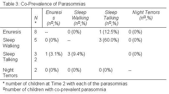
In Table 3 is shown the number and percent of parasomnias that occurred in association with other parasomnias at Time 2. Except for an association between sleep walking and sleep talking, parasomnias occurred independent of each other.  


Discussion 

The TuCASA study has documented the prevalence, incidence and remission of parasomnias in a population-based sample of 10-17 year old subjects. We found that incidence rates for parasomnias were very low for EN NT and SW and remission rates were high for all parasomnias. Furthermore, incidence and remission rates did not appear to be related to symptoms of sleep disturbances or learning problems, and except for sleep walking and sleep talking, they generally were not co-prevalent.

In this study, except for ST, prevalence rates for parasomnias in our cohort of adolescents were relatively low. Available data documenting the prevalence rates of various parasomnias in this age group are relatively sparse with previous reports largely restricted to preadolescents. 4, 9-12, 15 However, with respect to SW, 3 previous studies in adolescents have observed prevalence rates ranging from 3 to 15% which are higher than the 1.4% noted in our study.18-20 Inconsistent prevalence rates have been noted for NT with one study reporting rates <4%,20  but another reporting 10.2%.19 In contrast, the prevalence of enuresis 7, 18, 20 and the prevalence of ST 6,13, 20 in adolescents have been reported to be consistently low and high respectively. Our data are concordant with these previous reports. However, the relevance of these comparisons is unclear since no standard method of assessing the frequency of parasomnias exists. Our requirement that these events occurred more than three to five times per month are more stringent than those employed in most studies. Thus, it not surprising that our prevalence rates for SW and NT in adolescence are discordant with previous observations.

There is general consensus that childhood parasomnias remit as children develop from childhood to adolescence and that few adolescents develop them.5, 21 However, this impression is based primarily on empiric observations because there are few longitudinal studies.20, 22, 23 The largest longitudinal study prospectively interrogated parents of children at age 10 through age 13 years, but retrospectively questioned parents to determine if a parasomnia was present between ages 3 and 9 years.20 In this study, prevalence rates were noted to decline markedly by age 13 years to 3.3%, 1.2% and 2.0% for SW, NT and EN, respectively. In contrast, there was only a slight nonsignificant decrease in ST with 29.2% of children still having this condition at age 13 years. Our data are generally consistent with the results of this previous study although our prevalence of ST is somewhat lower. However, we extend these foregoing findings by documenting incidence and remission rates. Except for ST, very few adolescents developed new parasomnias. Nonetheless, although remission rates were high, some participants in this study had persistent symptoms which likely continue through into adulthood.24, 25

We previously examined the prevalence of parasomnias in the TuCASA cohort when the children were preadolescents.9 We found an associations between parasomnias, and SDB, symptoms of other sleep disturbances and learning problems. Unfortunately, because of the small numbers of children with parasomnias in this follow-up cohort, we were unable to determine whether these latter findings are still present.

In this study, standardized BMI increased as did the % of the cohort classified as obese. While these observations most likely are a reflection of the ongoing obesity epidemic in the United States, those with parasomnias did not have significantly higher standardized BMI.

Some evidence indicates that individuals with one parasomnia have a greater likelihood of having another one.20, 25 Consistent with these previous studies, we found a modest association between ST and SW, both of which are disorders of arousal. Otherwise, we found little evidence to support the contention that parasomnias are more likely to be co-prevalent.

Although our study is the first to prospectively document the incidence and remission of parasomnias in a large general population of children, it is not without some limitations. First, it is possible that parents underestimated the actual number of parasomnias that occurred. Depending on the bedtime of the child and the severity of the event, it is probable that parents are not awake during every occurrence. Second, recruitment may have incurred a selection bias so that parents who agreed to have their children participate might be more likely to have symptomatic children than those who did not. We think this unlikely because the focus of the study was SDB and it is unlikely that parents agreed to have their children participate based on the presence of parasomnias. Lastly, there were 153 subjects that participated at Time 1 but that did not participate at Time 2. There was no difference related to gender between those that participated at Time 2 and those that did not although slightly more Hispanic adolescents were lost to follow-up than Caucasians. In addition, the prevalence rates of parasomnias in the entire cohort and the restricted were the same. Thus, we do not believe that the restricted cohort of children who had data at both time points was markedly different than the larger group of children recruited at Time 1.

In conclusion, although parasomnias are relatively common in childhood, our study demonstrates that most remit, and that the development of parasomnias in older children is uncommon. Our findings provide objective data supporting the generally accepted perception that most parasomnias in children will resolve over time.


Acknowledgements         

This study was supported by Grant HL 62373 from the National Heart Lung and Blood Institute. All authors contributed to the writing and analyses contained in the study and had full access to the data. Dr. Quan is the Principal Investigator of the TuCASA study. Drs. Quan and Goodwin supervised the recruitment of participants and the operations of TuCASA.


References

1 . Matwiyoff G, Lee-Chiong T. Parasomnias: an overview. Indian J Med Res 2010; 131:333-337.

2 . Heussler HS. 9 Common causes of sleep disruption and daytime sleepiness: childhood sleep disorders II. Med J Aust 2005; 182(9):484-489.

3 . Chokroverty S. Overview of sleep & sleep disorders. Indian J Med Res 2010; 131:126-140.

4 . Agargun MY, Cilli AS, Sener S, et al. The prevalence of parasomnias in preadolescent school-aged children: a Turkish sample. Sleep 2004; 27(4):701-705.

5 . Avidan AY, Kaplish N. The parasomnias: epidemiology, clinical features, and diagnostic approach. Clin Chest Med 2010; 31(2):353-370.

6 . Arkin AM. Sleep-talking: a review. J Nerv Ment Dis 1966; 143(2):101-122.

7 . Hansakunachai T, Ruangdaraganon N, Udomsubpayakul U, Sombuntham T, Kotchabhakdi N. Epidemiology of enuresis among school-age children in Thailand. J Dev Behav Pediatr 2005; 26(5):356-360.

8 . Mikkelsen EJ, Rapoport JL, Nee L, Gruenau C, Mendelson W, Gillin JC. Childhood enuresis. I. Sleep patterns and psychopathology. Arch Gen Psychiatry 1980; 37(10):1139-1144.

9 . Goodwin JL, Kaemingk KL, Fregosi RF, et al. Parasomnias and sleep disordered breathing in Caucasian and Hispanic children - the Tucson children's assessment of sleep apnea study. BMC Med 2004; 2:14.

10 . Arruda MA, Guidetti V, Galli F, Albuquerque RC, Bigal ME. Childhood periodic syndromes: a population-based study. Pediatr Neurol 2010; 43(6):420-424.

11 . Liu X, Ma Y, Wang Y, et al. Brief report: An epidemiologic survey of the prevalence of sleep disorders among children 2 to 12 years old in Beijing, China. Pediatrics 2005; 115(1 Suppl):266-268.

12 . Simola P, Niskakangas M, Liukkonen K, et al. Sleep problems and daytime tiredness in Finnish preschool-aged children-a community survey. Child Care Health Dev 2010; 36(6):805-811.

13 . Garcia-Jimenez MA, Salcedo-Aguilar F, Rodriguez-Almonacid FM, et al. The prevalence of sleep disorders among adolescents in Cuenca, Spain. Rev Neurol 2004; 39(1):18-24.

14 . Ipsiroglu OS, Fatemi A, Werner I, Tiefenthaler M, Urschitz MS, Schwarz B. Prevalence of sleep disorders in school children between 11 and 15 years of age. Wien Klin Wochenschr 2001; 113(7-8):235-244.

15 . Petit D, Touchette E, Tremblay RE, Boivin M, Montplaisir J. Dyssomnias and parasomnias in early childhood. Pediatrics 2007; 119(5):e1016-25.

16 . Goodwin JL, Enright PL, Kaemingk KL, et al. Feasibility of using unattended polysomnography in children for research--report of the Tucson Children's Assessment of Sleep Apnea study (TuCASA). Sleep 2001; 24(8):937-944.

17 . Carroll JL, McColley SA, Marcus CL, Curtis S, Loughlin GM. Inability of clinical history to distinguish primary snoring from obstructive sleep apnea syndrome in children. Chest 1995; 108(3):610-618.

18 . Klackenberg G. Incidence of parasomnias in children in a general population. In: Sleep and its Disorders in Children. Guilleminault C, ed. New York: Raven Press, 1987; 99-113

19 . Ipsiroglu OS, Fatemi A, Werner I, Paditz E, Schwarz B. Self-reported organic and nonorganic sleep problems in schoolchildren aged 11 to 15 years in Vienna. J Adolesc Health 2002; 31(5):436-442.

20 . Laberge L, Tremblay RE, Vitaro F, Montplaisir J. Development of parasomnias from childhood to early adolescence. Pediatrics 2000; 106(1 Pt 1):67-74.

21 . Guilleminault G. Disorders of arousal in children: Somnambulism and night terros. In: Guilleminault C, ed. New York: Raven Press, 1987; 243-252

22 . DiMario FJ,Jr, Emery ES,3rd. The natural history of night terrors. Clin Pediatr (Phila) 1987; 26(10):505-511.

23 . Klackenberg G. Somnambulism in childhood--prevalence, course and behavioral correlations. A prospective longitudinal study (6-16 years). Acta Paediatr Scand 1982; 71(3):495-499.

24 . Oluwole OS. Lifetime prevalence and incidence of parasomnias in a population of young adult Nigerians. J Neurol 2010; 257(7):1141-1147.

25 . Bjorvatn B, Gronli J, Pallesen S. Prevalence of different parasomnias in the general population. Sleep Med 2010; 11(10):1031-1034.

Tuesday
Apr052011

A 45-YEAR-OLD MAN WITH EXCESSIVE DAYTIME SOMNOLENCE, AND WITNESSED APNEA AT ALTITUDE

Rebecca Keith MD

University of Colorado Denver, Denver, CO

rebecca.keith@ucdenver.edu


Carolyn H. Welsh MD

Veterans Affairs Medical Center, Denver, CO

Professor of Medicine

Division of Pulmonary Sciences and Critical Care Medicine, University of Colorado Denver, Denver, CO

carolyn.welsh@va.gov

Study performed at Denver Veterans Affairs Medical Center

Reference as: Keith R, Welsh CH. A 45-year-old man with excessive daytime somnolence, and witnessed apnea at altitude. Southwest J Pulm Crit Care 2011;2:53-57. (Click here for PDF version)


Abstract

A sleepy man without sleep apnea at 1609m (5280 feet) had disturbed sleep at his home altitude of 3200m (10500 feet).  In addition to common disruptors of sleep such as psychophysiologic insomnia, restless leg syndrome, alcohol and excessive caffeine use, central sleep apnea with periodic breathing can be a significant cause of disturbed sleep at altitude.  In symptomatic patients living at altitude, a sleep study at their home altitude should be considered to accurately diagnose the presence and magnitude of sleep disordered breathing as sleep studies performed at lower altitudes may miss this diagnosis. Treatments options differ from those to treat obstructive apnea.  Supplemental oxygen is considered by many to be first-line therapy.


Case Report 

A 45 year old man presents for evaluation of poor sleep, daytime somnolence, and morning headaches.  His bed partner reports that he has loud snoring, gasping arousals, and frequent episodes of apnea.  He is an obese man with diabetes, hypertension, rhinosinusitis, and gastroesophageal reflux disease who is a long time resident at 3200m (10500 feet).  Polysomnography performed at 1609m (5280 feet) showed a normal apnea-hypopnea index (AHI) of 1.4.  He spent less than one minute with an oxygen saturation (SaO2) less than 88%. 

Given the severity of his symptoms, a home cardiopulmonary sleep study was performed at 3200m (10500 feet).  AHI was 39 non-supine and 56 supine. A representative portion of his study is shown in Figure 1.  It demonstrates the absence of respiratory effort during apnea events, consistent with central sleep apnea from periodic breathing at altitude.

Figure 1. This is a representative example of breathing during sleep. The upper two channels show nasal (nasal flow) and oral air flow (thermistor).  Respiratory effort signals in the abdomen and thorax are shown in the next two channels and oxygen saturation in the bottom tracing. A ten minute period of recording is displayed. (Click here for JPEG image)

Discussion

Central apnea is characterized by recurrent episodes of apnea resulting from temporary loss of ventilatory effort.  These episodes generally result from a strong dependence on the metabolic control of ventilation during sleep.1   Conditions inducing a reduced partial pressure of carbon dioxide in arterial blood (PaCO2) can precipitate a central apnea, often associated with an arousal and leading to daytime somnolence.    

At higher altitudes, the fraction of inspired oxygen (FiO2) is the same as it is at sea level, but the barometric pressure and oxygen tension are less, so the partial pressure of oxygen in arterial blood (PaO2) and SaO2 tend to be reduced.  This tendency toward hypoxemia elicits a series of physiologic responses including increased alveolar ventilation, lowering the resting PaCO2, shrinking the gap between the resting PaO2 and the PaCO2 associated with apnea (the apnea threshold).  People with sensitive chemoreceptors are more likely to develop periodic breathing at altitude.  An increased ventilatory response to hypoxemia may reduce PaCO2 below the apnea threshold.  The resultant apnea will eventually result in worsened hypoxemia, which will trigger hyperventilation setting up the oscillating pattern of apnea and hyperventilation typical of periodic breathing.1  Of note, these responses seem primarily related to hypoxemia rather than low barometric pressure as they are present in simulated normobaric hypoxia equivalent to 2000m (6562 feet) altitude .2 

Central sleep apnea encompasses multiple disorders including narcotic-induced central apnea, obesity-hypoventilation syndrome, Cheyne-Stokes breathing often seen with heart failure or cerebrovascular disease, and high altitude-induced periodic breathing.  Periodic breathing at altitude has the pattern of apneic episodes with cycles of hypoxemia, subsequent hyperventilation, hypocapnia, and resulting apnea described above.  The skeletal muscle atonia and reduced respiratory drive3 that normally occurs during rapid eye movement (REM) sleep typically terminates the periodicity by eliminating hyperventilation and restoring regular breathing.  Thus, periodic breathing at altitude is seen primarily during non-REM sleep.4   This aberrant respiratory pattern may initially be beneficial at high altitude since the hyperventilation increases SaO2 by 2.9±1.5%, thus improving hypoxemia.4  In our patient, this potential benefit was overcome by a significant increase in sleep disturbance leading to sleep deprivation as well as mental and physical impairment.

Treatment options for periodic breathing at altitude include nighttime oxygen therapy, acetazolamide, and sedative-hypnotics.  Supplemental oxygen breaks the apnea cycle by eliminating the hypoxemia that results in hyperventilation, hypocapnia, and apnea.  Studies of supplemental oxygen at altitude have shown that subjects spend more time in deep sleep and have improvements in SaO2, tidal volume and AHI.5  

Acetazolamide, a carbonic anhydrase inhibitor, induces a metabolic acidosis by urinary elimination of bicarbonate. This is thought to increase ventilation, left-shift the hypercapnic ventilatory response and thus reduce apnea events.  Acetazolamide can significantly improve sleep disordered breathing and oxygen saturation during sleep.6 Sedative-hypnotic agents have also improved slow wave sleep at altitude without adversely affecting respiration or performance.  This improvement is thought to be secondary to a decrease in wakefulness after sleep onset and improved sleep efficiency.7

This patient presented with classic symptoms of sleep apnea with a negative sleep study at 1609m (5280 feet), arousing clinical suspicion for periodic breathing at altitude.  A repeat home sleep study revealed moderate to severe central sleep apnea.  After treatment with oxygen and acetazolamide his symptoms significantly improved and repeat home monitoring demonstrated an AHI of 6.  Treatment with oxygen alone resulted in improvement as well with an AHI of 13.

Central sleep apnea and periodic breathing are common causes of disturbed sleep at altitude.  Symptomatic patients living at altitude should have a sleep study at their home altitude to accurately diagnose the presence and magnitude of sleep disordered breathing.  Oxygen is considered by many to be first line therapy.


References

1.  Eckert DJ, Jordan AS, Merchia P, Malhotra A. Central sleep apnea: Pathophysiology and Treatment. Chest 2007; 131(2): 595-607.

2. Kinsman TA, Hahn AG, Gore CJ, Wilsmore BR, Martin DT, Chow CM. Respiratory events and periodic breathing in cyclists sleeping at 2,650-m simulated altitude. Journal of Applied Physiology 2002; 92 (5): 2114-8.

3. Schafer, T, Schlafke ME, Respiratory changes associated with rapid eye movements in normo- and hypercapnia during sleep. Journal of Applied Physiology 1998; 85: 2213-2219.

4. Salvaggio A, Insalaco G, Marrone O, et al. Effects of high-altitude periodic breathing on sleep and arterial oxyhaemoglobin saturation. European Respiratory Journal 1998; 12 (2): 408-13.

5. Windsor JS, Rodway GW. Supplemental oxygen and sleep at altitude. High Altitude Medicine & Biology 2006; 7 (4): 307-11.

6. Fischer R, Lang SM, Leitl M, Thiere M, Steiner U, Huber RM. Theophylline and acetazolamide reduce sleep-disordered breathing at high altitude. European Respiratory Journal 2004; 23 (1): 47-52.

7. Beaumont M, Batejat D, Pierard C, et al. Zaleplon and zolpidem objectively alleviate sleep disturbances in mountaineers at a 3,613 meter altitude. Sleep 2007; 30 (11): 1527-33.


Acknowledgments:

Rebecca Keith was the primary author writing the manuscript. Carolyn Welsh also contributed to content and manuscript preparation and revision.


Corresponding author: Rebecca Keith MD

                                     National Jewish Health

                                     1400 Jackson Street

                                     Denver, CO 80206

                                     Phone: 303-398-1511

                                     FAX: 303-398-1381

No financial support was provided for this manuscript and there is no off-label or investigational use of medications or devices.

Dr Keith has no conflict of interest to disclose

Dr. Welsh has no conflict of interest to disclose

Abbreviation List:  

AHI - Apnea hypopnea index, the number of apneas or hypopneas per hour of sleep or recording

m – Meters

FiO2 – Fraction of inspired oxygen

PaCO2 - Partial pressure of carbon dioxide in arterial blood

PaO2 – Partial pressure of oxygen in arterial blood

REM – Rapid eye movement

SaO2 – Percentage of available hemoglobin that is saturated with oxygen

Page 1 ... 4 5 6 7 8