
August 2014 Imaging Case of the Month
Michael B. Gotway, MD
Department of Radiology
Mayo Clinic Arizona
Scottsdale, AZ
Clinical History: A 42-year-old non-smoking woman presented with a history of relatively sudden onset left chest pain and shortness of breath. Her past medical history was remarkable for psoriasis, treated with Enbrel® (etanercept). She also had a history of partial hysterectomy for fibroids and right oophorectomy. Frontal and lateral chest radiography (Figure 1) were performed.
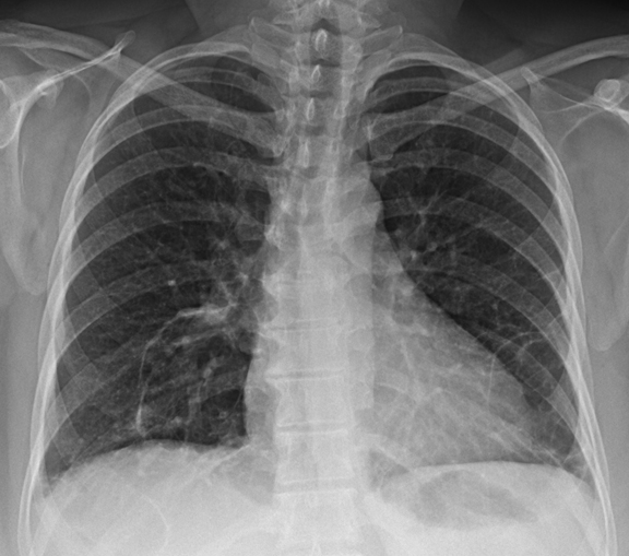
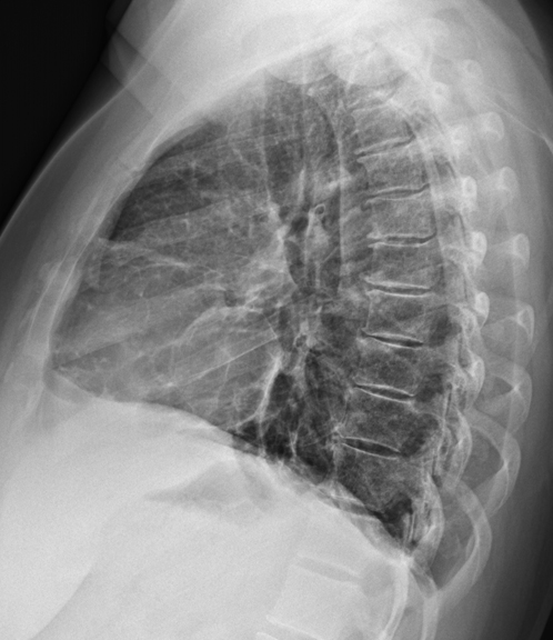
Figure 1. Frontal (A) and lateral (B) chest radiography.
Which of the following statements regarding the chest radiograph is most accurate? (Click on the correct answer to move to the next panel)
Reference as: Gotway MB. August 2014 imaging case of the month. Southwest J Pulm Crit Care. 2014;9(2):83-90. doi: http://dx.doi.org/10.13175/swjpcc104-14 PDF
 Post a Comment
Reader Comments