
Medical Image of the Week: Pneumocephalus
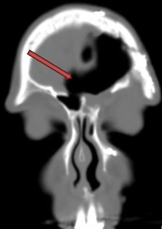
Figure 1. Coronal CT Scan Image (Brain Lab Protocol) showing air in continuity with the frontal sinuses where there is no bone between the frontal sinus and the brain.
A 53-year-old woman underwent left craniotomy at an outside hospital after a trauma related to a motor vehicle accident. She was recovering well from her surgery. Two months later she started having headache and noticed progressive difficulty with word finding. She also reported periodic rhinorrhea upon further questioning. She presented to the emergency room at an outside hospital where a CT head was done. She was subsequently transferred to our ICU for higher level of care. Her CT Head revealed pneumocephalus involving the left frontal lobe along with regional mass effect leading to left to right shift of the anterior interhemispheric fissure. A dedicated brain lab protocol showed air in continuity with the frontal sinuses with a bone defect between the frontal sinus and the brain. She was evaluated by ENT and Neurosurgery services. She was placed on 100% oxygen via non-rebreather mask, frequent neuro-checks and a redo of the left frontal craniotomy for repair of the skull base defect was performed.
Administration of postsurgical supplemental oxygen through a non-rebreather mask has shown to significantly increase the absorption rate of postcraniotomy pneumocephalus as compared with breathing room air (1). In a prospective study of thirteen patients with postoperative pneumocephalus that was estimated to be > 30 ml were alternately assigned to breathe 100% oxygen using a nonrebreather mask (treatment group) or to breathe room air (control group) for 24 hours. There was a significant difference (p = 0.009) between the mean rate of pneumocephalus volume reduction in the treatment (65%) and control groups (31%) per 24 hours.
Tauseef Afaq Siddiqi MD, Enas M. Al Zaghal MD, and Sairam Parthasarathy MD
Division of Pulmonary, Allergy, Critical Care and Sleep Medicine
The University of Arizona
Tucson, AZ
Reference
- Gore PA, Maan H, Chang S, Pitt AM, Spetzler RF, Nakaji P. Normobaric oxygen therapy strategies in the treatment of postcraniotomy pneumocephalus. J Neurosurg. 2008;108(5):926-9. [CrossRef] [PubMed]
Reference as: Siddiqi TA, Zaghal EM, Parthasarathy S. Medical image of the week: pneumocepahlus. Southwest J Pulm Crit Care. 2014;9(1):57-8. doi: http://dx.doi.org/10.13175/swjpcc095-14 PDF
 Post a Comment
Reader Comments