
November 2014 Imaging Case of the Month
Michael B. Gotway, MD
Department of Radiology
Mayo Clinic Arizona
Scottsdale, AZ
Clinical History: A 38-year-old non-smoking woman presented with complaints of intermittent dry cough, occasional vomiting, and dyspnea, occasionally with fever and chills. She indicated that she has suffered recurrent bouts of bronchitis and pneumonia annually over the previous 10 years. The patient had a history of upper arm localized melanoma resection 10 years earlier. She had smoked for 10 years, but quit one year prior to presentation. Her past medical and surgical histories were otherwise unremarkable.
Frontal and lateral chest radiography (Figure 1) was performed.
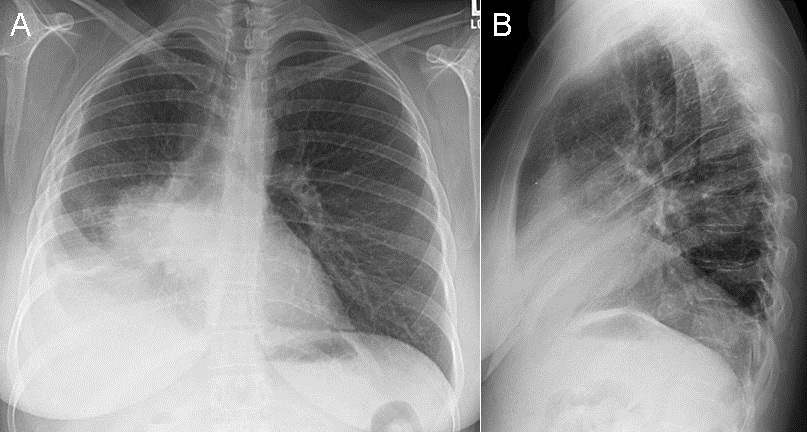
Figure 1. Frontal (A) and lateral (B) chest radiography.
Which of the following statements regarding the chest radiograph is most accurate? (click on the correct answer to proceed to the next panel)
Reference as: Gotway MB. November 2014 imaging case of the month. Southwest J Pulm Crit Care. 2014;9(5):264-77. doi: http://dx.doi.org/10.13175/swjpcc147-14 PDF
 Post a Comment
Reader Comments