
Saturday
May042013
May 2013 Imaging Case of the Month
Michael B. Gotway, MD
Department of Radiology
Mayo Clinic Arizona
Scottsdale, AZ
Clinical History
A 21-year-old woman presented with complaints of cough. Frontal and lateral chest radiography (Figures 1A & B) was performed. A detail comparison chest radiograph from several years prior (Figure 1C) is presented as well.

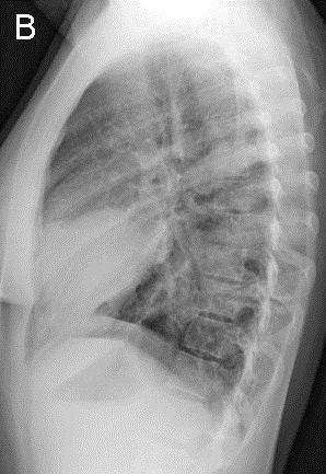
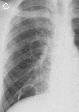
Figure 1. Frontal (A) and lateral (B) chest radiography at presentation and a radiograph from several years earlier (C).
Which of the following statements regarding the chest radiograph is most accurate?
- The chest radiograph predominantly shows bilateral linear and reticular abnormalities
- The chest radiograph shows a combination of nodules, masses and thin-walled cysts
- The chest radiograph shows multifocal consolidation with air bronchograms
- The chest radiograph shows multifocal pleural abnormalities
- The chest radiograph shows mediastinal widening & hilar lymphadenopathy
Reference as: Gotway MB. May 2013 imaging case of the month. Southwest J Pulm Crit Care.2013;6(5):218-30. PDF
 Post a Comment
Reader Comments