
Correct!
2. The chest radiograph shows a combination of nodules, masses and thin-walled cysts
The visible mediastinal and hilar contours are within normal limits; no lymphadenopathy is present. The right hilum appears enlarged on the frontal image, but the hilum is shown to be normal on the lateral projection. The appearance of hilar enlargement on the frontal chest radiograph is caused by a mass overlying this region, located slightly posterior to the right hilum, seen to advantage on the lateral radiograph. The interstitium is largely normal; linear or reticular abnormalities, or findings suggesting fibrotic lung disease, are not the dominant abnormalities present on the chest radiograph. The costophrenic angles are sharp and no pleural abnormalities are seen. A mass-like opacity is present in the right middle lobe and in the right suprahilar region, and at least one thin-walled cyst is seen in the left lower lobe, with another in the right mid-lung. No air bronchograms, however, are seen within the focal lung opacities.)
Several images from a thoracic CT scan (Figure 2) performed 1 year prior to the presentation chest radiograph were subsequently located.
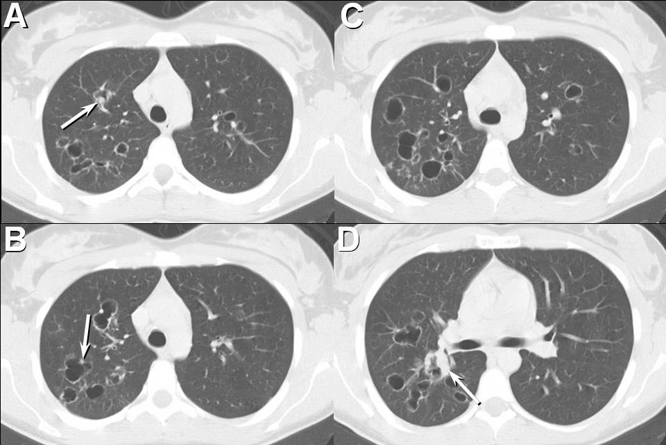
Figure 2. Thoracic CT performed 1 year prior to presentation.
Which of the following statements regarding this CT examination is most accurate?