
Sunday
Feb032013
February 2013 Imaging Case of the Month
Michael B. Gotway, MD
Associate Editor Imaging
Department of Radiology
Mayo Clinic Arizona
Scottsdale, AZ
Clinical History: A 50-year-old previously healthy woman presented with complaints of intermittent back pain. The patient’s physical examination was unremarkable. Conservative treatment for these complaints was unsuccessful and thoracic spine radiography was performed, which showed abnormal lung findings, prompting frontal chest radiography (Figure 1).
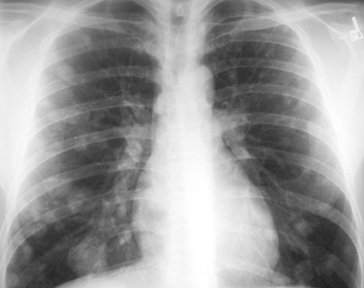
Figure 1. Frontal chest radiography.
Which of the following statements regarding the chest radiograph is most accurate?
- The chest radiograph shows multiple, bilateral cavitary nodules
- The chest radiograph shows nodular interstitial thickening
- The chest radiograph shows multiple, bilateral circumscribed nodules
- The chest radiograph shows mediastinal and hilar lymph node enlargement
- The chest radiograph shows multifocal nodular pulmonary consolidation
Reference as: Gotway MB. February 2013 imaging case of the month. Southwest J Pulm Crit Care. 2013;6(2):75-81. PDF
 Post a Comment
Reader Comments