Correct!
3. The chest radiograph shows multiple, bilateral circumscribed nodules
The chest radiograph shows multiple, bilateral circumscribed nodules, but the nodules do not contain internal lucency- there is no evidence of cavitation. The interstitium does not appear abnormally thickened. The circumscribed nature of the opacities, and the lack of air bronchograms, indicate that the chest radiographic abnormalities should not be characterized as consolidation. Finally, the hilar and mediastinal contours appear normal- no evidence of lymphadenopathy is seen.
The patient subsequently underwent thoracic CT (Figure 2) for further characterization of the pulmonary abnormalities seen at chest radiography.
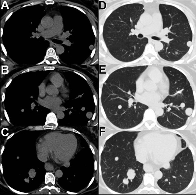
Figure 2. Thoracic CT displayed in soft tissue (A-C) and lung (D-F).
Regarding the thoracic CT, which of the following statements is most accurate?