
Saturday
Jun032017
June 2017 Imaging Case of the Month
Michael B. Gotway, MD
Department of Radiology
Mayo Clinic Arizona
Scottsdale, Arizona USA
Clinical History: A 30-year-old woman with no significant past medical history presented with complaints of chronic back pain, partially controlled with Ibuprofen. Recently she began to notice shortness of breath. Frontal and lateral chest radiography (Figure 1) was performed.
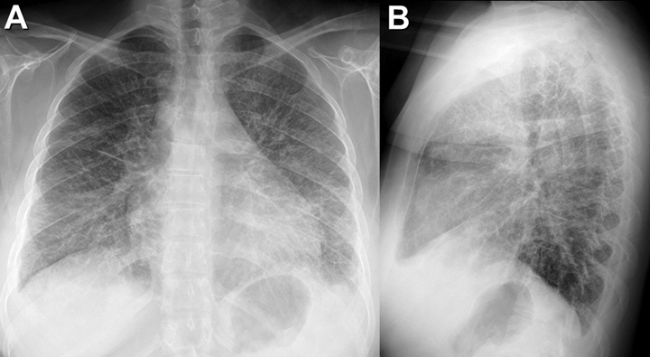
Figure 1. Frontal (A) and lateral (B) chest radiography
Which of the following statements regarding the chest radiograph is most accurate? (Click on the correct answer to proceed to the second of eight pages)
- The chest radiograph shows a diffuse linear, interstitial pattern
- The chest radiograph shows a large pleural effusion
- The chest radiograph shows a mediastinal mass
- The chest radiograph shows multifocal, bilateral consolidation
- The chest radiograph shows numerous small nodules
Cite as: Gotway MB. June 2017 imaging case of the month. Southwest J Pulm Crit Care. 2017;14(6):269-78. doi: https://doi.org/10.13175/swjpcc068-17 PDF
 Post a Comment
Reader Comments