
Tuesday
Jun032014
June 2014 Imaging Case of the Month
Michael B. Gotway, MD
Department of Radiology
Mayo Clinic Arizona
Scottsdale, AZ
Clinical History: A 63-year-old man with a history of early-stage Parkinson disease and coronary artery disease presented with a painful “lump” in the lower left neck. Frontal and lateral chest radiography (Figure 1) was performed.
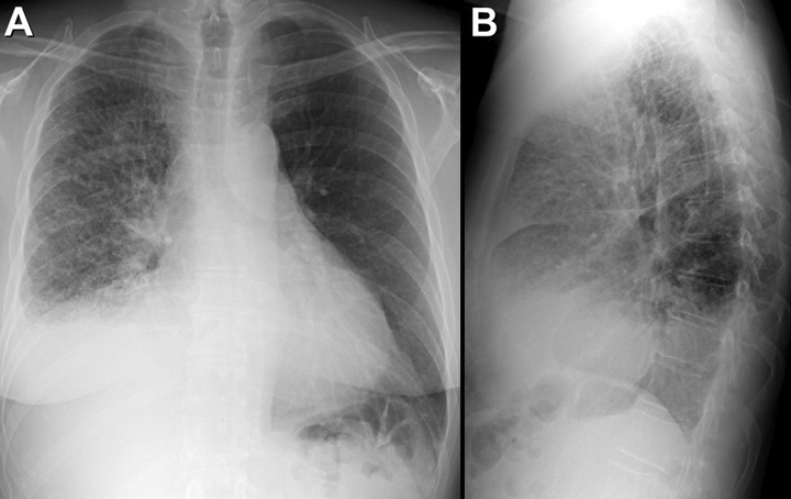
Figure 1. Frontal (panel A) and lateral (panel B) chest radiograph.
Which of the following statements regarding the chest radiograph is most accurate?
Reference as: Gotway MB. June 2014 imaging case of the month. Southwest J Pulm Crit Care. 2014;8(6):320-7. doi: http://dx.doi.org/10.13175/swjpcc074-14 PDF
 Post a Comment
Reader Comments