
Medical Image of the Week: Lepidic Growth
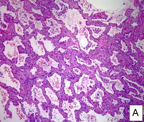
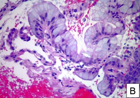
Figure 1. Two different tumors showing lepidic growth along the alveolar interstitium with preserved alveolar architecture.
Lepidic growth is most often seen in adenocarcinoma in situ (Figure A, 40x magnification). Adenocarcinoma in situ is formerly known as bronchoalveolar cell carcinoma (BAC). A similar growth pattern in a morphologically very different tumor (mucinous adenocarcinoma) is shown for comparison (Figure B, 400x). Mucinous adenocarcinoma growing on alveolar septae nearly always is invasive, so the entity of mucinous adencioarcinoma in situ practically doesn't exist, further differentiating this entity from BAC.
Ken Knox, MD and Richard Sobonya, MD
Departments of Medicine and Pathology
University of Arizona
Tucson, Arizona
Reference as: Knox KS, Sobonya RE. Medical image of the week: lepidic growth. Southwest J Pulm Crit Care. 2013;7(2):109. doi: http://dx.doi.org/10.13175/swjpcc111-13 PDF
 Post a Comment
Reader Comments