
March 2013 Imaging Case of the Month
Michael B. Gotway, MD*
Sudheer Penupolu, MD‡
Jasminder Mand, MD†
*Department of Radiology, Mayo Clinic, Arizona
‡Fellow, Pulmonary Medicine, Mayo Clinic Arizona
†Pulmonary and Critical Care Medicine, Maricopa Medical Center
Clinical History: A 54-year old Hispanic woman with no significant past medical history presented with complaints of cough and worsening dyspnea. She was in her usual state of health until 4-5 weeks prior to presentation when she started noticing gradually worsening dyspnea on exertion. She reported a dry cough initially which subsequently became productive of whitish, mucoid sputum. The patient denied chest pain, sore throat, sick contacts, or recent travel history. A chest x-ray was performed (Figure 1).
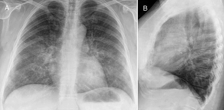
Figure 1. Frontal (A) and lateral (B) chest radiography.
Which of the following statements regarding the chest radiograph is most accurate?
- The chest radiograph shows bilateral linear and reticular abnormalities
- The chest radiograph shows nodular interstitial thickening
- The chest radiograph shows multiple, bilateral circumscribed nodules
- The chest radiograph shows mediastinal and hilar lymph node enlargement
- The chest radiograph shows mediastinal widening
Reference as: Gotway MB, Penupolu S, Mand J. March 2013 imaging case of the month. Southwest J Pulm Crit Care. 2013;6(3):112-24. PDF
 Post a Comment
Reader Comments