Search Journal-type in search term and press enter
Southwest Pulmonary and Critical Care Fellowships
In Memoriam
Social Media

Imaging

Last 50 Imaging Postings

(Most recent listed first. Click on title to be directed to the manuscript.)

May 2025 Medical Image of the Month: Aspirated Dental Screw
April 2025 Medical Image of the Month: An Unfortunate Case of Mimicry
March 2025 Medical Image of the Month: An Unusual Case of Pulmonary
   Infarction
February 2025 Medical Image of the Month: Unexpected Complications of
   Transjugular Intrahepatic Portosystemic Shunt (TIPS) 
February 2025 Imaging Case of the Month: A Wolf in Sheep’s Clothing
January 2025 Medical Image of the Month: Psoriasis with Pulmonary
   Involvement
December 2024 Medical Image of the Month: An Endobronchial Tumor
November 2024 Medical Image of the Month: A Case of Short Telomeres
November 2024 Imaging Case of the Month: A Recurring Issue
October 2024 Medical Image of the Month: Lofgren syndrome with Erythema
   Nodosum
September 2024 Medical Image of the Month: A Curious Case of Nasal
   Congestion
August 2024 Image of the Month: Lymphomatoid Granulomatosis
August 2024 Imaging Case of the Month: An Unexplained Pleural Effusion
July 2024 Medical Image of the Month: Vocal Cord Paralysis on PET-CT 
June 2024 Medical Image of the Month: A 76-year-old Man Presenting with
   Acute Hoarseness
May 2024 Medical Image of the Month: Hereditary Hemorrhagic
   Telangiectasia in a Patient on Veno-Arterial Extra-Corporeal Membrane
   Oxygenation
May 2024 Imaging Case of the Month: Nothing Is Guaranteed
April 2024 Medical Image of the Month: Wind Instruments Player Exhibiting
   Exceptional Pulmonary Function
March 2024 Medical Image of the Month: Sputum Cytology in Patients with
   Suspected Lung Malignancy Presenting with Acute Hypoxic Respiratory
   Failure
February 2024 Medical Image of the Month: Pulmonary Alveolar Proteinosis
   in Myelodysplastic Syndrome
February 2024 Imaging Case of the Month: Connecting Some Unusual Dots
January 2024 Medical Image of the Month: Polyangiitis Overlap Syndrome
   (POS) Mimicking Fungal Pneumonia 
December 2023 Medical Image of the Month: Metastatic Pulmonary
   Calcifications in End-Stage Renal Disease 
November 2023 Medical Image of the Month: Obstructive Uropathy
   Extremis
November 2023 Imaging Case of the Month: A Crazy Association
October 2023 Medical Image of the Month: Swyer-James-MacLeod
   Syndrome
September 2023 Medical Image of the Month: Aspergillus Presenting as a
   Pulmonary Nodule in an Immunocompetent Patient
August 2023 Medical Image of the Month: Cannonball Metastases from
   Metastatic Melanoma
August 2023 Imaging Case of the Month: Chew Your Food Carefully
July 2023 Medical Image of the Month: Primary Tracheal Lymphoma
June 2023 Medical Image of the Month: Solitary Fibrous Tumor of the Pleura
May 2023 Medical Image of the Month: Methamphetamine Inhalation
   Leading to Cavitary Pneumonia and Pleural Complications
April 2023 Medical Image of the Month: Atrial Myxoma in the setting of
   Raynaud’s Phenomenon: Early Echocardiography and Management of
   Thrombotic Disease
April 2023 Imaging Case of the Month: Large Impact from a Small Lesion
March 2023 Medical Image of the Month: Spontaneous Pneumomediastinum
   as a Complication of Marijuana Smoking Due to Müller's Maneuvers
February 2023 Medical Image of the Month: Reversed Halo Sign in the
   Setting of a Neutropenic Patient with Angioinvasive Pulmonary
   Zygomycosis
January 2023 Medical Image of the Month: Abnormal Sleep Study and PFT
   with Supine Challenge Related to Idiopathic Hemidiaphragmatic Paralysis
December 2022 Medical Image of the Month: Bronchoesophageal Fistula in
   the Setting of Pulmonary Actinomycosis
November 2022 Medical Image of the Month: COVID-19 Infection
   Presenting as Spontaneous Subcapsular Hematoma of the Kidney
November 2022 Imaging Case of the Month: Out of Place in the Thorax
October 2022 Medical Image of the Month: Infected Dasatinib Induced
   Chylothorax-The First Reported Case 
September 2022 Medical Image of the Month: Epiglottic Calcification
Medical Image of the Month: An Unexpected Cause of Chronic Cough
August 2022 Imaging Case of the Month: It’s All About Location
July 2022 Medical Image of the Month: Pulmonary Nodule in the
   Setting of Pyoderma Gangrenosum (PG) 
June 2022 Medical Image of the Month: A Hard Image to Swallow
May 2022 Medical Image of the Month: Pectus Excavatum
May 2022 Imaging Case of the Month: Asymmetric Apical Opacity–
   Diagnostic Considerations
April 2022 Medical Image of the Month: COVID Pericarditis
March 2022 Medical Image of the Month: Pulmonary Nodules in the
   Setting of Diffuse Idiopathic Pulmonary NeuroEndocrine Cell Hyperplasia
   (DIPNECH) 
February 2022 Medical Image of the Month: Multifocal Micronodular
   Pneumocyte Hyperplasia in the Setting of Tuberous Sclerosis
February 2022 Imaging Case of the Month: Between A Rock and a
   Hard Place
January 2022 Medical Image of the Month: Bronchial Obstruction
   Due to Pledget in Airway Following Foregut Cyst Resection
December 2021 Medical Image of the Month: Aspirated Dental Implant
Medical Image of the Month: Cavitating Pseudomonas
   aeruginosa Pneumonia
November 2021 Imaging Case of the Month: Let’s Not Dance
   the Twist
Medical Image of the Month: COVID-19-Associated Pulmonary
   Aspergillosis in a Post-Liver Transplant Patient


For complete imaging listings click here

Those who care for patients with pulmonary, critical care or sleep disorders rely heavily on chest radiology and pathology to determine diagnoses. The Southwest Journal of Pulmonary, Critical Care & Sleep publishes case-based articles with characteristic chest imaging and related pathology. The editor of this section will oversee and coordinate the publication of a core of the most important chest imaging topics. In doing so, they encourage the submission of unsolicited manuscripts. It cannot be overemphasized that both radiologic and pathologic images must be of excellent quality. As a rule, 600 DPI is sufficient for radiographic and pathologic images. Taking pictures of plain chest radiographs and CT scans with a digital camera is strongly discouraged. The figures should be cited in the text and numbered consecutively. The stain used for pathology specimens and magnification should be mentioned in the figure legend. Those who care for patients with pulmonary, critical care or sleep disorders rely heavily on chest radiology and pathology to determine diagnoses. The Southwest Journal of Pulmonary, Critical Care & Sleep publishes case-based articles with characteristic chest imaging and related pathology. The editor of this section will oversee and coordinate the publication of a core of the most important chest imaging topics. In doing so, they encourage the submission of unsolicited manuscripts. It cannot be overemphasized that both radiologic and pathologic images must be of excellent quality. As a rule, 600 DPI is sufficient for radiographic and pathologic images. Taking pictures of plain chest radiographs and CT scans with a digital camera is strongly discouraged. The figures should be cited in the text and numbered consecutively. The stain used for pathology specimens and magnification should be mentioned in the figure legend.

-------------------------------------------------------------------------------------------  

Wednesday
Nov012017

Medical Image of the Week: Erythema Nodosum


Figure 1. Panel A: photograph of legs at initial presentation. Panel B: 2 weeks later.

A healthy 43-year-old woman presented to the emergency room with one day of diarrhea, vomiting and severe left-sided pleuritic chest pain. Chest radiography revealed an infiltrate in the left lower lobe with a small pleural effusion. White count was 14,000 cells/mcL. Eosinophil count was 2%. She was thought to have pneumonia and treated with azithromycin.

Two weeks later she returned to the emergency room with painful raised erythematous lesions on both lower extremities and generalized joint pain. A thoracic CT scan was performed showing left lower lobe pneumonia and small bilateral pleural effusions. Eosinophilia was 14%. She was diagnosed again as having pneumonia. There was no diagnosis made of the skin lesions. A cocci serology was drawn and sent to Davis California. She was given doxycycline.

Subsequently the cocci serology was found to be positive for IgG and IgM and negative for complement fixation antibodies. She was seen by her primary care physician who diagnosed acute coccidioidomycosis and started her on Diflucan 200 mg daily and referred her for consultation. At her first visit 6 weeks into her illness she was still complaining of arthralgias, fatigue and cough.

Physical examination was negative except for innumerable red raised lesions on her thighs and anterior surfaces of her legs with confluence at the ankles (Figure 1). One month after her initial visit her legs were much improved although there was some peeling of the skin and residual erythema of the lower extremities (Figure 2).

Erythema Nodosum (EN) is a panniculitis of subcutaneous fat which can be associated with a variety of conditions including streptococcal pharyngitis, tuberculosis, sarcoidosis, inflammatory bowel disease, cancer, or bacterial infections (1). The usual presentation of this disease is the presence of painful raised erythematous nodules symmetrically on the anterior surfaces of the lower extremities. These lesions do not represent sites of infection but are most likely a result of type IV delayed hypersensitivity.

EN was first described as a benign form of coccidioidomycosis in 1936, and was further characterized by Charles Smith in 1940 when he described acute coccidioidomycosis as an illness characterized by an “influenza like initial phase followed in 2 to 18 days by the eruptive phase of erythema nodosum lasting from six days to three weeks with pigmented areas lasting for months” (2). He noted that recovery was invariable. This was in a time where the frequent presentation of acute coccidioidomycosis as an inapparent infection was not known and the mortality of acute cocci was as high as 50%. Twenty years later, Smith and Pappagiannis made the observation that EN was 2 to 10 times more frequent in females than males (3). Braverman (4) in 1999 observed the protective effect of EN by reporting on 60 pregnant women with coccidioidomycosis. Thirty of these women with EN had no dissemination, and of the 30 without EN, 11 disseminated and one died. The mechanism of this protective effect has yet to be characterized.

Physicians living in the Southwest have learned that “the bumps” (EN), desert rheumatism (polyarthralgia), eosinophilia associated with a flulike illness, and acute knifelike pleuritic chest pain in an otherwise healthy person are all signs and symptoms which lead to a rapid diagnosis of coccidioidomycosis (5). Because of the time sequence of the appearance of EN in this illness this rash is often felt incorrectly to be an allergic reaction to antibiotics given for the previously diagnosed pneumonia. Even though EN is associated with many varied conditions, its presence in the Southwestern United States should lead the physician to consider coccidioidomycosis as the most likely diagnosis.

Gerald F. Schwartzberg, MD

HonorHealth Pulmonology

Phoenix, AZ USA

References

  1. Blake T, Manahan M, Rodins K. Erythema nodosum - a review of an uncommon panniculitis. Dermatol Online J. 2014 Apr 16;20(4):22376. [PubMed]
  2. Smith CE. Epidemiology of acute coccidioidomycosis with erythema nodosum ("San Joaquin" or "Valley Fever"). Am J Public Health Nations Health. 1940 Jun;30(6):600-11. [CrossRef] [PubMed]
  3. Smith CE, Pappagianis D, Levine HB, Saito M. Human coccidioidomycosis. Bacteriol Rev. 1961 Sep;25:310-20. [PubMed]
  4. Braverman IM. Protective effects of erythema nodosum in coccidioidomycosis. Lancet. 1999 Jan 16;353(9148):168. [CrossRef] [PubMed]
  5. Stevens DA. Coccidioidomycosis. N Engl J Med. 1995 Apr 20;332(16):1077-82. [CrossRef] [PubMed]

Addendum: Another Erythema Nodosum

Figure 2. Another case of erythema nodosum.

While the above article was “in press”, another case of EN came into the office (Figure 2). Her history was similar to the first patient. Uncle Jun, the patriarch in the “Sopranos,” said in his heavy New York accent, “They come in threes.” Based on Uncle Jun, I am anticipating seeing another case of EN this week.

Gerald F. Schwartzberg, MD

HonorHealth Pulmonology

Phoenix, AZ USA

Cite as: Schwartzberg GF. Medical image of the week: erythema nodosum. Southwest J Pulm Crit Care. 2017;15(4):188-90. doi: https://doi.org/10.13175/swjpcc126-17 PDF 

Wednesday
Oct252017

Medical Image of the Week: Pulmonary Mycetoma

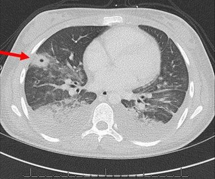
Figure 1. Thoracic CT scan showing mycetoma (arrow) in cavitary lesion in right upper lobe.

A 59 year-old woman presented with right sided chest pain and worsening shortness of breath. On CT of the chest she was found to have cavitary lesions in her right lung with one of them having a distinct opacity within the lesion concerning for a pulmonary mycetoma (Figure 1, arrow). Most literature describes pulmonary mycetomas occurring due to Aspergillus species. However, in our patient, neither the bronchoscopy with bronchoalveolar lavage (BAL) nor serological studies tested positive for Aspergillus. Cultures did however grow Candida albicans in 2 of the samples from the BAL. Mycetoma due to Candida has been described in the urinary tract in immunocompromised patients and, uncommonly, in the lung (1-3). Our patient had been treated for Stage III ovarian cancer with chemotherapy and at presentation her absolute neutrophil count was reduced at 860. In the hospital, she was treated for her shortness of breath with albuterol-ipratropium nebulizations to which she responded well. She was discharged once stable to follow up as outpatient for further treatment of her Candida albicans mycetoma.

Saud Khan, MD and Huzaifa A. Jaliawala, MD

Internal Medicine

University of Oklahoma Health Sciences Center

Oklahoma City, OK USA

References

  1. Praz V, Burruni R, Meid F, Wisard M, Jichlinski P, Tawadros T. Fungus ball in the urinary tract: A rare entity. Can Urol Assoc J. 2014 Jan-Feb;8(1-2):E118-20. [CrossRef] [PubMed]
  2. Song Z, Papanicolaou N, Dean S, Bing Z. Localized candidiasis in kidney presented as a mass mimicking renal cell carcinoma. Case Rep Infect Dis. 2012;2012:953590. [CrossRef] [PubMed]
  3. Bachh AA, Haq I, Gupta R, Varudkar H, Ram MB. Pulmonary candidiasis presenting as mycetoma. Lung India. 2008 Oct;25(4):165-7. [CrossRef] [PubMed]

Cite as: Khan S, Jaliawala HA. Medical image of the week: pulmonary mycetoma. Southwest J Pulm Crit Care. 2017;15(4):169-70. doi: https://doi.org/10.13175/swjpcc123-17 PDF