Imaging

Last 50 Imaging Postings

(Most recent listed first. Click on title to be directed to the manuscript.)

August 2025 Medical Image of the Month: Crazy Paving in a Case of 
   Eosinophilic Granulomatosis with Polyangiitis
July 2025 Medical Image of the Month: A Case of Severe Hiatal Hernia
   Presenting as Atypical Chest Pain
July 2025 Imaging Case of the Month: A Growing Lung Nodule in a
   Patient with Heart Disease
June 2025 Medical Image of the Month: Neurofibromatosis-Associated Diffuse
   Cystic Lung Disease
May 2025 Medical Image of the Month: Aspirated Dental Screw
April 2025 Medical Image of the Month: An Unfortunate Case of Mimicry
March 2025 Medical Image of the Month: An Unusual Case of Pulmonary
   Infarction
February 2025 Medical Image of the Month: Unexpected Complications of
   Transjugular Intrahepatic Portosystemic Shunt (TIPS) 
February 2025 Imaging Case of the Month: A Wolf in Sheep’s Clothing
January 2025 Medical Image of the Month: Psoriasis with Pulmonary
   Involvement
December 2024 Medical Image of the Month: An Endobronchial Tumor
November 2024 Medical Image of the Month: A Case of Short Telomeres
November 2024 Imaging Case of the Month: A Recurring Issue
October 2024 Medical Image of the Month: Lofgren syndrome with Erythema
   Nodosum
September 2024 Medical Image of the Month: A Curious Case of Nasal
   Congestion
August 2024 Image of the Month: Lymphomatoid Granulomatosis
August 2024 Imaging Case of the Month: An Unexplained Pleural Effusion
July 2024 Medical Image of the Month: Vocal Cord Paralysis on PET-CT 
June 2024 Medical Image of the Month: A 76-year-old Man Presenting with
   Acute Hoarseness
May 2024 Medical Image of the Month: Hereditary Hemorrhagic
   Telangiectasia in a Patient on Veno-Arterial Extra-Corporeal Membrane
   Oxygenation
May 2024 Imaging Case of the Month: Nothing Is Guaranteed
April 2024 Medical Image of the Month: Wind Instruments Player Exhibiting
   Exceptional Pulmonary Function
March 2024 Medical Image of the Month: Sputum Cytology in Patients with
   Suspected Lung Malignancy Presenting with Acute Hypoxic Respiratory
   Failure
February 2024 Medical Image of the Month: Pulmonary Alveolar Proteinosis
   in Myelodysplastic Syndrome
February 2024 Imaging Case of the Month: Connecting Some Unusual Dots
January 2024 Medical Image of the Month: Polyangiitis Overlap Syndrome
   (POS) Mimicking Fungal Pneumonia 
December 2023 Medical Image of the Month: Metastatic Pulmonary
   Calcifications in End-Stage Renal Disease 
November 2023 Medical Image of the Month: Obstructive Uropathy
   Extremis
November 2023 Imaging Case of the Month: A Crazy Association
October 2023 Medical Image of the Month: Swyer-James-MacLeod
   Syndrome
September 2023 Medical Image of the Month: Aspergillus Presenting as a
   Pulmonary Nodule in an Immunocompetent Patient
August 2023 Medical Image of the Month: Cannonball Metastases from
   Metastatic Melanoma
August 2023 Imaging Case of the Month: Chew Your Food Carefully
July 2023 Medical Image of the Month: Primary Tracheal Lymphoma
June 2023 Medical Image of the Month: Solitary Fibrous Tumor of the Pleura
May 2023 Medical Image of the Month: Methamphetamine Inhalation
   Leading to Cavitary Pneumonia and Pleural Complications
April 2023 Medical Image of the Month: Atrial Myxoma in the Setting of
   Raynaud’s Phenomenon: Early Echocardiography and Management of
   Thrombotic Disease
April 2023 Imaging Case of the Month: Large Impact from a Small Lesion
March 2023 Medical Image of the Month: Spontaneous Pneumomediastinum
   as a Complication of Marijuana Smoking Due to Müller's Maneuvers
February 2023 Medical Image of the Month: Reversed Halo Sign in the
   Setting of a Neutropenic Patient with Angioinvasive Pulmonary
   Zygomycosis
January 2023 Medical Image of the Month: Abnormal Sleep Study and PFT
   with Supine Challenge Related to Idiopathic Hemidiaphragmatic Paralysis
December 2022 Medical Image of the Month: Bronchoesophageal Fistula in
   the Setting of Pulmonary Actinomycosis
November 2022 Medical Image of the Month: COVID-19 Infection
   Presenting as Spontaneous Subcapsular Hematoma of the Kidney
November 2022 Imaging Case of the Month: Out of Place in the Thorax
October 2022 Medical Image of the Month: Infected Dasatinib Induced
   Chylothorax-The First Reported Case 
September 2022 Medical Image of the Month: Epiglottic Calcification
Medical Image of the Month: An Unexpected Cause of Chronic Cough
August 2022 Imaging Case of the Month: It’s All About Location
July 2022 Medical Image of the Month: Pulmonary Nodule in the
   Setting of Pyoderma Gangrenosum (PG) 
June 2022 Medical Image of the Month: A Hard Image to Swallow
May 2022 Medical Image of the Month: Pectus Excavatum
May 2022 Imaging Case of the Month: Asymmetric Apical Opacity–
   Diagnostic Considerations
April 2022 Medical Image of the Month: COVID Pericarditis
March 2022 Medical Image of the Month: Pulmonary Nodules in the
   Setting of Diffuse Idiopathic Pulmonary NeuroEndocrine Cell Hyperplasia
   (DIPNECH) 
February 2022 Medical Image of the Month: Multifocal Micronodular
   Pneumocyte Hyperplasia in the Setting of Tuberous Sclerosis
February 2022 Imaging Case of the Month: Between A Rock and a
   Hard Place


For complete imaging listings click here

Those who care for patients with pulmonary, critical care or sleep disorders rely heavily on chest radiology and pathology to determine diagnoses. The Southwest Journal of Pulmonary, Critical Care & Sleep publishes case-based articles with characteristic chest imaging and related pathology. The editor of this section will oversee and coordinate the publication of a core of the most important chest imaging topics. In doing so, they encourage the submission of unsolicited manuscripts. It cannot be overemphasized that both radiologic and pathologic images must be of excellent quality. As a rule, 600 DPI is sufficient for radiographic and pathologic images. Taking pictures of plain chest radiographs and CT scans with a digital camera is strongly discouraged. The figures should be cited in the text and numbered consecutively. The stain used for pathology specimens and magnification should be mentioned in the figure legend. Those who care for patients with pulmonary, critical care or sleep disorders rely heavily on chest radiology and pathology to determine diagnoses. The Southwest Journal of Pulmonary, Critical Care & Sleep publishes case-based articles with characteristic chest imaging and related pathology. The editor of this section will oversee and coordinate the publication of a core of the most important chest imaging topics. In doing so, they encourage the submission of unsolicited manuscripts. It cannot be overemphasized that both radiologic and pathologic images must be of excellent quality. As a rule, 600 DPI is sufficient for radiographic and pathologic images. Taking pictures of plain chest radiographs and CT scans with a digital camera is strongly discouraged. The figures should be cited in the text and numbered consecutively. The stain used for pathology specimens and magnification should be mentioned in the figure legend.

-------------------------------------------------------------------------------------------  

Tuesday
Jul012025

July 2025 Imaging Case of the Month: A Growing Lung Nodule in a Patient with Heart Disease

Michael T. Stib MD

Michael B. Gotway MD

Department of Radiology

Mayo Clinic, Arizona

Phoenix, Arizona USA

Clinical History: A 36-year-old woman with a history of unspecified anemia, treated with occasional iron infusion, and Hashimoto thyroiditis presented to the Emergency Room with complaints of chest pain, sharp and non-radiating worsening in the supine position and improving with sitting upright and leaning forward, blurred vision, bilateral upper extremity weakness and numbness, and intermittent subjective low-grade fever. These symptoms had been present for about 1 month prior to presentation in the Emergency Room.

The patient’s past medical history was otherwise unremarkable. She is a 20-pack-year smoker with no allergies. Her past surgical history was remarkable only for bilateral breast augmentation, tonsillectomy, and 2 C-sections. Her only medications included a multivitamin and vitamin D3 supplementation.

The patient’s vital signs included a blood pressure of 115/71 mmHg, a pulse rate of 95 / minute, a respiratory rate of 18 / minute and a temperature of 38.4°C. Pulse oximetry on room air was 96%. The patient’s weight was 83.4 kg. The physical examination was largely unremarkable aside from possible symmetric bilateral upper extremity weakness (3/5). Deep tendon reflexes were normal and symmetric bilaterally. In particular, the breath sounds were normal bilaterally.

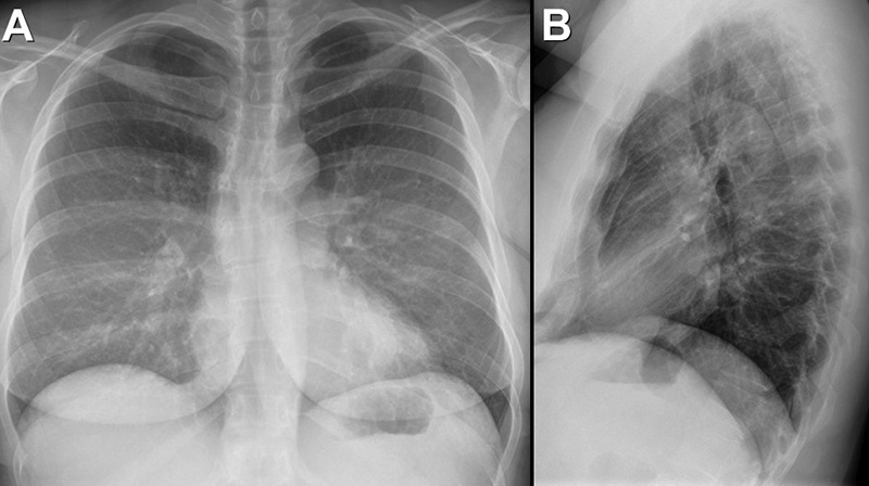
A complete blood count showed a mildly decreased white blood cell count at 3.3 x 10⁹/L (normal, 3.4 – 9.6 x 10⁹/L), with a mildly decreased absolute neutrophil count of 1.4 x 10⁹/L (normal, 1.5 – 7 x 10⁹/L). Her lymphocytes were decreased at 0.71 (normal, 1 – 3.4 x 10⁹/L), but peripheral eosinophilia (30%) was present, and her absolute eosinophil count was elevated at 1.07 x 10⁹/L (normal, 0.0 – 0.4 x 10⁹/L). Her hemoglobin and hematocrit values were mildly decreased at 11.4 gm/dL (normal, 13.5 – 17.5 gm/dL) and 32.3 % (normal, 38.8 – 50%). The platelet count was mildly decreased at 77 x 109/L (normal, 149 – 375 x 109/L). The patient’s serum chemistries, including glucose, electrolytes (including calcium), and liver function studies were normal. The urinary drug toxicity screen and pregnancy test were negative, and coagulation parameters were normal. The thyroid stimulating hormone level was within the normal range. The D-Dimer level was elevated at 2.04 mcg/mL (normal, 0 – 0.49 mcg/mL), and her fibrinogen level was elevated at 654 ng/dL (normal, 200-393 ng/dL).  Her troponin-T level was also elevated at 0.124 ng/mL (normal, ≤0.01 ng/mL). Frontal and lateral chest radiography (Figure 1) was performed.

Figure 1. Frontal and lateral chest radiography shows a normal heart size and normal lung volumes. To view Figure 1 in a separate, enlarged window click here.

Which of the following statements regarding this chest radiograph is most accurate? (Click on the correct answer to be directed to the 2nd of 17 pages)

  1. The frontal and lateral chest radiograph shows no abnormal findings
  2. The frontal and lateral chest radiograph shows basal predominant fibrotic abnormalities
  3. The frontal and lateral chest radiograph shows large lung volumes with a cystic appearance
  4. The frontal and lateral chest radiograph shows an ill-defined opacity projected over the left base
  5. The frontal and lateral chest radiograph shows abnormal mediastinal contours
Cite as: Stib MT, Gotway MB. July 2025 Imaging Case of the Month: A Growing Lung Nodule in a Patient with Heart Disease. Southwest J Pulm Crit Care Sleep. 2025;31(1):1-11. doi: https://doi.org/10.13175/swjpccs017-25 PDF
Monday
Jun022025

June 2025 Medical Image of the Month: Neurofibromatosis-Associated Diffuse Cystic Lung Disease

Figure 1. Photograph of patient taken ~5 years post double-ling transplant demonstrating multiple cutaneous neurofibromas and an old tracheostomy scar. To view Figure 1 in a separate, enlarged window click here.


Figure 2. Noncontrast chest CT obtained prior to double lung transplant.  Axial images from the upper (A) and lower (B) lungs as well as a coronal (C) reconstruction demonstrate multiple scattered pulmonary cysts, some being very large.  There are intervening areas of pulmonary ground glass, perhaps related to atelectasis. To view Figure 2 in a separate, enlarged window click here.


Figure 3. Noncontrast chest CT obtained 5 years after double lung transplant.  Axial images from the upper (A) and lower (B) lungs are normal-appearing. To view Figure 3 in a separate, enlarged window click here.


A 61-year-old man with a history of neurofibromatosis type 1 (NF1) and NF1-associated cystic lung disease presented for a routine follow-up visit 5 years post-bilateral lung transplantation.  The patient’s physical examination revealed multiple cutaneous neurofibromas, consistent with his diagnosis of NF1 (Figure 1).  Additionally, he had a prior tracheostomy scar; he temporarily required tracheostomy post lung transplant surgery.

Pre-Transplant History:

The patient had progressive chronic hypoxic respiratory failure, requiring home oxygen supplementation at up to 8 L/min. His medical history included a 15-pack-year smoking history, though he quit smoking 16 years prior to his lung transplant.  His diagnostic workup prior to transplant consisted of a high-resolution computed tomography (HRCT) of the chest, which  revealed diffuse cystic lung disease with intervening ground-glass opacities (Figure 2), an echocardiogram, which demonstrated severe pulmonary hypertension, and pulmonary function tests (PFTs), which  showed a combined restrictive and obstructive pattern with severely reduced DLCO at 25%. Given his progressive respiratory failure and severe pulmonary hypertension, the patient was referred for lung transplant evaluation. He underwent a successful bilateral lung transplant, with post-transplant imaging showing excellent graft function (Figure 3).  Post-transplant, the patient achieved an excellent clinical outcome. At 5 years post-transplant, he remains active and reports no significant limitations in his daily activities.

Pulmonary manifestations of NF1, though rare, are increasingly recognized and include upper lobe cystic changes, interstitial lung disease (ILD), and pulmonary hypertension [1, 2). The pathophysiology of NF1-associated diffuse lung disease (NF-DLD) is multifactorial and not fully understood, but several mechanisms have been proposed:

  1. Abnormal Mesenchymal Development: NF1 is caused by mutations in the NF1 gene, which encodes neurofibromin, a tumor suppressor protein that regulates cell growth and differentiation. Loss of neurofibromin leads to dysregulated mesenchymal cell proliferation, contributing to cystic and fibrotic changes in the lung (1,4).
  2. Mechanical Stress and Cyst Formation: The cystic changes observed in NF-DLD may result from mechanical stress on the lung parenchyma due to abnormal connective tissue development. This stress can lead to alveolar wall destruction and bullae formation, particularly in the upper lobes (2, 5).
  3. Nerve Growth Factor (NGF) and Fibrosis: Elevated levels of nerve growth factor (NGF) have been reported in NF1 patients. NGF promotes fibroblast proliferation and collagen deposition, creating a profibrotic environment that may contribute to interstitial lung disease and fibrosis (3, 6).
  4. Vascular Abnormalities and Pulmonary Hypertension: NF1 is associated with vascular dysplasia, which can lead to pulmonary arterial hypertension (PAH). Chronic hypoxemia secondary to parenchymal lung disease further exacerbates pulmonary hypertension, leading to right ventricular dysfunction, as seen in this patient (7, 8).
  5. Role of Smoking: While smoking is a known risk factor for lung disease, its role in NF-DLD progression remains unclear. In this case, the patient’s limited smoking history may have contributed to disease progression, but the primary driver was likely NF1-related parenchymal and vascular abnormalities (1, 2).

This case highlights the progressive nature of NF-DLD, which can lead to end-stage lung disease and severe pulmonary hypertension. Early recognition and monitoring of pulmonary complications in NF1 patients are critical. Multidisciplinary care, including referral for lung transplant evaluation, is essential for optimizing outcomes in advanced cases.

Abdulmonam Ali, MD

Interventional Pulmonologist

Pulmonary & Critical Care

Good Samaritan Hospital, SSM Health

Mount Vernon, IL USA

References

  1. Zamora AC, Collard HR, Wolters PJ, Webb WR, King TE. Neurofibromatosis-associated lung disease: a case series and literature review. Eur Respir J. 2007 Jan;29(1):210-4. [CrossRef] [PubMed]
  2. Oikonomou A, Vadikolias K, Birbilis T, et al. Neurofibromatosis type 1 presenting with diffuse lung disease. Respir Med Case Rep. 2015;16:1-3.
  3. Fabricant RN, Todaro GJ. Increased serum levels of nerve growth factor in von Recklinghausen's disease. Arch Neurol. 1981 Jul;38(7):401-5. [CrossRef] [PubMed]
  4. Riccardi VM. Neurofibromatosis: phenotype, natural history, and pathogenesis. Johns Hopkins University Press. 1992.
  5. Ryu JH, Parambil JG, McGrann PS, Aughenbaugh GL. Lack of evidence for an association between neurofibromatosis and pulmonary fibrosis. Chest. 2005 Oct;128(4):2381-6. [CrossRef] [PubMed]
  6. Li Y, O’Connell MP, O’Connell MJ, et al. Nerve growth factor promotes fibroblast migration and collagen production in neurofibromatosis 1. J Invest Dermatol. 2011;131(4):857-864.
  7. Stewart DR, Cogan JD, Kramer MR, Miller WT Jr, Christiansen LE, Pauciulo MW, Messiaen LM, Tu GS, Thompson WH, Pyeritz RE, Ryu JH, Nichols WC, Kodama M, Meyrick BO, Ross DJ. Is pulmonary arterial hypertension in neurofibromatosis type 1 secondary to a plexogenic arteriopathy? Chest. 2007 Sep;132(3):798-808. [CrossRef] [PubMed]
  8. Montani D, Coulet F, Girerd B, et al. Pulmonary hypertension in patients with neurofibromatosis type I. Medicine (Baltimore). 2011 May;90(3):201-211. [CrossRef] [PubMed]

Cite as: Ali A. June 2025 Medical Image of the Month: Neurofibromatosis-Associated Diffuse Cystic Lung Disease. Southwest J Pulm Crit Care Sleep. 2025;30(6):63-65. doi: https://doi.org/10.13175/swjpccs006-25 PDF