Imaging

Last 50 Imaging Postings

(Most recent listed first. Click on title to be directed to the manuscript.)

August 2025 Medical Image of the Month: Crazy Paving in a Case of 
   Eosinophilic Granulomatosis with Polyangiitis
July 2025 Medical Image of the Month: A Case of Severe Hiatal Hernia
   Presenting as Atypical Chest Pain
July 2025 Imaging Case of the Month: A Growing Lung Nodule in a
   Patient with Heart Disease
June 2025 Medical Image of the Month: Neurofibromatosis-Associated Diffuse
   Cystic Lung Disease
May 2025 Medical Image of the Month: Aspirated Dental Screw
April 2025 Medical Image of the Month: An Unfortunate Case of Mimicry
March 2025 Medical Image of the Month: An Unusual Case of Pulmonary
   Infarction
February 2025 Medical Image of the Month: Unexpected Complications of
   Transjugular Intrahepatic Portosystemic Shunt (TIPS) 
February 2025 Imaging Case of the Month: A Wolf in Sheep’s Clothing
January 2025 Medical Image of the Month: Psoriasis with Pulmonary
   Involvement
December 2024 Medical Image of the Month: An Endobronchial Tumor
November 2024 Medical Image of the Month: A Case of Short Telomeres
November 2024 Imaging Case of the Month: A Recurring Issue
October 2024 Medical Image of the Month: Lofgren syndrome with Erythema
   Nodosum
September 2024 Medical Image of the Month: A Curious Case of Nasal
   Congestion
August 2024 Image of the Month: Lymphomatoid Granulomatosis
August 2024 Imaging Case of the Month: An Unexplained Pleural Effusion
July 2024 Medical Image of the Month: Vocal Cord Paralysis on PET-CT 
June 2024 Medical Image of the Month: A 76-year-old Man Presenting with
   Acute Hoarseness
May 2024 Medical Image of the Month: Hereditary Hemorrhagic
   Telangiectasia in a Patient on Veno-Arterial Extra-Corporeal Membrane
   Oxygenation
May 2024 Imaging Case of the Month: Nothing Is Guaranteed
April 2024 Medical Image of the Month: Wind Instruments Player Exhibiting
   Exceptional Pulmonary Function
March 2024 Medical Image of the Month: Sputum Cytology in Patients with
   Suspected Lung Malignancy Presenting with Acute Hypoxic Respiratory
   Failure
February 2024 Medical Image of the Month: Pulmonary Alveolar Proteinosis
   in Myelodysplastic Syndrome
February 2024 Imaging Case of the Month: Connecting Some Unusual Dots
January 2024 Medical Image of the Month: Polyangiitis Overlap Syndrome
   (POS) Mimicking Fungal Pneumonia 
December 2023 Medical Image of the Month: Metastatic Pulmonary
   Calcifications in End-Stage Renal Disease 
November 2023 Medical Image of the Month: Obstructive Uropathy
   Extremis
November 2023 Imaging Case of the Month: A Crazy Association
October 2023 Medical Image of the Month: Swyer-James-MacLeod
   Syndrome
September 2023 Medical Image of the Month: Aspergillus Presenting as a
   Pulmonary Nodule in an Immunocompetent Patient
August 2023 Medical Image of the Month: Cannonball Metastases from
   Metastatic Melanoma
August 2023 Imaging Case of the Month: Chew Your Food Carefully
July 2023 Medical Image of the Month: Primary Tracheal Lymphoma
June 2023 Medical Image of the Month: Solitary Fibrous Tumor of the Pleura
May 2023 Medical Image of the Month: Methamphetamine Inhalation
   Leading to Cavitary Pneumonia and Pleural Complications
April 2023 Medical Image of the Month: Atrial Myxoma in the Setting of
   Raynaud’s Phenomenon: Early Echocardiography and Management of
   Thrombotic Disease
April 2023 Imaging Case of the Month: Large Impact from a Small Lesion
March 2023 Medical Image of the Month: Spontaneous Pneumomediastinum
   as a Complication of Marijuana Smoking Due to Müller's Maneuvers
February 2023 Medical Image of the Month: Reversed Halo Sign in the
   Setting of a Neutropenic Patient with Angioinvasive Pulmonary
   Zygomycosis
January 2023 Medical Image of the Month: Abnormal Sleep Study and PFT
   with Supine Challenge Related to Idiopathic Hemidiaphragmatic Paralysis
December 2022 Medical Image of the Month: Bronchoesophageal Fistula in
   the Setting of Pulmonary Actinomycosis
November 2022 Medical Image of the Month: COVID-19 Infection
   Presenting as Spontaneous Subcapsular Hematoma of the Kidney
November 2022 Imaging Case of the Month: Out of Place in the Thorax
October 2022 Medical Image of the Month: Infected Dasatinib Induced
   Chylothorax-The First Reported Case 
September 2022 Medical Image of the Month: Epiglottic Calcification
Medical Image of the Month: An Unexpected Cause of Chronic Cough
August 2022 Imaging Case of the Month: It’s All About Location
July 2022 Medical Image of the Month: Pulmonary Nodule in the
   Setting of Pyoderma Gangrenosum (PG) 
June 2022 Medical Image of the Month: A Hard Image to Swallow
May 2022 Medical Image of the Month: Pectus Excavatum
May 2022 Imaging Case of the Month: Asymmetric Apical Opacity–
   Diagnostic Considerations
April 2022 Medical Image of the Month: COVID Pericarditis
March 2022 Medical Image of the Month: Pulmonary Nodules in the
   Setting of Diffuse Idiopathic Pulmonary NeuroEndocrine Cell Hyperplasia
   (DIPNECH) 
February 2022 Medical Image of the Month: Multifocal Micronodular
   Pneumocyte Hyperplasia in the Setting of Tuberous Sclerosis
February 2022 Imaging Case of the Month: Between A Rock and a
   Hard Place


For complete imaging listings click here

Those who care for patients with pulmonary, critical care or sleep disorders rely heavily on chest radiology and pathology to determine diagnoses. The Southwest Journal of Pulmonary, Critical Care & Sleep publishes case-based articles with characteristic chest imaging and related pathology. The editor of this section will oversee and coordinate the publication of a core of the most important chest imaging topics. In doing so, they encourage the submission of unsolicited manuscripts. It cannot be overemphasized that both radiologic and pathologic images must be of excellent quality. As a rule, 600 DPI is sufficient for radiographic and pathologic images. Taking pictures of plain chest radiographs and CT scans with a digital camera is strongly discouraged. The figures should be cited in the text and numbered consecutively. The stain used for pathology specimens and magnification should be mentioned in the figure legend. Those who care for patients with pulmonary, critical care or sleep disorders rely heavily on chest radiology and pathology to determine diagnoses. The Southwest Journal of Pulmonary, Critical Care & Sleep publishes case-based articles with characteristic chest imaging and related pathology. The editor of this section will oversee and coordinate the publication of a core of the most important chest imaging topics. In doing so, they encourage the submission of unsolicited manuscripts. It cannot be overemphasized that both radiologic and pathologic images must be of excellent quality. As a rule, 600 DPI is sufficient for radiographic and pathologic images. Taking pictures of plain chest radiographs and CT scans with a digital camera is strongly discouraged. The figures should be cited in the text and numbered consecutively. The stain used for pathology specimens and magnification should be mentioned in the figure legend.

-------------------------------------------------------------------------------------------  

Wednesday
Dec102014

Medical Image of the Week: PE with Infarct and Pulmonary Cavitation

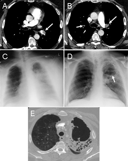
Figure 1. Panels A & B: thoracic CT scan showing multiple pulmonary emboli (arrows). Panel C: frontal chest radiograph showed extensive left lung opacification most dense in the left upper lobe. Panel D: frontal chest radiograph taken 3 weeks later showing mild volume loss of the left upper lobe with a large lucency suggestive of cavitation (arrow). Panel E: thoracic CT scan confirming the cavitation.

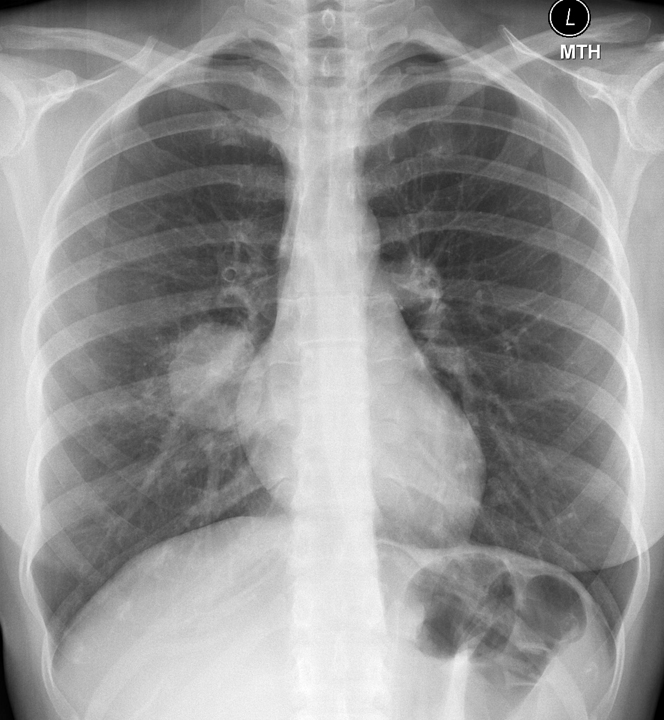
A 49 year old man with a history of COPD presented to the ER with the sudden onset of chest pain at 3:30 AM waking him from sleep. His pain was left sided, felt like broken ribs, and was worse with deep inspiration. He acknowledged some shortness of breath which was worse over baseline for the past couple days without cough or hemoptysis. The patient was tachycardic but comfortable with SpO2 saturation 98% on 2 liters. He had trace edema and pleurisy. Laboratory evaluation was unremarkable except for a WBC count 13,000 X 106 cells/L. Chest x-ray was unremarkable but thoracic CT scan showed pulmonary emboli (PE) involving left upper and lower lobar arteries (Figure 1A and 1B, arrows). Anticoagulation was started and the patient experienced increasing shortness of breath, worsening oxygenation and fever to 102ºF. On Day 2, frontal chest radiograph showed extensive left lung opacification most dense in the left upper lobe (Figure 1C). Hemoglobin dropped from 12 to 9.8 g/dL suggesting alveolar hemorrhage. He improved over the next week but low grade fevers persisted and a chest x-ray taken 3 weeks later showed mild volume loss of the left upper lobe with a large lucency suggestive of cavitation (Figure 1D, Arrow). Thoracic CT confirmed a cavitary lesion in the left apex in the region of prior thrombus with adjacent consolidated atelectasis within a background of emphysema (Figure 1E).  The patient was lost to follow up after 6 months of anticoagulation.

Pulmonary infarction is relatively uncommon, occurring in less than 10% of PE, due to dual and collateral blood supply to the lung. Cavitary infarcts are even less common (4% in autopsy studies) and are more likely in those with pulmonary venous hypertension (1). Cavitary infarcts are more likely to occur when the infarct size in larger than 4 cm and most often occurs in the mid and upper lung zones. Despite alveolar hemorrhage, anticoagulation should be continued.

Kenneth S. Knox, MD and Veronica A. Arteaga, MD

Divisions of Pulmonary and Critical Care Medicine and Thoracic Imaging

University of Arizona

Tucson, AZ

Reference 

  1. Libby LS, King TE, LaForce FM, Schwarz MI. Pulmonary cavitation following pulmonary infarction. Medicine (Baltimore). 1985;64(5):342-8. [CrossRef] [PubMed]

Reference as: Knox KS, Arteaga VA. Medical image of the week: PE with infarct and pulmonary cavitation. Southwest J Pulm Crit Care. 2014;9(6):333-4. doi: http://dx.doi.org/10.13175/swjpcc158-14 PDF 

Thursday
Dec042014

December 2014 Imaging Case of the Month

Michael B. Gotway, MD


Department of Radiology

Mayo Clinic Arizona

Scottsdale, AZ


Clinical History: A 34-year-old non-smoking woman presented to her physician as an outpatient with complaints of intermittent chest pain and intermittent mild hemoptysis. Her previous medical history was otherwise unremarkable.

Frontal chest radiography (Figure 1) was performed.

Figure 1. Frontal chest radiography.

Which of the following statements regarding the chest radiograph is most accurate? (Click on the correct answer to proceed to the second of 6 panels)

  1. The chest radiograph shows a circumscribed pulmonary mass
  2. The chest radiograph shows asymmetric pulmonary vascularity
  3. The chest radiograph shows bilateral linear and reticular opacities and diminished lung volumes suggesting fibrotic lung disease
  4. The chest radiograph shows mild streaky central opacities, possibly reflecting airway thickening
  5. The chest radiograph shows numerous small nodules

Reference as: Gotway MB. December 2014 imaging case of the month. Southwest J Pulm Crit Care. 2014;9(6):311-9. doi: http://dx.doi.org/10.13175/swjpcc157-14 PDF