Imaging

Last 50 Imaging Postings

(Most recent listed first. Click on title to be directed to the manuscript.)

August 2025 Medical Image of the Month: Crazy Paving in a Case of 
   Eosinophilic Granulomatosis with Polyangiitis
July 2025 Medical Image of the Month: A Case of Severe Hiatal Hernia
   Presenting as Atypical Chest Pain
July 2025 Imaging Case of the Month: A Growing Lung Nodule in a
   Patient with Heart Disease
June 2025 Medical Image of the Month: Neurofibromatosis-Associated Diffuse
   Cystic Lung Disease
May 2025 Medical Image of the Month: Aspirated Dental Screw
April 2025 Medical Image of the Month: An Unfortunate Case of Mimicry
March 2025 Medical Image of the Month: An Unusual Case of Pulmonary
   Infarction
February 2025 Medical Image of the Month: Unexpected Complications of
   Transjugular Intrahepatic Portosystemic Shunt (TIPS) 
February 2025 Imaging Case of the Month: A Wolf in Sheep’s Clothing
January 2025 Medical Image of the Month: Psoriasis with Pulmonary
   Involvement
December 2024 Medical Image of the Month: An Endobronchial Tumor
November 2024 Medical Image of the Month: A Case of Short Telomeres
November 2024 Imaging Case of the Month: A Recurring Issue
October 2024 Medical Image of the Month: Lofgren syndrome with Erythema
   Nodosum
September 2024 Medical Image of the Month: A Curious Case of Nasal
   Congestion
August 2024 Image of the Month: Lymphomatoid Granulomatosis
August 2024 Imaging Case of the Month: An Unexplained Pleural Effusion
July 2024 Medical Image of the Month: Vocal Cord Paralysis on PET-CT 
June 2024 Medical Image of the Month: A 76-year-old Man Presenting with
   Acute Hoarseness
May 2024 Medical Image of the Month: Hereditary Hemorrhagic
   Telangiectasia in a Patient on Veno-Arterial Extra-Corporeal Membrane
   Oxygenation
May 2024 Imaging Case of the Month: Nothing Is Guaranteed
April 2024 Medical Image of the Month: Wind Instruments Player Exhibiting
   Exceptional Pulmonary Function
March 2024 Medical Image of the Month: Sputum Cytology in Patients with
   Suspected Lung Malignancy Presenting with Acute Hypoxic Respiratory
   Failure
February 2024 Medical Image of the Month: Pulmonary Alveolar Proteinosis
   in Myelodysplastic Syndrome
February 2024 Imaging Case of the Month: Connecting Some Unusual Dots
January 2024 Medical Image of the Month: Polyangiitis Overlap Syndrome
   (POS) Mimicking Fungal Pneumonia 
December 2023 Medical Image of the Month: Metastatic Pulmonary
   Calcifications in End-Stage Renal Disease 
November 2023 Medical Image of the Month: Obstructive Uropathy
   Extremis
November 2023 Imaging Case of the Month: A Crazy Association
October 2023 Medical Image of the Month: Swyer-James-MacLeod
   Syndrome
September 2023 Medical Image of the Month: Aspergillus Presenting as a
   Pulmonary Nodule in an Immunocompetent Patient
August 2023 Medical Image of the Month: Cannonball Metastases from
   Metastatic Melanoma
August 2023 Imaging Case of the Month: Chew Your Food Carefully
July 2023 Medical Image of the Month: Primary Tracheal Lymphoma
June 2023 Medical Image of the Month: Solitary Fibrous Tumor of the Pleura
May 2023 Medical Image of the Month: Methamphetamine Inhalation
   Leading to Cavitary Pneumonia and Pleural Complications
April 2023 Medical Image of the Month: Atrial Myxoma in the Setting of
   Raynaud’s Phenomenon: Early Echocardiography and Management of
   Thrombotic Disease
April 2023 Imaging Case of the Month: Large Impact from a Small Lesion
March 2023 Medical Image of the Month: Spontaneous Pneumomediastinum
   as a Complication of Marijuana Smoking Due to Müller's Maneuvers
February 2023 Medical Image of the Month: Reversed Halo Sign in the
   Setting of a Neutropenic Patient with Angioinvasive Pulmonary
   Zygomycosis
January 2023 Medical Image of the Month: Abnormal Sleep Study and PFT
   with Supine Challenge Related to Idiopathic Hemidiaphragmatic Paralysis
December 2022 Medical Image of the Month: Bronchoesophageal Fistula in
   the Setting of Pulmonary Actinomycosis
November 2022 Medical Image of the Month: COVID-19 Infection
   Presenting as Spontaneous Subcapsular Hematoma of the Kidney
November 2022 Imaging Case of the Month: Out of Place in the Thorax
October 2022 Medical Image of the Month: Infected Dasatinib Induced
   Chylothorax-The First Reported Case 
September 2022 Medical Image of the Month: Epiglottic Calcification
Medical Image of the Month: An Unexpected Cause of Chronic Cough
August 2022 Imaging Case of the Month: It’s All About Location
July 2022 Medical Image of the Month: Pulmonary Nodule in the
   Setting of Pyoderma Gangrenosum (PG) 
June 2022 Medical Image of the Month: A Hard Image to Swallow
May 2022 Medical Image of the Month: Pectus Excavatum
May 2022 Imaging Case of the Month: Asymmetric Apical Opacity–
   Diagnostic Considerations
April 2022 Medical Image of the Month: COVID Pericarditis
March 2022 Medical Image of the Month: Pulmonary Nodules in the
   Setting of Diffuse Idiopathic Pulmonary NeuroEndocrine Cell Hyperplasia
   (DIPNECH) 
February 2022 Medical Image of the Month: Multifocal Micronodular
   Pneumocyte Hyperplasia in the Setting of Tuberous Sclerosis
February 2022 Imaging Case of the Month: Between A Rock and a
   Hard Place


For complete imaging listings click here

Those who care for patients with pulmonary, critical care or sleep disorders rely heavily on chest radiology and pathology to determine diagnoses. The Southwest Journal of Pulmonary, Critical Care & Sleep publishes case-based articles with characteristic chest imaging and related pathology. The editor of this section will oversee and coordinate the publication of a core of the most important chest imaging topics. In doing so, they encourage the submission of unsolicited manuscripts. It cannot be overemphasized that both radiologic and pathologic images must be of excellent quality. As a rule, 600 DPI is sufficient for radiographic and pathologic images. Taking pictures of plain chest radiographs and CT scans with a digital camera is strongly discouraged. The figures should be cited in the text and numbered consecutively. The stain used for pathology specimens and magnification should be mentioned in the figure legend. Those who care for patients with pulmonary, critical care or sleep disorders rely heavily on chest radiology and pathology to determine diagnoses. The Southwest Journal of Pulmonary, Critical Care & Sleep publishes case-based articles with characteristic chest imaging and related pathology. The editor of this section will oversee and coordinate the publication of a core of the most important chest imaging topics. In doing so, they encourage the submission of unsolicited manuscripts. It cannot be overemphasized that both radiologic and pathologic images must be of excellent quality. As a rule, 600 DPI is sufficient for radiographic and pathologic images. Taking pictures of plain chest radiographs and CT scans with a digital camera is strongly discouraged. The figures should be cited in the text and numbered consecutively. The stain used for pathology specimens and magnification should be mentioned in the figure legend.

-------------------------------------------------------------------------------------------  

Monday
Jan052015

January 2015 Imaging Case of the Month

Michael B. Gotway, MD


Department of Radiology

Mayo Clinic Arizona

Scottsdale, AZ


Clinical History: A 68-year-old woman with a history of myelodysplastic syndrome associated with transfusion-dependent anemia and thrombocytopenia presented with recent onset left chest pain and fever. The patient had a remote history of total right knee arthroplasty, hypertension, asthma, and schizoaffective disorder. Several months earlier the patient was hospitalized with methicillin-sensitive Staphylococcus aureus infection involving the right knee arthroplasty, associated with bacteremia and a septic right elbow. This infection was treated with incision and drainage of the elbow, antibiotic bead placement about the right knee arthroplasty with an antibiotic-impregnated spacer, and antibiotics (6 weeks intravenous cefazolin followed by chronic doxycycline suppression therapy, the former later switched to nafcillin and rifampin). The patient had been discharged from the hospital with only compression hose for deep venous thrombosis prophylaxis, owing to her episodes of epistaxis in the setting of transfusion-dependent anemia.

Upon presentation, the patient was hypotensive, tachycardic, and hypotensive. Laboratory data showed a white cell count of 3.9 cells x 109 / L, a platelet count of 7000 x 109 / L, and a hemoglobin level of 7 g/dL.

Frontal chest radiography (Figure 1A) was performed (a baseline chest radiograph- Figure 1B- is presented for comparison).


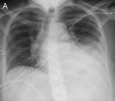
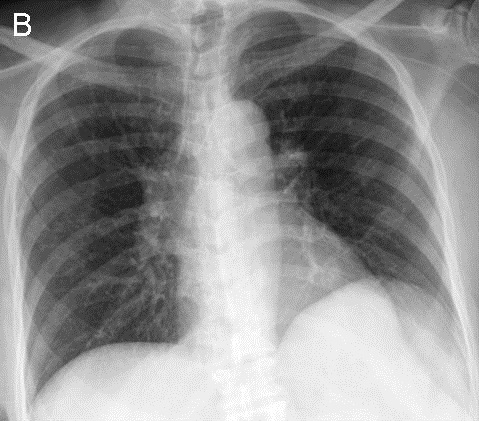
Figure 1. Panel A: Frontal chest radiography Panel B: Frontal chest radiograph obtained 3 months to presentation.

Which of the following statements regarding the chest radiograph is most accurate? (Click on the correct answer to proceed to the 2nd of 7 panels)

Reference as: Gotway MB. January 2015 imaging case of the month. Southwest J Pulm Crit Care. 2015;10(1):21-31. doi: http://dx.doi.org/10.13175/swjpcc003-15 PDF


Wednesday
Dec312014

Medical Image of the Week: Metastatic Collecting Duct Carcinoma

C

Figure 1. Transverse section of CT chest and abdomen shows enhancing pleural nodularity (yellow arrows) with a pleural effusion.


Figure 2.  Transverse section of CT abdomen shows heterogeneous enhancing mass in the right kidney (red arrow).


Figure 3. Coronal section of CT chest and abdomen showing a large right pleural effusion (yellow arrow) and atelectatic lung with mediastinal shift to the left. Red arrow points to the heterogeneous mass in the right kidney.


A 40-year-old woman home health nurse presented to the ED with intermittent right sided sharp chest pain and progressive dyspnea for 2 weeks. On admission she was found to be in respiratory distress. Chest x-ray revealed a massive right sided pleural effusion. Thoracic CT scan with contrast confirmed a large right pleural effusion with associated enhancing pleural nodularity also involving the diaphragmatic surface (Figure 1).  The visualized part of the abdomen revealed a mass in the midpole of right kidney (Figure 2). Subsequent CT scan of the abdomen with contrast revealed a heterogeneous enhancing mass in the right kidney suspicious for malignancy (Figure 3) and multiple paracaval lymph nodes. Thoracentesis revealed a hemorrhagic pleural effusion and during subsequent right video-assisted thoracoscopy showed disseminated tumorlets along the diaphragm and pleura. Pleural biopsy and fluid cytology was consistent with metastatic poorly differentiated collecting duct carcinoma of the kidney. The patient is currently getting outpatient chemotherapy. Collecting duct carcinoma of the kidney is an unusual variant of renal cell carcinoma and accounts for about 1% of all renal cell carcinomas (1). This variant has a poor prognosis and frequently metastasizes to the lung and liver.

Chandramohan Meenakshisundaram, MD

Nanditha Malakkla, MD

St. Francis Hospital.

Evanston, IL

Reference

  1. Wang X, Hao J, Zhou R, Zhang X, Yan T, Ding D, Shan L, Liu Z. Collecting duct carcinoma of the kidney: a clinicopathological study of five cases. Diagn Pathol. 2013;8:96. [CrossRef] [PubMed]

Reference as: Meenakshisundaram C, Malakkla N. Medical image of the week: metastatic collecting duct carcinoma. Southwest J Pulm Crit Care. 2014;9(6):348-9. doi: http://dx.doi.org/10.13175/swjpcc160-14 PDF