
Medical Image Of The Week: Tricuspid Valve Vegetation with Septic Pulmonary Emboli

Figure 1. Chest radiograph on presentation consistent with septic pulmonary embolic and cavitation.
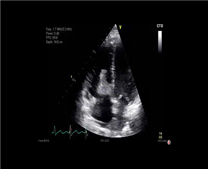
Figure 2. Echocardiogram demonstrating a highly mobile echo-dense vegetation attached to the atrial side of the tricuspid valve.
A 28-year-old woman with a history of extensive intravenous heroin use presented to the hospital with generalized chest and abdominal pain. Vital signs were remarkable for hypotension, tachypnea, and tachycardia. Laboratory studies revealed leukocytosis, hyponatremia, acute kidney injury, and lactic acidosis. A radiograph of the chest demonstrated multiple airspace opacities throughout the bilateral lungs with associated cavitary lesions and a small right-sided pleural effusion (Figure 1). A transthoracic echocardiogram was obtained, which demonstrated a 3.6 cm x 2.0 cm tricuspid valve vegetation (Figure 2). Blood cultures identified methicillin-sensitive Staphylococcus aureus.
Infective endocarditis, valvular vegetation, and septic pulmonary emboli are common complications of intravenous drug use. Staphylococcus aureus is the most common bacterial cause of infective endocarditis among intravenous drug users (1). Like endocarditis, patients with septic pulmonary emboli often present with non-specific clinical manifestations such as fever (86%), dyspnea (48%), and chest pain (49%) (2). Management may be surgical or medical, and determining the best course is complicated by social and psychiatric factors affecting adherence to treatment. Cardiac valve surgery has been advocated early for large right-sided vegetations but carries high morbidity and expense, as well as risk of compromised recovery, in the setting of ongoing IV drug use. Even for patients with valvular vegetations ≥ 1cm, medical therapy alone may be a safe option under some circumstances in the absence of other surgical indications (3).
Sarah Harris BA1, Kady Goldlist MD2, Maria Tumanik DO2, Cameron Hypes MD MPH3,4
1 University of Arizona College of Medicine
2 Department of Internal Medicine, Banner University Medical Center – South Campus
3 Department of Medicine, Division of Pulmonary, Allergy, Critical Care, and Sleep Medicine
4Department of Emergency Medicine
University of Arizona
Tucson, AZ USA
References
- Ortiz-Bautista C, López J, García-Granja PE, et al. Current profile of infective endocarditis in intravenous drug users: The prognostic relevance of the valves involved. Int J Cardiol. 2015;187:472-4. [CrossRef] [PubMed]
- Ye R, Zhao L, Wang C, Wu X, Yan H. Clinical characteristics of septic pulmonary embolism in adults: a systematic review. Respir Med. 2014 Jan;108(1):1-8. [CrossRef] [PubMed]
- Otome O, Guy S, Tramontana A, Lane G, Karunajeewa H. A retrospective review: significance of vegetation size in injection drug users with right-sided infective endocarditis. Heart Lung Circ. 2016 May;25(5):466-70. [CrossRef] [PubMed]
Cite as: Harris S, Goldlist K, Tumanik M, Hypes C. Medical image of the week: tricuspid valve vegetation with septic pulmonary emboli. Southwest J Pulm Crit Care. 2016:12(6):253-4. doi: http://dx.doi.org/10.13175/swjpcc042-16 PDF
 Post a Comment