Search Journal-type in search term and press enter
Southwest Pulmonary and Critical Care Fellowships
In Memoriam

Critical Care

Last 50 Critical Care Postings

April 2024 Critical Care Case of the Month: A 53-year-old Man Presenting
   with Fatal Acute Intracranial Hemorrhage and Cryptogenic Disseminated
   Intravascular Coagulopathy 
Delineating Gastrointestinal Dysfunction Variants in Severe Burn Injury
   Cases: A Retrospective Case Series with Literature Review
Doggonit! A Classic Case of Severe Capnocytophaga canimorsus Sepsis
January 2024 Critical Care Case of the Month: I See Tacoma
October 2023 Critical Care Case of the Month: Multi-Drug Resistant
   K. pneumoniae
May 2023 Critical Care Case of the Month: Not a Humerus Case
Essentials of Airway Management: The Best Tools and Positioning for 
   First-Attempt Intubation Success (Review)
March 2023 Critical Care Case of the Month: A Bad Egg
The Effect of Low Dose Dexamethasone on the Reduction of Hypoxaemia
   and Fat Embolism Syndrome After Long Bone Fractures
Unintended Consequence of Jesse’s Law in Arizona Critical Care Medicine
Impact of Cytomegalovirus DNAemia Below the Lower Limit of
Quantification: Impact of Multistate Model in Lung Transplant Recipients
October 2022 Critical Care Case of the Month: A Middle-Aged Couple “Not
   Acting Right”
Point-of-Care Ultrasound and Right Ventricular Strain: Utility in the
   Diagnosis of Pulmonary Embolism
Point of Care Ultrasound Utility in the Setting of Chest Pain: A Case of
   Takotsubo Cardiomyopathy
A Case of Brugada Phenocopy in Adrenal Insufficiency-Related Pericarditis
Effect Of Exogenous Melatonin on the Incidence of Delirium and Its 
   Association with Severity of Illness in Postoperative Surgical ICU Patients
Pediculosis As a Possible Contributor to Community-Acquired MRSA
Bacteremia and Native Mitral Valve Endocarditis
April 2022 Critical Care Case of the Month: Bullous Skin Lesions in
   the ICU
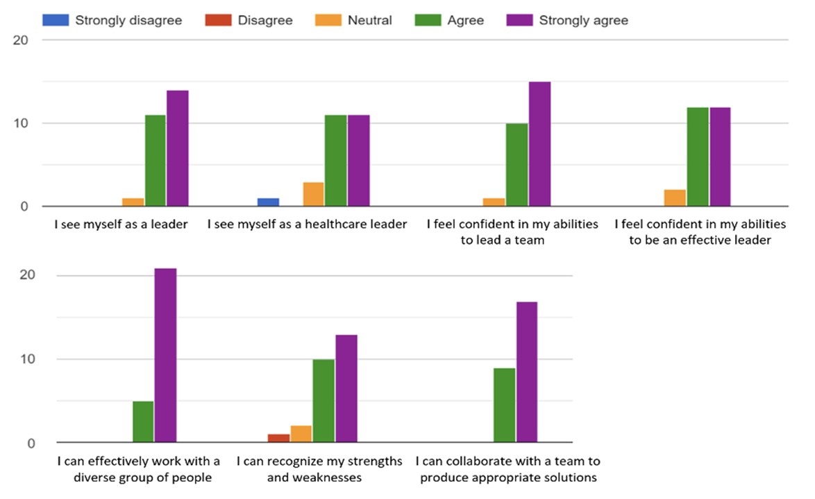
Leadership in Action: A Student-Run Designated Emphasis in
   Healthcare Leadership
MSSA Pericarditis in a Patient with Systemic Lupus
   Erythematosus Flare
January 2022 Critical Care Case of the Month: Ataque Isquémico
   Transitorio in Spanish 
Rapidly Fatal COVID-19-associated Acute Necrotizing
   Encephalopathy in a Previously Healthy 26-year-old Man 
Utility of Endobronchial Valves in a Patient with Bronchopleural Fistula in
   the Setting of COVID-19 Infection: A Case Report and Brief Review
October 2021 Critical Care Case of the Month: Unexpected Post-
   Operative Shock 
Impact of In Situ Education on Management of Cardiac Arrest after
   Cardiac Surgery
A Case and Brief Review of Bilious Ascites and Abdominal Compartment
   Syndrome from Pancreatitis-Induced Post-Roux-En-Y Gastric Remnant
   Leak
Methylene Blue Treatment of Pediatric Patients in the Cardiovascular
   Intensive Care Unit
July 2021 Critical Care Case of the Month: When a Chronic Disease
   Becomes Acute
Arizona Hospitals and Health Systems’ Statewide Collaboration Producing a
   Triage Protocol During the COVID-19 Pandemic
Ultrasound for Critical Care Physicians: Sometimes It’s Better to Be Lucky
   than Smart
High Volume Plasma Exchange in Acute Liver Failure: A Brief Review
April 2021 Critical Care Case of the Month: Abnormal Acid-Base Balance
   in a Post-Partum Woman
First-Attempt Endotracheal Intubation Success Rate Using A Telescoping
   Steel Bougie 
January 2021 Critical Care Case of the Month: A 35-Year-Old Man Found
   Down on the Street
A Case of Athabaskan Brainstem Dysgenesis Syndrome and RSV
   Respiratory Failure
October 2020 Critical Care Case of the Month: Unexplained
   Encephalopathy Following Elective Plastic Surgery
Acute Type A Aortic Dissection in a Young Weightlifter: A Case Study with
   an In-Depth Literature Review
July 2020 Critical Care Case of the Month: Not the Pearl You Were
   Looking For...
Choosing Among Unproven Therapies for the Treatment of Life-Threatening
   COVID-19 Infection: A Clinician’s Opinion from the Bedside
April 2020 Critical Care Case of the Month: Another Emerging Cause
   for Infiltrative Lung Abnormalities
Further COVID-19 Infection Control and Management Recommendations for
   the ICU
COVID-19 Prevention and Control Recommendations for the ICU
   Loperamide Abuse: A Case Report and Brief Review
Single-Use Telescopic Bougie: Case Series
Safety and Efficacy of Lung Recruitment Maneuvers in Pediatric Post-
   Operative Cardiac Patients
January 2020 Critical Care Case of the Month: A Code Post Lung 
   Needle Biopsy
October 2019 Critical Care Case of the Month: Running Naked in the
   Park
Severe Accidental Hypothermia in Phoenix? Active Rewarming Using 
   Thoracic Lavage
Left Ventricular Assist Devices: A Brief Overview


For complete critical care listings click here.

The Southwest Journal of Pulmonary and Critical Care publishes articles directed to those who treat patients in the ICU, CCU and SICU including chest physicians, surgeons, pediatricians, pharmacists/pharmacologists, anesthesiologists, critical care nurses, and other healthcare professionals. Manuscripts may be either basic or clinical original investigations or review articles. Potential authors of review articles are encouraged to contact the editors before submission, however, unsolicited review articles will be considered.

------------------------------------------------------------------------------------

Saturday
Jan012022

January 2022 Critical Care Case of the Month: Ataque Isquémico Transitorio in Spanish

Mohammad Abdelaziz Mahmoud DO MD

Bo Gu MD

Benito Armenta BA

Nikita Samra

Doctors Medical Center of Modesto and Emanuel Medical Center

Modesto and Turlock, CA USA


History of Present Illness:

The patient is a previously healthy 61-year-old Spanish-speaking woman who was unable to speak after awakening. Per Emergency Medical Service she was found to be aphasic upon their arrival. While in the Emergency Room the patient was able to speak, alert and oriented x4, with all her symptoms spontaneously resolved. The patient denied fever, chills, blurred vision, headache or any history of migraines, TIA, or stroke.

The patient had a similar event about two weeks earlier which also spontaneously resolved. During that time, the patient had a non-contrast CT head and an MRI of the brain, both of which were unremarkable. Her home medications include aspirin 81 mg and atorvastatin 40 mg daily.

Past Medical History, Family History and Social History

The patient denies tobacco use or use of illicit drugs.  She reports that she will occasionally drink alcohol. There is no family history of strokes.

Physical Examination

  • Vitals:  BP 123/80 mm Hg, T-max of 36.5° C, heart rate 72 bpm, SpO2 97%
  • HEENT: scleral icterus.
  • Lungs: clear
  • Heart: regular rhythm
  • Abdomen: soft without organomegaly, masses or tenderness
  • Skin: jaundiced 
  • Neurological examination:
    • Alert and oriented x4 with no focal neurological deficit observed
    • Cranial nerves II to XII were intact
    • Normal motor function
    • Normal speech
    • No facial asymmetry or facial droop
    • Normal mood and affect

Which of the following laboratory tests should be ordered? (click on the correct answer to be directed to the second of eight pages)

  1. None. She should be sent home
  2. Serum calcium/phosphorus 
  3. Liver function studies
  4. 1 and 3
  5. All of the above

Cite as: Mahmoud MA, Gu B, Armenta B, Samra N. January 2022 Critical Care Case of the Month: Ataque Isquémico Transitorio in Spanish. Southwest J Pulm Crit Care. 2022;24(1):1-5. doi: https://doi.org/10.13175/swjpcc051-21 PDF 

Wednesday
Nov172021

Rapidly Fatal COVID-19-associated Acute Necrotizing Encephalopathy in a Previously Healthy 26-year-old Man

Robert A. Raschke MD and Cristian Jivcu MD

HonorHealth Scottsdale Osborn Medical Center

Scottsdale, AZ USA

Case Presentation

A 26-year-old man presented to our Emergency Department at 0200 on the day of admission with chief complaints of subjective fever, leg myalgias, and progressive dyspnea of one week duration. An oropharyngeal swab PCR had revealed SARS-CoV-2 RNA three days previously. He had not received a SARS CoV-2 vaccination, but had made an appointment to receive it just a few days prior to the onset of his symptoms.

The patient had no significant past medical history, was taking no medications except for ibuprofen and acetaminophen over the past week, and did not take recreational drugs. He specifically denied headache and had no prior history of seizure.

On admission, his HR was 150 bpm (sinus), RR 22, BP 105/46 mmHg, temp 40.2° C. and SpO2 92% on room air. He was ill-appearing, but alert and oriented, his neck was supple and lung auscultation revealed bilateral rhonchi, but physical examination was otherwise unremarkable.

A CBC showed WBC 17.3 103/uL, hemoglobin 13.9 g/dl, and platelet count 168 K/uL. A complete metabolic profile was normal except for the following: Na 135 mmol/L, creatinine 1.7 mg/dL, AST 95 and ALT 134 IU/L. D-dimer was 1.08 ug/ml (normal range 0.00-0.50 ug/ml), and ferritin 783 ng/ml. A urine drug screen was negative. Chest radiography showed subtle bilateral pulmonary infiltrates. CT angiography of the chest was negative for pulmonary embolism but showed bilateral patchy infiltrates consistent with COVID19 pneumonia. One liter NS bolus and dexamethasone 10mg were given intravenously, acetaminophen administered orally, and the patient was admitted to telemetry.

Shortly thereafter, the patient experienced a brief generalized seizure associated with urinary incontinence. He was stuporous post-ictally, exhibiting only arm flexion to painful stimuli. A stroke alert was called and radiographic studies emergently obtained. CT of the brain was normal and CT angiography of the head and neck showed no large vessel occlusion or flow-limiting stenosis, and a CT perfusion study (Figure 1) showed patchy Tmax prolongation in the right cerebellum and bilateral parietal occipital lobes “which may reflect artifact or relative ischemia” with no matching core infarct.

Figure 1. CT perfusion study showing mild bilateral posterior distribution ischemia (Tmax > 6 secs) without matching core infarct (CBF<30%), interpreted by a neuroradiologist as possible artifact.

The patient was transferred to the ICU at 10:00, and experienced a 40-second generalized tonic-clonic seizure shortly thereafter. Lorazepam 2mg was administered intravenously. The HR was 104, RR 21, BP 105/61, temp 36.5 C. and SpO2 96% on 2L /min nasal canula oxygen. On neurological examination, the Glasgow Coma Scale was 3, right pupil was 3mm, left pupil 2mm - both reactive, the gaze was disconjugate and directed downward, there was no blink to visual threat, and glabellar ridge pressure did not elicit grimace, but minimal arm flexion. The gag reflex was positive. Peripheral reflexes were 2+ with down-going toes bilaterally. Levetiracetam 1000mg bolus was administered intravenously. Glucose was 147 mg/dL. An EEG obtained at 12:00 showed diffuse bilateral slowing without seizure activity. A presumptive diagnosis of post-ictal encephalopathy was made. The patient seemed to be protecting his airway and nasal canula oxygen was continued.

The patient’s condition was not noted to significantly change over the next 12 hours. There were no episodes of hypoxia, hypotension or hypoglycemia. Around 0100 on the second day of hospitalization, the patient exhibited extensor-posturing and appeared to be choking on his oral secretions. HR rose to 135, BP 155/99, RR 12 and temp 37.8 C. His SpO2 fell into the mid 80% range. He no longer had a gag or cough reflex and he was emergently intubated without complication. MRI (Figure 2) and MRV of the brain were emergently obtained. 

Figure 2. A: T2-weighted image demonstrating bilateral thalamic and L occipital white matter hypoattenuation. B: DWI and GRE images showing bilateral thalamic infarctions with hemorrhage. C: Representative DWI images of cerebrum and cerebellum and pons showing widespread diffusion restriction.

The MRI showed extensive diffusion restriction involving bilateral thalami, cerebellar hemispheres, pons, and cerebral hemispheres with scattered hemorrhage most obvious/confluent in the bilateral thalami.

Normal flow voids were present in intracranial arteries and venous structures. Partial effacement of the lateral and third ventricles was noted, with early uncal herniation. The MRV showed no evidence of dural venous sinus thrombosis.

At 05:00 of the second hospital day, it was noted that the patient’s pupils were dilated and unreactive and his respiratory rate was 16 – equal to the respiratory rate set on the ventilator. BP fell to 85/45 and norepinephrine infusion was started to maintain MAP >65 mmHg. STAT CT brain (Figure 3) showed hemorrhagic infarcts of the bilateral thalami with surrounding edema, interval development of low attenuation of the bilateral cerebrum and cerebellum, and mass effect with total effacement of fourth ventricle, basal cisterns and cerebral sulci consistent with severe cerebral edema.

Figure 3. STAT CT brain from 05:30 on the second hospital day showing bilateral thalamic infarctions and diffuse cerebral edema with effacement of the sulci and loss of grey/white differentiation.

Two neurologists confirmed the clinical diagnosis of brain death, including an apnea test. A venous ammonia level ordered that morning was not drawn. An autopsy was requested by the physicians, but not able to be obtained.

Discussion

Acute necrotizing encephalopathy (ANE) is a rarely-reported clinical-radiographic syndrome lacking pathopneumonic laboratory test or histological findings (1-3). It is characterized by an acute febrile viral prodrome, most commonly due to influenza or HHV-6, followed by rapidly progressive altered mental status and seizures. The most specific finding of ANE is necrosis of the bilateral thalami, appearing on MRI as hypoattenuated lesions on T2 and FLAIR images with diffusion restriction on DWI, and often with hemorrhage demonstrated on GRE images (as shown in figure 2 above). Symmetric multifocal lesions are typically seen throughout various other locations in the brain including the cerebral periventricular white matter, cerebellum, brainstem and spinal cord. Mizuguchi (who first described ANE in 1995) proposed elevation of serum aminotransferase without hyperammonemia, and cerebrospinal albuminocytologic dissociation (elevated CSF protein without leukocytosis) as laboratory criteria supporting the diagnosis of ANE (1,2). These were only partially evaluated in our patient. The mortality of ANE is 30% and significant neurological sequelae are common in survivors (2).

The clinical, radiographic and laboratory findings in our case are all characteristic of ANE, but our work-up was abbreviated by the patient’s fulminant presentation. The differential diagnosis includes hyper-acute forms of acute disseminated encephalomyelitis (ADEM) or acute hemorrhagic leukoencephalitis that may also occur after a viral prodrome and may be associated with diffuse white matter lesions (4,5), although bilateral thalamic necrosis is not characteristic of either of these entities. Examination of cerebral spinal fluid (CSF) for pleocytosis, oligoclonal bands, and testing for the myelin oligodendrocyte glycoprotein IgG autoantibody and the aquaporin-4 IgG serum autoantibody would have been indicated to further evaluate for the initial presentation of a relapsing CNS demyelinating disease (5,6). CSF examination would also have been helpful in ruling out viral encephalitis affecting the thalami, such as that caused by West Nile Virus (WNV) (7). An acute metabolic encephalopathy with diffuse brain edema, such as that caused by severe hyperammonemia associated with late-onset ornithine transcarbamylase deficiency (8) was not ruled out. Arterial or venous thromboembolism associated with COVID-19 were effectively ruled out by CT angiogram, CT perfusion and MRI and MRV findings.    

We found five previous case reports of ANE as a complication of COVID-19, ranging 33-59 years of age (9-13). The onset of altered mental status occurred 3, 4, 7,10 and 21 days after onset of COVID-19 symptoms and rapidly progressed to coma. Two had generalized seizures, one myoclonus and another “rhythmic movements” of an upper extremity. All had bilateral hypoattenuation of the thalami on CT and MRI with variable involvement of temporal lobes, subinsular regions, cerebellum, brainstem and supratentorial grey and white matter. Two patients had EEGs that showed generalized slow waves. All underwent examination of CSF with negative PCR tests for various common encephalopathy viruses including herpes simplex virus 1&2 and WNV - four reported CSF protein and cell counts, three of which demonstrated albuminocytologic dissociation. Three patients received IVIG. Two patients died on days 5 and 8 after onset of neurological symptoms. Two recovered after prolonged ICU care and the outcome of the third patient was not reported. ANE may be less rare than these few case reports suggest. A retrospective study carried out at 11 hospitals in Europe describes radiographic findings of 64 COVID-19 patients with neurological symptoms (14). The most common finding was ischemic stroke, but 8 patients had MRI findings consistent with encephalitis and two had findings characteristic of ANE.

The pathogenesis of ANE is unknown. Ten cases of fatal ANE with brain biopsy are reported (1,15-19). These showed diffuse cerebral edema, and hemorrhagic necrosis invariably involving the thalami. An exudative small vessel vasculopathy with endothelial necrosis was found in 7/10 patients (This could perhaps explain the early CT perfusion findings interpreted as artifactual in our patient). Demyelination or inflammatory infiltration of the brain or leptomeninges was absent. There has been conjecture that these pathological findings might be due to disruption of the blood brain barrier caused by hypercytokinemia but there is scant supportive evidence (20). 

There is no proven treatment for ANE. Corticosteroids, IVIg and plasma exchange have been previously used (3,9-11,21). Clinical trials are unlikely given the rarity of the disorder.

It was unfortunate that this young man had not availed himself of SARS CoV-2 vaccination. We did not make a pre-mortem diagnosis of ANE between his first abnormal CT brain at 0100 and his death at 06:00. We would have performed an LP, measured serum ammonia and given a trial of corticosteroids and IVIg if we had had more time.

References

  1. Mizuguchi M, Abe J, Mikkaichi K, Noma S, Yoshida K, Yamanaka T, Kamoshita S. Acute necrotising encephalopathy of childhood: a new syndrome presenting with multifocal, symmetric brain lesions. J Neurol Neurosurg Psychiatry. 1995 May;58(5):555-61. [CrossRef] [PubMed]
  2. Mizuguchi M. Acute necrotizing encephalopathy of childhood: a novel form of acute encephalopathy prevalent in Japan and Taiwan. Brain Dev. 1997 Mar;19(2):81-92. [CrossRef] [PubMed]
  3. Wu X, Wu W, Pan W, Wu L, Liu K, Zhang HL. Acute necrotizing encephalopathy: an underrecognized clinicoradiologic disorder. Mediators Inflamm. 2015;2015:792578. [CrossRef] [PubMed]
  4. Marchioni E, Ravaglia S, Montomoli C, et al. Postinfectious neurologic syndromes: a prospective cohort study. Neurology. 2013 Mar 5;80(10):882-9. [CrossRef] [PubMed]
  5. Manzano GS, McEntire CRS, Martinez-Lage M, Mateen FJ, Hutto SK. Acute Disseminated Encephalomyelitis and Acute Hemorrhagic Leukoencephalitis Following COVID-19: Systematic Review and Meta-synthesis. Neurol Neuroimmunol Neuroinflamm. 2021 Aug 27;8(6):e1080. [CrossRef] [PubMed]
  6. López-Chiriboga AS, Majed M, et al. Association of MOG-IgG Serostatus With Relapse After Acute Disseminated Encephalomyelitis and Proposed Diagnostic Criteria for MOG-IgG-Associated Disorders. JAMA Neurol. 2018 Nov 1;75(11):1355-1363. [CrossRef] [PubMed]
  7. Guth JC, Futterer SA, Hijaz TA, Liotta EM, Rosenberg NF, Naidech AM, Maas MB. Pearls & oy-sters: bilateral thalamic involvement in West Nile virus encephalitis. Neurology. 2014 Jul 8;83(2):e16-7. [CrossRef] [PubMed]
  8. Cavicchi C, Donati M, Parini R, et al. Sudden unexpected fatal encephalopathy in adults with OTC gene mutations-Clues for early diagnosis and timely treatment. Orphanet J Rare Dis. 2014 Jul 16;9:105. [CrossRef] [PubMed]
  9. Poyiadji N, Shahin G, Noujaim D, Stone M, et al.  COVID19-associated acute necrotizing encephalopathy: CT and MRI features.  Radiology. 2020;296:E119-E120. [CrossRef]
  10. Virhammar J, Kumlien E, Fällmar D,et al. Acute necrotizing encephalopathy with SARS-CoV-2 RNA confirmed in cerebrospinal fluid. Neurology. 2020 Sep 8;95(10):445-449. [CrossRef] [PubMed]
  11. Delamarre L, Galion C, Goudeau G, et al. COVID-19-associated acute necrotising encephalopathy successfully treated with steroids and polyvalent immunoglobulin with unusual IgG targeting the cerebral fibre network. J Neurol Neurosurg Psychiatry. 2020 Sep;91(9):1004-1006. [CrossRef] [PubMed]
  12. Dixon L, Varley J, Gontsarova A, Mallon D, Tona F, Muir D, Luqmani A, Jenkins IH, Nicholas R, Jones B, Everitt A. COVID-19-related acute necrotizing encephalopathy with brain stem involvement in a patient with aplastic anemia. Neurol Neuroimmunol Neuroinflamm. 2020 May 26;7(5):e789. [CrossRef] [PubMed]
  13. Elkady A, Rabinstein AA. Acute necrotizing encephalopathy and myocarditis in a young patient with COVID-19. Neurol Neuroimmunol Neuroinflamm Sep 2020, 7 (5) e801. [CrossRef]
  14. Kremer S, Lersy F, Anheim M, et al. Neurologic and neuroimaging findings in patients with COVID-19: A retrospective multicenter study. Neurology. 2020 Sep 29;95(13):e1868-e1882. [CrossRef] [PubMed]
  15. Kirton A, Busche K, Ross C, Wirrell E. Acute necrotizing encephalopathy in caucasian children: two cases and review of the literature. J Child Neurol. 2005 Jun;20(6):527-32. [CrossRef] [PubMed]
  16. Mastroyianni SD, Gionnis D, Voudris K, Skardoutsou A, Mizuguchi M. Acute necrotizing encephalopathy of childhood in non-Asian patients: report of three cases and literature review. J Child Neurol. 2006 Oct;21(10):872-9. [CrossRef] [PubMed]
  17. Nakano I, Otsuki N, Hasegawa A. Acute Stage Neuropathology of a Case of Infantile Acute Encephalopathy with Thalamic Involvement: Widespread Symmetrical Fresh Necrosis of the Brain. Neuropathology 1993;13: 315-25. [CrossRef]
  18. Yagishita A, Nakano I, Ushioda T, Otsuki N, Hasegawa A. Acute encephalopathy with bilateral thalamotegmental involvement in infants and children: imaging and pathology findings. AJNR Am J Neuroradiol. 1995 Mar;16(3):439-47. [PubMed]
  19. San Millan B, Teijeira S, Penin C, Garcia JL, Navarro C. Acute necrotizing encephalopathy of childhood: report of a Spanish case. Pediatr Neurol. 2007 Dec;37(6):438-41. [CrossRef] [PubMed]
  20. Wang GF, Li W, Li K. Acute encephalopathy and encephalitis caused by influenza virus infection. Curr Opin Neurol. 2010 Jun;23(3):305-11. [CrossRef] [PubMed]
  21. Okumura A, Mizuguchi M, Kidokoro H, et al. Outcome of acute necrotizing encephalopathy in relation to treatment with corticosteroids and gammaglobulin. Brain Dev. 2009 Mar;31(3):221-7. [CrossRef] [PubMed]

Cite as: Raschke RA, Jivcu C. Rapidly Fatal COVID-19-associated Acute Necrotizing Encephalopathy in a Previously Healthy 26-year-old Man. Southwest J Pulm Crit Care. 2021;23(5):138-43. doi: https://doi.org/10.13175/swjpcc039-21 PDF

Friday
Oct292021

Utility of Endobronchial Valves in a Patient with Bronchopleural Fistula in the Setting of COVID-19 Infection: A Case Report and Brief Review

Nazanin Sheikhan, MD1, Elizabeth J. Benge, MD1, Amanpreet Kaur, MD1, Jerome K Hruska, DO2, Yi McWhorter DO3, Arnold Chung MD4

1Department of Internal Medicine, HCA Healthcare; MountainView Hospital, Las Vegas, NV, USA

2Department of Pulmonology, HCA Healthcare; MountainView Hospital, Las Vegas, NV, USA

3Department of Anesthesiology Critical Care Medicine, HCA Healthcare; MountainView Hospital, Las Vegas, NV, USA

4MountainView Cardiovascular and Thoracic Surgery Associates, HCA Healthcare; MountainView Hospital, Las Vegas, NV, USA

Abstract

Patients with COVID-19 pneumonia frequently develop acute respiratory distress syndrome (ARDS), and in severe cases, require invasive mechanical ventilation. One complication that can develop in patients with ARDS who are mechanically ventilated is a bronchopleural fistula (BPF). Although rare, the frequency of BPF in patients with COVID-19 pneumonia is increasingly recognized. Here, we present a 48-year old man with BPF associated with COVID-19 pneumonia. Treatment with a commercial endobronchial valve (EBV) system resulted in reduced air leak allowing for tracheostomy placement. Our case adds to a growing body of evidence suggesting that the presence of COVID-19 pneumonia does not hinder the utility of EBV’s in the treatment of BPF’s.

Abbreviation List

  • ARDS = acute respiratory distress syndrome
  • BIPAP = Bilevel Positive Airway Pressure
  • BPF = Bronchopleural Fistula
  • COVID-19 = Coronavirus Disease-2019
  • CT = Computed Tomography
  • CTA = Computed Tomography Angiography
  • EBV = Endobronchial Valve
  • HFNC = High Flow Nasal Cannula
  • ICU = Intensive Care Unit
  • RML = Right Middle Lobe
  • RUL = Right Upper Lobe
  • SARS-CoV-2 = Severe Acute Respiratory Syndrome Coronavirus-2
  • VATS = Video-Assisted Thoracoscopic Surgery

Introduction

The COVID-19 pandemic has resulted in over one hundred million infections worldwide, in addition to millions of deaths (1). A less common sequelae of COVID-19 is bronchopleural fistula (2). A bronchopleural fistula is an abnormal sinus tract that forms between the lobar, main stem, or segmental bronchus, and the pleural space (3). BPF is typically treated by surgical repair, via a video-assisted thoracoscopic surgical approach (VATS) (3). Bronchoscopic approach with placement of airway stents, coils or transcatheter occlusion devices can be considered for those who are not suitable for surgical intervention (3).  A newer therapeutic modality for bronchopleural fistulae are endobronchial valves, which have been used successfully to treat COVID-19 patients diagnosed concurrently with bronchopleural fistulae (4). 

Here, we present a case of a critically ill patient developing a bronchopleural fistula with a concurrent COVID-19 infection, whose respiratory status was stabilized with an endobronchial valve.  To our knowledge, this is one of four case reports of a bronchopleural fistula arising in the setting of COVID-19.

Brief Review of Endobronchial Valves in COVID-19

Several other studies report success using endobronchial valves to treat bronchopleural fistulae in patients with COVID-19 pneumonia. One case series documents two cases of COVID-19 pneumonia complicated by bacterial super-infections, in which both patients experienced pneumothorax and persistent air leaks after mechanical invasive ventilation.  Both patients were successfully treated via EBV positioning. These researchers speculate that the severe inflammation associated with COVID-19 related ARDS induces inflammatory-related tissue frailty, pre-disposing lung tissue to damage via barotrauma, and the subsequent development of BPF (5).  

Another case documents the treatment of a 49-year-old male with COVID-19 pneumonia who was treated with steroids and tocilizumab. He also had a 3-week history of persistent air leak, which was successfully treated with an EBV. This team emphasizes that the thick, copious sections evident in patients afflicted by COVID-19 pose a risk for EBV occlusion. They highlight the importance of medically optimizing the patient and draining the air leak to mitigate the potential of this procedural complication developing (4).

In conjunction with the treatment course presented in our case, these case reports provide compelling evidence indicating that endobronchial valves can be successfully used to treat persistent air leaks in patients with COVID-19 pneumonia.

Case Presentation

Our patient is a 48-year-old male with a medical history significant for essential hypertension and Type 1 diabetes mellitus who presented to the emergency department complaining of acute onset generalized weakness, shortness of breath, and a near-syncopal event that had occurred the day prior. Vital signs on admission showed an oxygen saturation of 86% on ambient air, respiratory rate of 18 breaths per min, heart rate of 111 beats per min with a temperature of 37.6°C. He was tested for SARS-CoV-2 on admission and was found to be positive.

Initial computed tomography (CT) chest showed diffuse bilateral ground-glass opacities compatible with COVID-19 pneumonia. On admission, his inflammatory markers were elevated, with C-reactive protein 4.48 mg/dL, ferritin 1230 ng/ml, lactate dehydrogenase 281 IU/L, and D-dimer 0.76 mg/L. He received 1 dose of tocilizumab, convalescent plasma, as well as 5-day course of Remdesivir. His oxygen requirement increased as well as his work of breathing requiring High Flow Nasal Cannula (HFNC) and subsequently Bilevel Positive Airway Pressure (BiPAP); patient was transferred to the medical intensive care unit (ICU) 17 days after admission requiring intubation. Computed tomography angiography (CTA) chest could not be obtained to rule out pulmonary embolism as patient was too unstable. Patient was started on Heparin drip empirically which had to be discontinued due to gastrointestinal bleeding. He had worsening oxygenation, ventilator asynchrony, with P:F ratio of 47, requiring high-dose sedation and neuromuscular blockade, as well as prone positioning. Repeat CT chest on day 21 demonstrated bilateral pneumothoraces and pneumomediastinum as well as interval worsening of diffuse ground glass infiltrates (Figure 1), requiring bilateral chest tube placement.

Figure 1. Computed tomography chest showing pneumomediastinum, bilateral pneumothoraces, and diffuse ground glass attenuation of the lungs bilaterally.

On the 34th day of admission, he developed a right-sided tension pneumothorax likely secondary to ongoing severe ARDS, requiring replacement of dislodged right chest tube. Patient subsequently had worsening of right pneumothorax requiring an additional second chest tube placement. Patient developed persistent air leak concerning for right bronchopleural fistula. On hospital day 42, patient underwent intrathoracic autologous blood patch with persistence of large air leak. After interdisciplinary conference with cardiothoracic surgery, pulmonary, and the ICU team, it was decided that patient is not a surgical candidate hence interventional pulmonology was consulted for EBV placement to facilitate chest tube removal and ventilator weaning.

Patient underwent fiberoptic bronchoscopy on hospital day 52; pulmonary balloon was used to sequentially block the right mainstem, bronchus intermedius, and basilar segments. The air leak was recognized to be coming from right middle lobe (RML) and the apex of the right upper lobe (RUL) status post placement of two endobronchial valves in the medial and lateral segments of the RML (Figure 2).

Figure 2. Bronchoscopic view of endobronchial valves.

The RUL could not be entered secondary to angulation and technical inability of the instruments to achieve a sharp bend. Post-bronchoscopy, patient had 50 mL reduction in air leak resulting in improvement of his ventilator settings such that a tracheostomy could be safely performed. Left-sided chest tube was removed with resolution of pneumothorax. Repeat CT chest on hospital day 115 demonstrated persistent right bronchopleural fistula (Figure 3).

Figure 3. Computed tomography chest showing bronchopleural fistula in the right middle lobe and collapsed and shrunken right middle lobe with endobronchial occlusion stents at the central airway. Yellow arrow showing endobronchial valves and red arrows showing bronchopleural fistula

The patient is currently pending transfer to a long-term acute care hospital for aggressive physical therapy and eventual transfer to a tertiary center for lung transplantation evaluation.

Discussion

Scientific research has moved at an unprecedented speed in an attempt to shed light on the manifestations of COVID-19. The most common presentation of COVID-19 includes cough, fever, shortness of breath, and new onset anosmia and ageusia (6).

Common complications include coagulopathy, pulmonary emboli, and in severe cases, acute respiratory distress syndrome (7). Bronchopleural fistulae have emerged as a rare but known complication of COVID-19. This pathology is traditionally seen as a post-surgical complication arising from lobectomy or pneumonectomy (8). All cause mortality secondary to bronchopleural fistulae are high; with mortality rates ranging from 18-67% (8).

A relatively novel therapeutic modality for bronchopleural fistulae are endobronchial valves, which have been used in patients who are not candidates for surgery, such as our patient (9). They work as a one-way valve that allow the pathologically trapped air to exit the respiratory system, but not enter (4).

Differential diagnoses for bronchopleural fistulae include alveolar pleural fistulas and empyema (11). Alveolar pleural fistulas are abnormal communications between the pulmonary parenchyma, distal to a segmental bronchus, and the pleural space, while bronchopleural fistulas are more proximal; representing abnormal connections between a mainstem, lobar, or segmental bronchus and the pleural space (12). These pathologies are differentiated with direct visualization on bronchoscopy, as was demonstrated in our patient (12).

There are currently no official statistics on the epidemiology of bronchopleural fistulae in COVID-19. A disappointing aspect of our case was the lack of complete resolution of the patient’s air leak after the placement of the endobronchial valve. While the patient’s condition did improve after the valve was placed, he continued to suffer from respiratory illness related to his bronchopleural fistula. Although complete remission was not achieved, the endobronchial valve placement did facilitate respiratory recovery sufficient enough to facilitate a tracheostomy. The patient was then stabilized for eventual transfer to a long-term acute care facility, where he will undergo physical therapy and await lung transplantation. It is important to emphasize that while the endobronchial valve was not curative, it stabilized the patient for possible future curative treatments.  

Conclusion

Despite their rarity, bronchopleural fistulas are a pulmonary complication of COVID-19. Although the insertion of the endobronchial valve in our patient resulted in a reduction of the air leak as opposed to complete resolution, this case still emphasizes a therapeutic benefit of endobronchial valves in such instances. Overall, our case demonstrates the importance of clinical vigilance in the face of unusual pulmonary complications related to COVID-19, and that treatment of these complications requires flexibility and creativity.

References

  1. WHO Coronavirus (COVID-19) Dashboard [Internet]. World Health Organization. World Health Organization; [cited 2021May31]. Available from: https://covid19.who.int/ 
  2. Hopkins C, Surda P, Kumar N. Presentation of new onset anosmia during the COVID-19 pandemic. Rhinology. 2020 Jun 1;58(3):295-298. [CrossRef] [PubMed]
  3. Miesbach W, Makris M. COVID-19: Coagulopathy, Risk of Thrombosis, and the Rationale for Anticoagulation. Clin Appl Thromb Hemost. 2020 Jan-Dec;26:1076029620938149. [CrossRef] [PubMed]
  4. Talon A, Arif MZ, Mohamed H, Khokar A, Saeed AI. Bronchopleural Fistula as a Complication in a COVID-19 Patient Managed With Endobronchial Valves. J Investig Med High Impact Case Rep. 2021 Jan-Dec;9:23247096211013215. [CrossRef] [PubMed]
  5. Donatelli P, Trenatacosti F, Pellegrino MR, et al. Endobronchial valve positioning for alveolar-pleural fistula following ICU management complicating COVID-19 pneumonia. BMC Pulm Med. 2021 Sep 27;21(1):307. [CrossRef] [PubMed]
  6. Salik I, Vashisht R, Abramowicz AE. Bronchopleural fistula. StatPearls [Internet]. 2020 Aug 27. [CrossRef]
  7. Cardillo G, Carbone L, Carleo F, Galluccio G, Di Martino M, Giunti R, Lucantoni G, Battistoni P, Batzella S, Dello Iacono R, Petrella L, Dusmet M. The Rationale for Treatment of Postresectional Bronchopleural Fistula: Analysis of 52 Patients. Ann Thorac Surg. 2015 Jul;100(1):251-7. [CrossRef] [PubMed]
  8. Sarkar P, Chandak T, Shah R, Talwar A. Diagnosis and management bronchopleural fistula. Indian J Chest Dis Allied Sci. 2010 Apr-Jun;52(2):97-104. [PubMed]
  9. Pathak V, Waite J, Chalise SN. Use of endobronchial valve to treat COVID-19 adult respiratory distress syndrome-related alveolopleural fistula. Lung India. 2021 Mar;38(Supplement):S69-S71. [CrossRef] [PubMed]
  10. Musani AI, Dutau H. Management of alveolar-pleural fistula: a complex medical and surgical problem. Chest. 2015 Mar;147(3):590-592. [CrossRef] [PubMed]
  11. Mehta HJ, Malhotra P, Begnaud A, Penley AM, Jantz MA. Treatment of alveolar-pleural fistula with endobronchial application of synthetic hydrogel. Chest. 2015 Mar;147(3):695-699. [CrossRef]  [PubMed]

Acknowlegements

This research was supported (in whole or in part) by HCA Healthcare and/or an HCA Healthcare affiliated entity. The views expressed in this publication represent those of the author(s) and do not necessarily represent the official views of HCA Healthcare or any of its affiliated entities. 

Cite as: Sheikhan N, Benge EJ, Kaur A, Hruska JK, McWhorter Y, Chung A. Utility of Endobronchial Valves in a Patient with Bronchopleural Fistula in the Setting of COVID-19 Infection: A Case Report and Brief Review. Southwest J Pulm Crit Care. 2021;23(4):109-14. doi: https://doi.org/10.13175/swjpcc046-21 PDF 

Friday
Oct012021

October 2021 Critical Care Case of the Month: Unexpected Post-Operative Shock

Sharanyah Srinivasan MBBS

Sooraj Kumar MBBS

Benjamin Jarrett MD

Janet Campion MD

University of Arizona College of Medicine, Department of Internal Medicine and Division of Pulmonary, Allergy, Critical Care and Sleep Medicine, Tucson, AZ USA


History of Present Illness 

A 55-year-old man with a past medical history significant for endocarditis secondary to intravenous drug use, osteomyelitis of the right lower extremity was admitted for ankle debridement. Pre-operative assessment revealed no acute illness complaints and no significant findings on physical examination except for the ongoing right lower extremity wound. He did well during the approximate one-hour “incision and drainage of the right lower extremity wound”, but became severely hypotensive just after the removal of the tourniquet placed on his right lower extremity. Soon thereafter he experienced pulseless electrical activity (PEA) cardiac arrest and was intubated with return of spontaneous circulation being achieved rapidly after the addition of vasopressors. He remained intubated and on pressors when transferred to the intensive care unit for further management.

PMH, PSH, SH, and FH

  • S/P Right lower extremity incision and drainage for suspected osteomyelitis as above
  • Distant history of endocarditis related to IVDA
  • Not taking any prescription medications
  • Current smoker, occasional alcohol use
  • Former IVDA
  • No pertinent family history including heart disease

Physical Exam

  • Vitals: 100/60, 86, 16, afebrile, 100% on ACVC 420, 15, 5, 100% FiO2
  • Sedated well appearing male, intubated on fentanyl and norepinephrine
  • Pupils reactive, nonicteric, no oral lesions or elevated JVP
  • CTA, normal chest rise, not overbreathing the ventilator
  • Heart: Regular, normal rate, no murmur or rubs
  • Abdomen: Soft, nondistended, bowel sounds present
  • No left lower extremity edema, right calf dressed with wound vac draining serosanguious fluid, feet warm with palpable pedal pulses
  • No cranial nerve abnormality, normal muscle bulk and tone

Clinically, the patient is presenting with post-operative shock with PEA cardiac arrest and has now been resuscitated with 2 liters emergent infusion and norepinephrine at 70 mcg/minute.

What type of shock is most likely with this clinical presentation?

  1. Cardiogenic shock
  2. Hemorrhagic shock
  3. Hypovolemic shock
  4. Obstructive shock
  5. Septic / distributive shock

Cite as: Srinivasan S, Kumar S, Jarrett B, Campion J. October 2021 Critical Care Case of the Month: Unexpected Post-Operative Shock. Southwest J Pulm Crit Care. 2021;23(4):93-7. doi: https://doi.org/10.13175/swjpcc041-21 PDF 

Monday
Aug162021

Impact of In Situ Education on Management of Cardiac Arrest after Cardiac Surgery

Bhargavi Gali, M.D.1

Grace M. Arteaga, M.D.2

Glen Au, R.N., C.C.R.N.3

Vitaly Herasevich, M.D., Ph.D.1

1Division of Anesthesia-Critical Care Medicine, Department of Anesthesiology and Perioperative Medicine

2Division of Pediatric Critical Care Medicine, Department of Pediatric and Adolescent Medicine

3Department of Nursing

Mayo Clinic

Rochester, Minnesota USA


Abstract

Background:  Advanced life support interventions have been modified for patients who have recently undergone sternotomy for cardiac surgery and have new suture lines. We aimed to determine whether the use of in-situ simulation increased adherence to the cardiac surgery unit-advanced life support algorithm (CSU-ALS) for patients with cardiac arrest after cardiac surgery (CAACS).

Methods:  This was a retrospective chart review of cardiac arrest management of patients who sustained CAACS before and after implementation of in-situ simulation scenarios utilizing CSU-ACLS in place of traditional advanced cardiac life support.  We utilized classroom education of CSU-ACLS followed by in-situ high-fidelity simulated scenarios of patients with CAACS..  Interprofessional learners (n = 210) participated in 18 in-situ simulations of CAACS.  Two groups of patients with CAACS were retrospectively compared before and after in situ training (preimplementation, n=22 vs postimplementation, n=38).  Outcomes included adherence to CSU-ALS for resuscitation, delay in initiation of chest compressions, use of defibrillation and pacing before external cardiac massage, and time to initial medication.

Results:  Chest compressions were used less often in the postimplementation vs the preimplementation period (11/22 [29%] vs 13/38 [59%], P = 0.02).  Time to initial medication administration, use of defibrillation and pacing, return to the operating room, and survival were similar between periods.  

Conclusion:  In this pilot, adherence to a key component of the CSU-ALS algorithm—delaying initiation of chest compressions—improved after classroom combined with in-situ simulation education.

Abbreviations

  • ACLS, advanced cardiac life support
  • CAACS, cardiac arrest after cardiac surgery
  • CPR, cardiopulmonary resuscitation
  • CSICU, cardiac surgical intensive care unit
  • CSU-ALS, cardiac surgical unit–advanced life support EACTS, European Association for Cardio-Thoracic Surgery
  • IQR, interquartile range
  • STS, Society of Thoracic Surgeons

Introduction 

Immediate and appropriate resuscitation of patients with cardiac arrest has been called “the formula for survival” (1). Patient-specific and cause-specific resuscitation algorithms have been developed to optimize management and outcome measures (2). Advanced cardiac life support (ACLS) interventions are modified for special causes, environments, and patient populations. Patients who have recently undergone sternotomy for cardiac surgery and have new suture lines is one of these groups.

Because of their unique circumstances and physiologic conditions, patients who have recently undergone cardiac surgery benefit from modified cardiac-arrest management protocols. A recent consensus guideline by The Society of Thoracic Surgeons (STS) recommends use of a postcardiac surgery–specific resuscitation protocol prepared by the European Association for Cardio-Thoracic Surgery (EACTS), hereafter called the STS/EACTS protocol (3). In contrast to ACLS guidelines(4), the STS/EACTS protocol is based on recent sternotomy and increased risks of cardiac tamponade and cardiac ventricular rupture. The STS/EACTS protocol recommends sequential attempts at defibrillation before administration of chest compressions, administration of low-dose epinephrine, use of pacing to manage severe bradycardia or asystole, and immediate consideration of resternotomy (Table 1).

Because poststernotomy patients have new suture lines, they are at risk for comorbid conditions (e.g., cardiac tamponade, ventricular rupture) if external chest compressions are used (4). The cardiac surgical unit–advanced life support (CSU-ALS) protocol emphasizes use of defibrillation and delayed use of chest compressions (Table 1). In-situ simulation-based education has been shown to be an effective method for training in high-risk, low-frequency resuscitation situations (5). During in-situ simulation-based education, health care providers receive training in their clinical work environment.

A systematic review and meta-analysis of 182 studies reported that simulation-based training was highly effective in improving knowledge and process skill (6).

The STS/EACTS protocol was introduced to the CSICU in April 2014.  The CSICU team members, who all had background training in ACLS, received classroom-based education on the application of the cardiac surgery unit–advanced life support (CSU-ALS) algorithm. In-situ simulation-based training with resuscitation scenarios offered the team members the experimental application of the STS/EACTS resuscitation protocol-CSU-ALS protocol.  We hypothesized that adherence to the CSU-ALS protocol for the treatment of patients with CAACS would improve after a pilot implementing in-situ simulations with our CSICU team members.

Methods

After obtaining approval from the Mayo Clinic Institutional Review Board, we performed a single-center, retrospective review of the electronic health records of patients with CAACS. Only the records of patients that had consented to have their data utilized for research were included. The CONSORT 2010 Checklist was utilized in preparation of this manuscript. We identified patients who were treated before (October 2013 through March 2014; preimplementation period) or after simulation training (October 2015 through March 2016; postimplementation period). technicians; in total, 210 participants, took part in 18 simulations.  All participants, except the pharmacists, respiratory therapists, and phlebotomists, had participated in CSU-ALS classroom education.  No repeat participants were included in these sessions.  A combined 35% of our CSICU staff participated.

Included patients were those admitted to the CSICU after sternotomy for cardiac surgery, specifically patients who had undergone sternotomy and a cardiac surgical procedure (including those who underwent initiation of central extracorporeal membrane oxygenation). Patients from the above group who had cardiac arrest within the first 14 days after sternotomy for cardiac surgery were included. We excluded inpatients who were in the CSICU 14 days after their original sternotomy at time of cardiac arrest.

The educational in-situ simulations portrayed adult patients with cardiac arrest immediately after cardiac surgery. The details of the simulation have been previously published (7). Briefly, the learning objectives were established according to the CSU-ALS protocol. Before the simulation, a facilitator familiar with the CSU-ALS protocol reviewed it with the participants and discussed the differences compared to ACLS. Cardiopulmonary resuscitation (CPR) was defined as basic life support with use of the ACLS algorithm, airway management, greater epinephrine doses, and chest compressions initiated immediately after rhythm check; ACLS included all algorithms used in resuscitation, as recommended by the American Heart Association (4). In contrast to ACLS, CSU-ALS emphasizes the need to initially defibrillate rather than to perform chest compressions.  A patient room inside the CSICU was used as the scenario set-up. A high-fidelity mannequin was endotracheally intubated and mechanically ventilated. The simulation timeline involved 10 to 15 minutes for the case development and followed by a reflective debriefing period of 10 to 15 minutes.

The participating interprofessional team included critical care nurses, critical care fellows, cardiac surgical fellows, critical care physicians, pharmacists, nurse practitioners, respiratory therapists, and phlebotomy participants were included in these sessions. A combined 35% of our CSICU staff participated.

We collected data on patient demographic characteristics, surgical procedures and dates, specific cardiac arrest characteristics (initial cardiac rhythm and presumed cause), and resuscitation characteristics (return to the operating room for resternotomy [yes or no], intubation [yes or no], and survival of event [yes or no]).

The primary outcome measure in our scenarios was the use of defibrillation with successive “stacked” shocks prior to the standard ACLS, which recommends immediate initiation of chest compressions (7). Secondary outcome measures included time to initiation of chest compressions, time to use of ventricular defibrillation and pacing, and time to initial medication administration.

Statistical Analysis

Results are reported with descriptive statistics. All continuous variables are summarized as median (interquartile range [IQR]) or mean (SD) as appropriate, and we used the Wilcoxon rank sum test to compare the means and medians of continuous variables. Categorical data are summarized as number (percentage), and we used the Fisher’s exact test to compare categorical variables. Two-tailed hypothesis testing was used, and P < 0.05 was considered significant. Analysis was performed with JMP Pro 14.1.0 (SAS Institute Inc; Cary, North Carolina) and Microsoft Excel 2010 version 14 (Microsoft Corp; Redmond, Washington).

Results

Sixty patients met the inclusion criteria. We identified 22 patients in the preimplementation period (10 women, 45%) and 38 patients in the postimplementation period (12 women, 32%). In the preimplementation group, 6/22 patients (27%) received extracorporeal membrane oxygenation, compared with 8/38 patients (21%) in the postimplementation group.  Initial presentation and etiology of the arrests in the pre- and postimplementation period are presented in Table 2.

The use of chest compressions was 59% (preimplementation: 13/22 patients) vs 29% (the postimplementation phase 12/38 patients) (P = 0.02) and standard CPR (22/22 patients [100%] vs 27/38 patients [71%], P < 0.001) respectively (Table 2).  Median (IQR) time from onset of cardiac arrest to initiation of chest compressions was 1 minute (1-1.5 minutes) in the preimplementation period and 1.5 minutes (1-5 minutes) in the postimplementation period; these findings were statistically similar (P = 0.11) (Figure 1).

Median time to initial medication administration was similar between periods (P = 0.11).  However, in the preimplementation period, one patient was administered medication 47 minutes after cardiac arrest.  This result was an outlier (Figure 2). 

Similar percentages of patients received defibrillation to manage ventricular fibrillation or tachycardia (14/22 patients [64%] in the preimplementation period vs 20/38 patients [53%] in the postimplementation period, P = 0.40), returned to the operating room for resternotomy (2/22 patients [9%] vs 3/38 patients [8%], P = 0.80), and survived the event (19/22 patients [86%] vs 32/33 patients [84%], P = 0.80) (Table 3).


Discussion

The findings in this pilot study revealed an increase in adherence to CUS-ALS principles in CAACS when online courses are followed by in-situ simulation-based education.  Our preliminary data show a decrease in the use of standard CPR and chest compressions to manage CAACS.  These results suggest that in situ simulation–based training may potentially increase adherence to alternative resuscitation protocols for special patient populations and circumstances.

Mundell et al. (6) described how team training, including practice of interactions during resuscitation with provision of feedback, positively affected trainee satisfaction, knowledge, time to action, and process skill outcomes.  In addition, a recent systematic review and meta-analysis of observational studies reported a positive association between participation in ACLS courses and patient outcomes, including return of spontaneous circulation (8).

The current study provides preliminary evidence that in situ simulation-based training improves clinical performance.  Participation in simulation-based training allowed our CSICU team members to apply classroom-based knowledge in an experiential-learning environment, thereby improving their clinical performance of CSU-ALS protocol when they managed high-risk events.

We were able to educate our team members about a key component of the CSU-ALS protocol-namely, delay initiation of chest compressions and standard CPR. Our study did not find significant differences between groups for time to medication administration, use of defibrillation, return to the operating room, or survival.  Because this study was retrospective, we were unable to determine whether our CSICU team members who participated in simulation-based training subsequently resuscitated patients after the CSU-ALS protocol was implemented at our institution.  This could have affected our ability to assess the effects of in situ simulation–based training on clinical management.

Limitations

Our study has limitations is its retrospective design and involvement of 35% of staff with the in-situ simulations.  Documentation of cardiac arrest has improved at our institution, but one patient in the preimplementation period had a long-documented time from cardiac arrest to initial medication administration (47 minutes); this result was an outlier and was most likely a charting error.  

Another limitation was our inability to exactly determine which CSICU team members who treated patients in the postimplementation period had participated in in situ simulation-based training. based on de-identified data collection, one-third of our CSICU staff participated in this educational experience. 

Due to our limited number of arrests, alterations in outcomes based on in situ simulation would not likely be noted.  In situ simulation–based training improves cardiac arrest management and provides health care personnel a safe environment to practice interventions, which subsequently improves patient safety.[6, 12-14]  Further prospective studies of  the use of in situ simulation–based training may help determine the true effectiveness of this tool in educational and clinical practices that use specific resuscitation algorithms and highlight the relationship to patient outcomes and patient safety.

Conclusions

Analysis of the effects of in situ simulation-based training in the clinical setting showed a significant beneficial decrease in the use of chest compressions for the management of CAACS in patients who recently had undergone sternotomy.  Increased adherence to the CSU-ALS protocol could improve the outcome measures of patients with CAACS and decrease the deleterious effects of chest compressions after recent sternotomy with the expectation of decreased complications and ultimately, improved clinical outcomes.  As this was a small pilot study, further investigation with use of in-situ simulation in special circumstances would help determine its utility as an educational tool for high risk low frequency events.

References

  1. Søreide E, Morrison L, Hillman K, Monsieurs K, Sunde K, Zideman D, Eisenberg M, Sterz F, Nadkarni VM, Soar J, Nolan JP; Utstein Formula for Survival Collaborators. The formula for survival in resuscitation. Resuscitation. 2013 Nov;84(11):1487-93. [CrossRef] [PubMed]
  2. Truhlář A, Deakin CD, Soar J, et al. European Resuscitation Council Guidelines for Resuscitation 2015: Section 4. Cardiac arrest in special circumstances. Resuscitation. 2015 Oct;95:148-201. [CrossRef] [PubMed]
  3. Advanced Cardiovascular Life Support (ACLS) American Heart Association 2020 Guidelines for CPR and ECC Available at: https://cpr.heart.org/en/resuscitation-science/cpr-and-ecc-guidelines , Accessed July1, 2021.
  4. Society of Thoracic Surgeons Task Force on Resuscitation After Cardiac Surgery. The Society of Thoracic Surgeons Expert Consensus for the Resuscitation of Patients Who Arrest After Cardiac Surgery. Ann Thorac Surg. 2017 Mar;103(3):1005-1020. [CrossRef] [PubMed]
  5. Greif R, Lockey AS, Conaghan P, Lippert A, De Vries W, Monsieurs KG.  European Resuscitation Council Guidelines for Resuscitation 2015: Section 10. Education and implementation of resuscitation. Resuscitation. 2015;95:288-301. [CrossRef] [PubMed]
  6. Mundell WC, Kennedy CC, Szostek JH, Cook DA. Simulation technology for resuscitation training: a systematic review and meta-analysis. Resuscitation. 2013 Sep;84(9):1174-83. [CrossRef] [PubMed]
  7. Gali B, Au G, Rosenbush KA. Simulation Incorporating Cardiac Surgery Life Support Algorithm Into Cardiac Intensive Care Unit Practice. Simul Healthc. 2016 Dec;11(6):419-424. [CrossRef] [PubMed]
  8. Lockey A, Lin Y, Cheng A. Impact of adult advanced cardiac life support course participation on patient outcomes-A systematic review and meta-analysis. Resuscitation. 2018 Aug;129:48-54. [CrossRef] [PubMed]
  9. Fernández Lozano I, Urkía C, Lopez Mesa JB, Escudier JM, Manrique I, de Lucas García N, Pino Vázquez A, Sionis A, Loma Osorio P, Núñez M, López de Sá E. European Resuscitation Council Guidelines for Resuscitation 2015: Key Points. Rev Esp Cardiol (Engl Ed). 2016 Jun;69(6):588-94. [CrossRef] [PubMed]
  10. Dunning J, Fabbri A, Kolh PH, Levine A, Lockowandt U, Mackay J, Pavie AJ, Strang T, Versteegh MI, Nashef SA; EACTS Clinical Guidelines Committee. Guideline for resuscitation in cardiac arrest after cardiac surgery. Eur J Cardiothorac Surg. 2009 Jul;36(1):3-28. [CrossRef] [PubMed]
  11. Dunning J, Nandi J, Ariffin S, Jerstice J, Danitsch D, Levine A. The Cardiac Surgery Advanced Life Support Course (CALS): delivering significant improvements in emergency cardiothoracic care. Ann Thorac Surg. 2006 May;81(5):1767-72. [CrossRef] [PubMed]
  12. Haffner L, Mahling M, Muench A, et al. Improved recognition of ineffective chest compressions after a brief Crew Resource Management (CRM) training: a prospective, randomised simulation study. BMC Emerg Med. 2017 Mar 3;17(1):7. [CrossRef] [PubMed]
  13. Edwards FH, Ferraris VA, Kurlansky PA, et al. Failure to Rescue Rates After Coronary Artery Bypass Grafting: An Analysis From The Society of Thoracic Surgeons Adult Cardiac Surgery Database. Ann Thorac Surg. 2016 Aug;102(2):458-64. [CrossRef] [PubMed]
  14. Mahramus TL, Penoyer DA, Waterval EM, Sole ML, Bowe EM. Two Hours of Teamwork Training Improves Teamwork in Simulated Cardiopulmonary Arrest Events. Clin Nurse Spec. 2016 Sep-Oct;30(5):284-91. [CrossRef] [PubMed]

Acknowledgements

We would like to acknowledge Robin Williams for her work on editing and formatting the manuscript.

Cite as: Gali B, Arteaga GM, Au B, Herasevich V. Impact of In Situ Education on Management of Cardiac Arrest after Cardiac Surgery. Southwest J Pulm Crit Care. 2021;23(2):54-61. doi: https://doi.org/10.13175/swjpcc028-21 PDF 

Page 1 ... 3 4 5 6 7 ... 40 Next 5 Entries »