Search Journal-type in search term and press enter
Southwest Pulmonary and Critical Care Fellowships
In Memoriam
Social Media

Imaging

Last 50 Imaging Postings

(Most recent listed first. Click on title to be directed to the manuscript.)

May 2025 Medical Image of the Month: Aspirated Dental Screw
April 2025 Medical Image of the Month: An Unfortunate Case of Mimicry
March 2025 Medical Image of the Month: An Unusual Case of Pulmonary
   Infarction
February 2025 Medical Image of the Month: Unexpected Complications of
   Transjugular Intrahepatic Portosystemic Shunt (TIPS) 
February 2025 Imaging Case of the Month: A Wolf in Sheep’s Clothing
January 2025 Medical Image of the Month: Psoriasis with Pulmonary
   Involvement
December 2024 Medical Image of the Month: An Endobronchial Tumor
November 2024 Medical Image of the Month: A Case of Short Telomeres
November 2024 Imaging Case of the Month: A Recurring Issue
October 2024 Medical Image of the Month: Lofgren syndrome with Erythema
   Nodosum
September 2024 Medical Image of the Month: A Curious Case of Nasal
   Congestion
August 2024 Image of the Month: Lymphomatoid Granulomatosis
August 2024 Imaging Case of the Month: An Unexplained Pleural Effusion
July 2024 Medical Image of the Month: Vocal Cord Paralysis on PET-CT 
June 2024 Medical Image of the Month: A 76-year-old Man Presenting with
   Acute Hoarseness
May 2024 Medical Image of the Month: Hereditary Hemorrhagic
   Telangiectasia in a Patient on Veno-Arterial Extra-Corporeal Membrane
   Oxygenation
May 2024 Imaging Case of the Month: Nothing Is Guaranteed
April 2024 Medical Image of the Month: Wind Instruments Player Exhibiting
   Exceptional Pulmonary Function
March 2024 Medical Image of the Month: Sputum Cytology in Patients with
   Suspected Lung Malignancy Presenting with Acute Hypoxic Respiratory
   Failure
February 2024 Medical Image of the Month: Pulmonary Alveolar Proteinosis
   in Myelodysplastic Syndrome
February 2024 Imaging Case of the Month: Connecting Some Unusual Dots
January 2024 Medical Image of the Month: Polyangiitis Overlap Syndrome
   (POS) Mimicking Fungal Pneumonia 
December 2023 Medical Image of the Month: Metastatic Pulmonary
   Calcifications in End-Stage Renal Disease 
November 2023 Medical Image of the Month: Obstructive Uropathy
   Extremis
November 2023 Imaging Case of the Month: A Crazy Association
October 2023 Medical Image of the Month: Swyer-James-MacLeod
   Syndrome
September 2023 Medical Image of the Month: Aspergillus Presenting as a
   Pulmonary Nodule in an Immunocompetent Patient
August 2023 Medical Image of the Month: Cannonball Metastases from
   Metastatic Melanoma
August 2023 Imaging Case of the Month: Chew Your Food Carefully
July 2023 Medical Image of the Month: Primary Tracheal Lymphoma
June 2023 Medical Image of the Month: Solitary Fibrous Tumor of the Pleura
May 2023 Medical Image of the Month: Methamphetamine Inhalation
   Leading to Cavitary Pneumonia and Pleural Complications
April 2023 Medical Image of the Month: Atrial Myxoma in the setting of
   Raynaud’s Phenomenon: Early Echocardiography and Management of
   Thrombotic Disease
April 2023 Imaging Case of the Month: Large Impact from a Small Lesion
March 2023 Medical Image of the Month: Spontaneous Pneumomediastinum
   as a Complication of Marijuana Smoking Due to Müller's Maneuvers
February 2023 Medical Image of the Month: Reversed Halo Sign in the
   Setting of a Neutropenic Patient with Angioinvasive Pulmonary
   Zygomycosis
January 2023 Medical Image of the Month: Abnormal Sleep Study and PFT
   with Supine Challenge Related to Idiopathic Hemidiaphragmatic Paralysis
December 2022 Medical Image of the Month: Bronchoesophageal Fistula in
   the Setting of Pulmonary Actinomycosis
November 2022 Medical Image of the Month: COVID-19 Infection
   Presenting as Spontaneous Subcapsular Hematoma of the Kidney
November 2022 Imaging Case of the Month: Out of Place in the Thorax
October 2022 Medical Image of the Month: Infected Dasatinib Induced
   Chylothorax-The First Reported Case 
September 2022 Medical Image of the Month: Epiglottic Calcification
Medical Image of the Month: An Unexpected Cause of Chronic Cough
August 2022 Imaging Case of the Month: It’s All About Location
July 2022 Medical Image of the Month: Pulmonary Nodule in the
   Setting of Pyoderma Gangrenosum (PG) 
June 2022 Medical Image of the Month: A Hard Image to Swallow
May 2022 Medical Image of the Month: Pectus Excavatum
May 2022 Imaging Case of the Month: Asymmetric Apical Opacity–
   Diagnostic Considerations
April 2022 Medical Image of the Month: COVID Pericarditis
March 2022 Medical Image of the Month: Pulmonary Nodules in the
   Setting of Diffuse Idiopathic Pulmonary NeuroEndocrine Cell Hyperplasia
   (DIPNECH) 
February 2022 Medical Image of the Month: Multifocal Micronodular
   Pneumocyte Hyperplasia in the Setting of Tuberous Sclerosis
February 2022 Imaging Case of the Month: Between A Rock and a
   Hard Place
January 2022 Medical Image of the Month: Bronchial Obstruction
   Due to Pledget in Airway Following Foregut Cyst Resection
December 2021 Medical Image of the Month: Aspirated Dental Implant
Medical Image of the Month: Cavitating Pseudomonas
   aeruginosa Pneumonia
November 2021 Imaging Case of the Month: Let’s Not Dance
   the Twist
Medical Image of the Month: COVID-19-Associated Pulmonary
   Aspergillosis in a Post-Liver Transplant Patient


For complete imaging listings click here

Those who care for patients with pulmonary, critical care or sleep disorders rely heavily on chest radiology and pathology to determine diagnoses. The Southwest Journal of Pulmonary, Critical Care & Sleep publishes case-based articles with characteristic chest imaging and related pathology. The editor of this section will oversee and coordinate the publication of a core of the most important chest imaging topics. In doing so, they encourage the submission of unsolicited manuscripts. It cannot be overemphasized that both radiologic and pathologic images must be of excellent quality. As a rule, 600 DPI is sufficient for radiographic and pathologic images. Taking pictures of plain chest radiographs and CT scans with a digital camera is strongly discouraged. The figures should be cited in the text and numbered consecutively. The stain used for pathology specimens and magnification should be mentioned in the figure legend. Those who care for patients with pulmonary, critical care or sleep disorders rely heavily on chest radiology and pathology to determine diagnoses. The Southwest Journal of Pulmonary, Critical Care & Sleep publishes case-based articles with characteristic chest imaging and related pathology. The editor of this section will oversee and coordinate the publication of a core of the most important chest imaging topics. In doing so, they encourage the submission of unsolicited manuscripts. It cannot be overemphasized that both radiologic and pathologic images must be of excellent quality. As a rule, 600 DPI is sufficient for radiographic and pathologic images. Taking pictures of plain chest radiographs and CT scans with a digital camera is strongly discouraged. The figures should be cited in the text and numbered consecutively. The stain used for pathology specimens and magnification should be mentioned in the figure legend.

-------------------------------------------------------------------------------------------  

Wednesday
Aug092017

Medical Image of the Week: Tortuosity of Thoracic Aorta Mimicking a Lung Mass

Figure 1. PA (A) and lateral (B) chest X-ray showing a 5x4 cm round mass with sharp margins in retrocardiac area.


Figure 2. A-C: Initial CT image showing thoracic aorta acutely angulated above the diaphragm and crossing to the right side of the chest. Then the aorta acutely angulates again and descends into the abdomen on the right. D: Follow-up CT image after 2 years showing saccular dilatation of transverse area of thoracic aorta.


An 83-year-old female presented with epigastric discomfort and nausea for 1 month. Her past medical history included hypertension and osteoarthritis. Her vital signs at were unremarkable. Her electrocardiogram revealed only atrial premature beats. Laboratory examination, including complete blood count, liver function test, blood urea nitrogen, creatinine, and electrolytes were normal.

Esophagogastroduodenoscopy revealed minimal changes of reflux esophagitis, erosive gastritis, and extrinsic compression of lower esophagus. Her chest x-ray (Figure 1) showed a 5x4 cm sized round retrocardiac mass with sharp margin. Chest CT was ordered to evaluate the lung mass and it revealed that acutely angulated lower thoracic aorta which crossed from left to right above the left diaphragm (Figure 2). After treatment with a proton pump inhibitor and a gastrointestinal pro-motility agent, her symptoms gradually decreased. Follow-up CT after 2 years shows saccular dilatation of the transverse area of thoracic aorta (Figure 2D), however, she has no specific symptoms.

Abnormal vascular structures like a severe tortuous thoracic aorta occasionally can be confused with a lung mass or neoplasm. The most common cause of aortic disease mimicking lung mass on CXR is an aortic aneurysm (1). Some cases have reported an intervention or even an operation being performed. The symptoms of tortuosity of thoracic aorta are varied from asymptomatic to dysphagia, gastroesophageal reflux, nausea and vomiting (2). Therefore, clinical symptom is not helpful to diagnose the underlying cause. As in this case, chest computed tomography (CT) can be beneficial for the differential diagnosis between vascular lesion and lung mass. Chest CT also gives additional information for communication of the aneurysm with the aorta, relationship of vascular structure to mediastinal organs. In children, Loeys-Dietz syndrome or arterial tortuosity syndrome should be considered (3). If aortic aneurysm or tortuosity of aorta is diagnosed as a cause in older age, close observation should be performed because of the possibility of progression to aortic aneurysm, dissection or compression of adjacent organs.

Jong Seol Park, MD and Yong Sung Kim, MD, PhD

Department of Internal Medicine

Wonkwang University Sanbon Hospital

Gunpo, Korea

References

  1. Wixson D, Baltaxe HA, Sos TA. Pitfalls in the plain film evaluation of the thoracic aorta: the mimicry of aneurysms and adjacent masses and the value of aortography. Part I. Transverse aortic arch. Cardiovasc Radiol. 1979 Apr 27;2(2):69-76. [CrossRef] [PubMed]
  2. Badila E, Bartos D, Balahura C, Daraban AM. A rare cause of Dysphagia - Dysphagia aortica - complicated with intravascular disseminated coagulopathy. Maedica (Buchar). 2014 Mar;9(1):83-7. [PubMed]
  3. Na KJ, Park KH. Multiple aortic operations in loeys-dietz syndrome: report of 2 cases. Korean J Thorac Cardiovasc Surg. 2014 Dec;47(6):536-40. [CrossRef] [PubMed] 

Cite as: Park JS, Kim YS. Medical image of the week: tortuosity of thoracic aorta mimicking a lung mass. Southwest J Pulm Crit Care. 2017;15(2):80-1. doi: https://doi.org/10.13175/swjpcc086-17 PDF 

Thursday
Aug032017

August 2017 Imaging Case of the Month

Brandon T. Larsen, MD, PhD1

Michael B. Gotway, MD2

Departments of Pathology1 and Radiology2

Mayo Clinic Arizona

Scottsdale, Arizona USA


Clinical History: A 67-year-old man with a 23 pack-year history of smoking, stopping 6 years earlier, presented with a year-long history of intermittent hemoptysis consisting of small specs of blood particularly in the morning after he awoke. No sputum discoloration was reported and the patient denied shortness of breath, fever, shortness of breath, and chills. The patient also denied rash, joint pain, and night sweats. His past surgical history was remarkable only for an appendectomy, tonsillectomy, and repair of an ankle fracture, all as a young man. The patient did report some asbestos exposure in the past. He takes a multivitamin and occasional over-the counter pain relievers, but was not taking prescription medications.

Physical examination: unremarkable and the patient’s oxygen saturation was 98% on room air.

Laboratory evaluation: largely unremarkable.  Quantiferon testing for Mycobacterium tuberculosis was negative. An outside otolaryngology examination was reported to show no abnormalities. Frontal chest radiography (Figure 1) was performed.
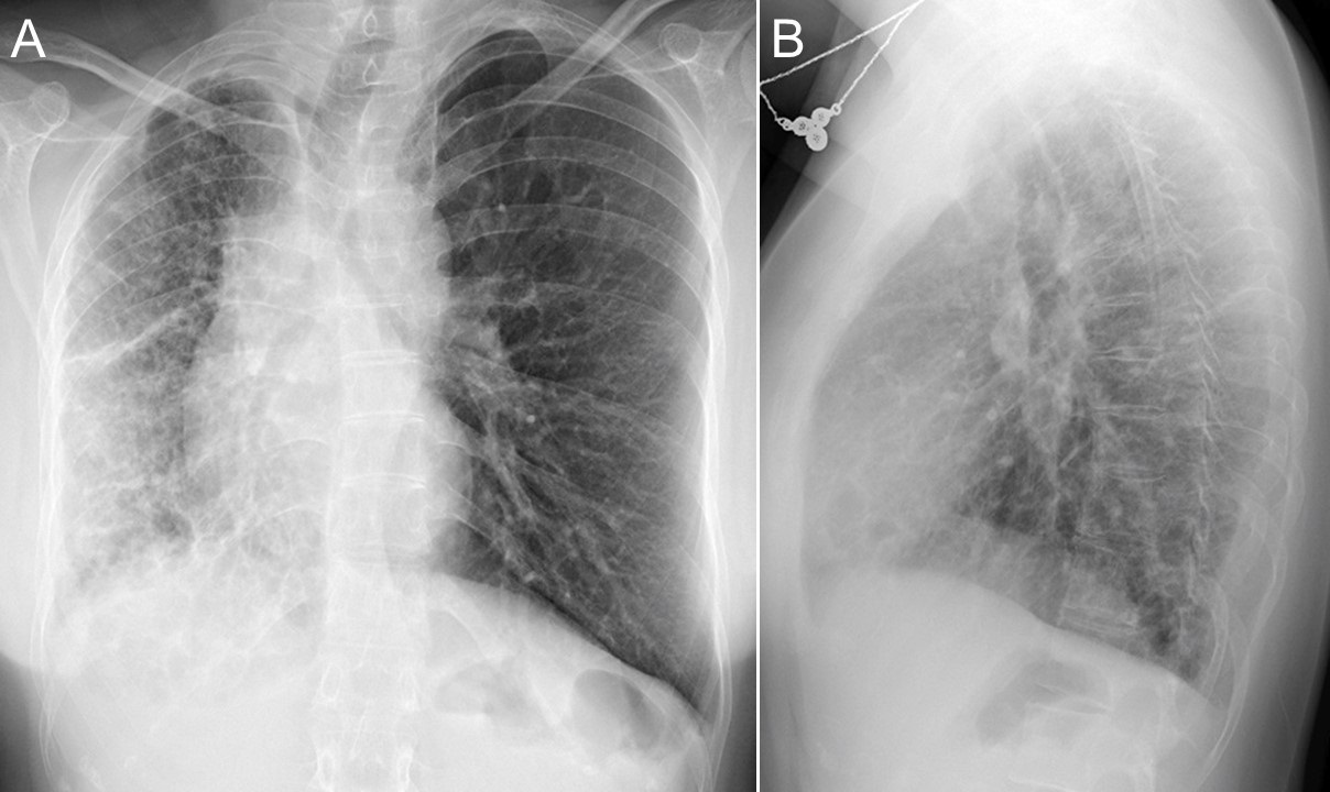
Figure 1.  Frontal chest radiography.

Which of the following statements regarding the chest radiograph is most accurate? (Click on the correct answer to proceed to the second of nine pages)

  1. The chest radiograph shows a mediastinal mass
  2. The chest radiograph shows multifocal consolidation and pleural effusion
  3. The chest radiograph shows multifocal smooth interlobular septal thickening
  4. The chest radiograph shows a possible focal air space opacity
  5. The chest radiograph shows small cavitary pulmonary nodules

Cite as: Larsen BT, Gotway MB. August 2017 imaging case of the month. Southwest J Pulm Crit Care. 2017;15(2):69-79. doi: https://doi.org/10.13175/swjpcc098-17 PDF