Wednesday
Apr032013
Medical Image of the Week: Septic Emboli

Figure 1. Photograph showing septic emboli to distal digits.
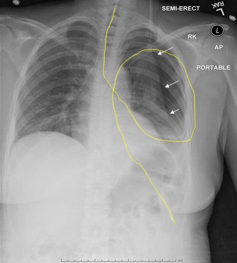
A 34 year old woman was admitted for a vasculitis workup after presenting with painful fingers, chest pain, and diffuse joint pain. Her blood cultures grew Staphyloccccus aureus and she was diagnosed with mitral and aortic valve endocarditis. She had widespread joint involvement as well as a thoracic epidural abscess.
Jarrod Mosier, MD and Nathaniel Reyes, MD
Departments of Medicine and Emergency Medicine
University of Arizona
Tucson, Arizona
Reference as: Mosier J, Reyes N. Medical image of the week: septic emboli. Southwest J Pulm Crit Care. 2013;6(4):170. PDF
 Post a Comment