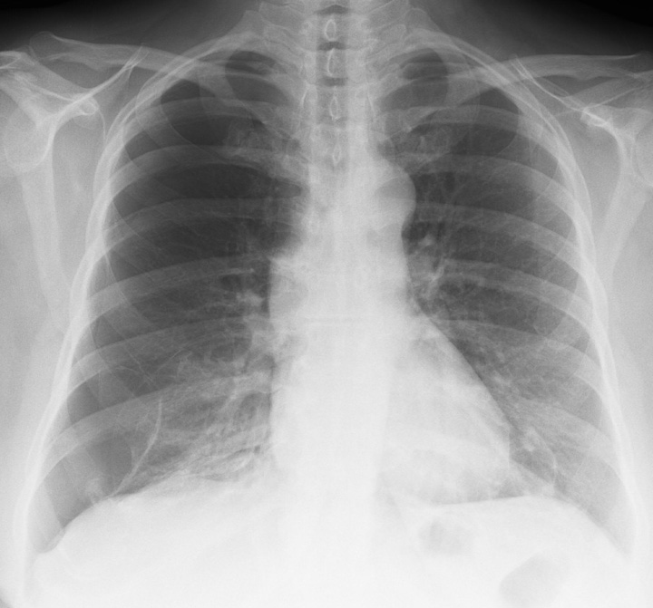
Search Journal-type in search term and press enter
Southwest Pulmonary and Critical Care Fellowships
Social Media

Imaging

Last 50 Imaging Postings

(Most recent listed first. Click on title to be directed to the manuscript.)

June 2025 Medical Image of the Month: Neurofibromatosis-Associated Diffuse
   Cystic Lung Disease
May 2025 Medical Image of the Month: Aspirated Dental Screw
April 2025 Medical Image of the Month: An Unfortunate Case of Mimicry
March 2025 Medical Image of the Month: An Unusual Case of Pulmonary
   Infarction
February 2025 Medical Image of the Month: Unexpected Complications of
   Transjugular Intrahepatic Portosystemic Shunt (TIPS) 
February 2025 Imaging Case of the Month: A Wolf in Sheep’s Clothing
January 2025 Medical Image of the Month: Psoriasis with Pulmonary
   Involvement
December 2024 Medical Image of the Month: An Endobronchial Tumor
November 2024 Medical Image of the Month: A Case of Short Telomeres
November 2024 Imaging Case of the Month: A Recurring Issue
October 2024 Medical Image of the Month: Lofgren syndrome with Erythema
   Nodosum
September 2024 Medical Image of the Month: A Curious Case of Nasal
   Congestion
August 2024 Image of the Month: Lymphomatoid Granulomatosis
August 2024 Imaging Case of the Month: An Unexplained Pleural Effusion
July 2024 Medical Image of the Month: Vocal Cord Paralysis on PET-CT 
June 2024 Medical Image of the Month: A 76-year-old Man Presenting with
   Acute Hoarseness
May 2024 Medical Image of the Month: Hereditary Hemorrhagic
   Telangiectasia in a Patient on Veno-Arterial Extra-Corporeal Membrane
   Oxygenation
May 2024 Imaging Case of the Month: Nothing Is Guaranteed
April 2024 Medical Image of the Month: Wind Instruments Player Exhibiting
   Exceptional Pulmonary Function
March 2024 Medical Image of the Month: Sputum Cytology in Patients with
   Suspected Lung Malignancy Presenting with Acute Hypoxic Respiratory
   Failure
February 2024 Medical Image of the Month: Pulmonary Alveolar Proteinosis
   in Myelodysplastic Syndrome
February 2024 Imaging Case of the Month: Connecting Some Unusual Dots
January 2024 Medical Image of the Month: Polyangiitis Overlap Syndrome
   (POS) Mimicking Fungal Pneumonia 
December 2023 Medical Image of the Month: Metastatic Pulmonary
   Calcifications in End-Stage Renal Disease 
November 2023 Medical Image of the Month: Obstructive Uropathy
   Extremis
November 2023 Imaging Case of the Month: A Crazy Association
October 2023 Medical Image of the Month: Swyer-James-MacLeod
   Syndrome
September 2023 Medical Image of the Month: Aspergillus Presenting as a
   Pulmonary Nodule in an Immunocompetent Patient
August 2023 Medical Image of the Month: Cannonball Metastases from
   Metastatic Melanoma
August 2023 Imaging Case of the Month: Chew Your Food Carefully
July 2023 Medical Image of the Month: Primary Tracheal Lymphoma
June 2023 Medical Image of the Month: Solitary Fibrous Tumor of the Pleura
May 2023 Medical Image of the Month: Methamphetamine Inhalation
   Leading to Cavitary Pneumonia and Pleural Complications
April 2023 Medical Image of the Month: Atrial Myxoma in the Setting of
   Raynaud’s Phenomenon: Early Echocardiography and Management of
   Thrombotic Disease
April 2023 Imaging Case of the Month: Large Impact from a Small Lesion
March 2023 Medical Image of the Month: Spontaneous Pneumomediastinum
   as a Complication of Marijuana Smoking Due to Müller's Maneuvers
February 2023 Medical Image of the Month: Reversed Halo Sign in the
   Setting of a Neutropenic Patient with Angioinvasive Pulmonary
   Zygomycosis
January 2023 Medical Image of the Month: Abnormal Sleep Study and PFT
   with Supine Challenge Related to Idiopathic Hemidiaphragmatic Paralysis
December 2022 Medical Image of the Month: Bronchoesophageal Fistula in
   the Setting of Pulmonary Actinomycosis
November 2022 Medical Image of the Month: COVID-19 Infection
   Presenting as Spontaneous Subcapsular Hematoma of the Kidney
November 2022 Imaging Case of the Month: Out of Place in the Thorax
October 2022 Medical Image of the Month: Infected Dasatinib Induced
   Chylothorax-The First Reported Case 
September 2022 Medical Image of the Month: Epiglottic Calcification
Medical Image of the Month: An Unexpected Cause of Chronic Cough
August 2022 Imaging Case of the Month: It’s All About Location
July 2022 Medical Image of the Month: Pulmonary Nodule in the
   Setting of Pyoderma Gangrenosum (PG) 
June 2022 Medical Image of the Month: A Hard Image to Swallow
May 2022 Medical Image of the Month: Pectus Excavatum
May 2022 Imaging Case of the Month: Asymmetric Apical Opacity–
   Diagnostic Considerations
April 2022 Medical Image of the Month: COVID Pericarditis
March 2022 Medical Image of the Month: Pulmonary Nodules in the
   Setting of Diffuse Idiopathic Pulmonary NeuroEndocrine Cell Hyperplasia
   (DIPNECH) 
February 2022 Medical Image of the Month: Multifocal Micronodular
   Pneumocyte Hyperplasia in the Setting of Tuberous Sclerosis
February 2022 Imaging Case of the Month: Between A Rock and a
   Hard Place
January 2022 Medical Image of the Month: Bronchial Obstruction
   Due to Pledget in Airway Following Foregut Cyst Resection
December 2021 Medical Image of the Month: Aspirated Dental Implant
   Medical Image of the Month: Cavitating Pseudomonas
   aeruginosa Pneumonia
November 2021 Imaging Case of the Month: Let’s Not Dance
   the Twist


For complete imaging listings click here

Those who care for patients with pulmonary, critical care or sleep disorders rely heavily on chest radiology and pathology to determine diagnoses. The Southwest Journal of Pulmonary, Critical Care & Sleep publishes case-based articles with characteristic chest imaging and related pathology. The editor of this section will oversee and coordinate the publication of a core of the most important chest imaging topics. In doing so, they encourage the submission of unsolicited manuscripts. It cannot be overemphasized that both radiologic and pathologic images must be of excellent quality. As a rule, 600 DPI is sufficient for radiographic and pathologic images. Taking pictures of plain chest radiographs and CT scans with a digital camera is strongly discouraged. The figures should be cited in the text and numbered consecutively. The stain used for pathology specimens and magnification should be mentioned in the figure legend. Those who care for patients with pulmonary, critical care or sleep disorders rely heavily on chest radiology and pathology to determine diagnoses. The Southwest Journal of Pulmonary, Critical Care & Sleep publishes case-based articles with characteristic chest imaging and related pathology. The editor of this section will oversee and coordinate the publication of a core of the most important chest imaging topics. In doing so, they encourage the submission of unsolicited manuscripts. It cannot be overemphasized that both radiologic and pathologic images must be of excellent quality. As a rule, 600 DPI is sufficient for radiographic and pathologic images. Taking pictures of plain chest radiographs and CT scans with a digital camera is strongly discouraged. The figures should be cited in the text and numbered consecutively. The stain used for pathology specimens and magnification should be mentioned in the figure legend.

-------------------------------------------------------------------------------------------  

Wednesday
Oct212015

Medical Image of the Week: Acute Amiodarone Pulmonary Toxicity

Figure 1. Chest X-ray showing diffuse interstitial markings, right upper lobe consolidation, small pleural effusions, thoracotomy wires and external leads.


Figure 2. Axial image of the thoracic CT scan showing increased interstitial markings, ground glass opacities and bilateral pleural effusions.


A 71 year old man with a medical history significant for chronic obstructive pulmonary disease, coronary artery disease with post-operative status coronary artery bypass grafting, heart failure with reduced ejection fraction (25%) and atrial fibrillation/flutter underwent an elective ablation of the tachyarrhythmia at another facility and was prescribed amiodarone post procedure. He started complaining of cough and dyspnea one day post procedure and was empirically treated with 2 weeks of broad spectrum antibiotics. He subsequently was transferred to our facility due to worsening symptoms. He also complained of nausea, anorexia with resultant weight loss since starting amiodarone, which was stopped 5 days prior to transfer. Infectious work up was negative.

On arrival to our facility, he was diagnosed with small sub-segmental pulmonary emboli, pulmonary edema and possible acute amiodarone toxicity. His was profoundly hypoxic requiring high flow nasal cannula or 100% non-rebreather mask at all times. His symptoms persisted despite antibiotics, diuresis, anticoagulation and heart rate control. Steroid therapy was then initiated for acute amiodarone toxicity. Although he reported some improvement in symptoms 2-3 days after initiation of steroids, his oxygen requirement did not improve. Unfortunately he suffered a cardiac arrest on day 10 of admission and did not survive.

Amiodarone is a class B anti-arrhythmic used to treat multiple supraventricular and ventricular tachyarrhythmias. Its adverse effects are usually dose and duration dependent. Amiodarone pulmonary toxicity (APT) has been shown to correlate with total cumulative dose; however acute reactions to amiodarone toxicity have previously been reported. Men are at increased risk for APT, and this risk increases with age and those with pre-existing lung conditions. Diagnosis of APT is predominantly a diagnosis of exclusion; however laboratory tests may show leukocytosis with neutrophil predominance (as in our patient) and imaging may provide a clue for diagnosis. Chest x-ray reveals patchy or diffuse infiltrates, which may have predominance in the upper lobes, particularly the right upper lobe (as in our patient). A thoracic CT scan may show bilateral alveolar or interstitial infiltrates with higher attenuation, secondary to the iodine component of the drug. The current mainstay of treatment is discontinuation of the drug permanently along with steroid therapy typically, 40-60 mg of prednisone a day for an extended period of time.  

Konstantin Mazursky DO1, Bhupinder Natt MD2, Laura Meinke MD1,2

1Department of Internal Medicine.

2Division of Pulmonary, Critical Care, Allergy and Sleep

Banner-University Medical Center

Tucson AZ

Reference

  1. Wolkove N, Baltzan M. Amiodarone pulmonary toxicity. Can Respir J. 2009;16(2):43-8. [PubMed] 

Cite as: Mazursky K, Natt B, Meinke L. Medical image of the week: acute amiodarone pulmonary toxicity. Southwest J Pulm Crit Care. 2015;11(4):189-90. doi: http://dx.doi.org/10.13175/swjpcc099-15 PDF

Wednesday
Oct142015

Medical Image of the Week: PSG Sweat Artifact

Figure 1. A 30 second epoch suggestive of sweat artifact and incidentally noted snore artifact on the M1 channels.


Figure 2: Sweat artifact as seen in a 10 second epoch.


Figure 3.  30 second epoch after removal of the M1 channels.


A 61-year-old man, with a past medical history significant for hypertension, COPD and morbid obesity with a body mass index (BMI) of 45.81 is referred for an overnight sleep study for suspicion of obstructive sleep apnea. Artifact was noted on the polysomnogram recording as shown above (Figures 1-3).

Sweat artifact is characterized by slow undulating movement of the baseline recording in the affected channels due to perspiration altering the potential of the involved electrodes (1). Sweat artifact may mimic delta waves and scored as non-rapid eye movement (NREM) stage 3 sleep. Lowering the room temperature, using a fan on the scalp or replacing the conductive paste on the electrodes may help eliminate the artifact.

Safal Shetty, MD1 and John Roehrs, MD2

1Banner University Medical Center Tucson, AZ

2Southern Arizona VA Health Care System

Tucson, AZ

Reference

  1. Siddiqui F, Osuna E, Walters AS, Chokroverty S. Sweat artifact and respiratory artifact occurring simultaneously in polysomnogram. Sleep Med. 2006;7(2):197-9. [CrossRef] [PubMed] 

Cite as: Shetty S, Roehrs J. Medical image of the week: PSG sweat artifact. Southwest J Pulm Crit Care. 2015;11(4):171-2. doi: http://dx.doi.org/10.13175/swjpcc097-15 PDF