Imaging

Last 50 Imaging Postings

(Most recent listed first. Click on title to be directed to the manuscript.)

August 2025 Medical Image of the Month: Crazy Paving in a Case of 
   Eosinophilic Granulomatosis with Polyangiitis
July 2025 Medical Image of the Month: A Case of Severe Hiatal Hernia
   Presenting as Atypical Chest Pain
July 2025 Imaging Case of the Month: A Growing Lung Nodule in a
   Patient with Heart Disease
June 2025 Medical Image of the Month: Neurofibromatosis-Associated Diffuse
   Cystic Lung Disease
May 2025 Medical Image of the Month: Aspirated Dental Screw
April 2025 Medical Image of the Month: An Unfortunate Case of Mimicry
March 2025 Medical Image of the Month: An Unusual Case of Pulmonary
   Infarction
February 2025 Medical Image of the Month: Unexpected Complications of
   Transjugular Intrahepatic Portosystemic Shunt (TIPS) 
February 2025 Imaging Case of the Month: A Wolf in Sheep’s Clothing
January 2025 Medical Image of the Month: Psoriasis with Pulmonary
   Involvement
December 2024 Medical Image of the Month: An Endobronchial Tumor
November 2024 Medical Image of the Month: A Case of Short Telomeres
November 2024 Imaging Case of the Month: A Recurring Issue
October 2024 Medical Image of the Month: Lofgren syndrome with Erythema
   Nodosum
September 2024 Medical Image of the Month: A Curious Case of Nasal
   Congestion
August 2024 Image of the Month: Lymphomatoid Granulomatosis
August 2024 Imaging Case of the Month: An Unexplained Pleural Effusion
July 2024 Medical Image of the Month: Vocal Cord Paralysis on PET-CT 
June 2024 Medical Image of the Month: A 76-year-old Man Presenting with
   Acute Hoarseness
May 2024 Medical Image of the Month: Hereditary Hemorrhagic
   Telangiectasia in a Patient on Veno-Arterial Extra-Corporeal Membrane
   Oxygenation
May 2024 Imaging Case of the Month: Nothing Is Guaranteed
April 2024 Medical Image of the Month: Wind Instruments Player Exhibiting
   Exceptional Pulmonary Function
March 2024 Medical Image of the Month: Sputum Cytology in Patients with
   Suspected Lung Malignancy Presenting with Acute Hypoxic Respiratory
   Failure
February 2024 Medical Image of the Month: Pulmonary Alveolar Proteinosis
   in Myelodysplastic Syndrome
February 2024 Imaging Case of the Month: Connecting Some Unusual Dots
January 2024 Medical Image of the Month: Polyangiitis Overlap Syndrome
   (POS) Mimicking Fungal Pneumonia 
December 2023 Medical Image of the Month: Metastatic Pulmonary
   Calcifications in End-Stage Renal Disease 
November 2023 Medical Image of the Month: Obstructive Uropathy
   Extremis
November 2023 Imaging Case of the Month: A Crazy Association
October 2023 Medical Image of the Month: Swyer-James-MacLeod
   Syndrome
September 2023 Medical Image of the Month: Aspergillus Presenting as a
   Pulmonary Nodule in an Immunocompetent Patient
August 2023 Medical Image of the Month: Cannonball Metastases from
   Metastatic Melanoma
August 2023 Imaging Case of the Month: Chew Your Food Carefully
July 2023 Medical Image of the Month: Primary Tracheal Lymphoma
June 2023 Medical Image of the Month: Solitary Fibrous Tumor of the Pleura
May 2023 Medical Image of the Month: Methamphetamine Inhalation
   Leading to Cavitary Pneumonia and Pleural Complications
April 2023 Medical Image of the Month: Atrial Myxoma in the Setting of
   Raynaud’s Phenomenon: Early Echocardiography and Management of
   Thrombotic Disease
April 2023 Imaging Case of the Month: Large Impact from a Small Lesion
March 2023 Medical Image of the Month: Spontaneous Pneumomediastinum
   as a Complication of Marijuana Smoking Due to Müller's Maneuvers
February 2023 Medical Image of the Month: Reversed Halo Sign in the
   Setting of a Neutropenic Patient with Angioinvasive Pulmonary
   Zygomycosis
January 2023 Medical Image of the Month: Abnormal Sleep Study and PFT
   with Supine Challenge Related to Idiopathic Hemidiaphragmatic Paralysis
December 2022 Medical Image of the Month: Bronchoesophageal Fistula in
   the Setting of Pulmonary Actinomycosis
November 2022 Medical Image of the Month: COVID-19 Infection
   Presenting as Spontaneous Subcapsular Hematoma of the Kidney
November 2022 Imaging Case of the Month: Out of Place in the Thorax
October 2022 Medical Image of the Month: Infected Dasatinib Induced
   Chylothorax-The First Reported Case 
September 2022 Medical Image of the Month: Epiglottic Calcification
Medical Image of the Month: An Unexpected Cause of Chronic Cough
August 2022 Imaging Case of the Month: It’s All About Location
July 2022 Medical Image of the Month: Pulmonary Nodule in the
   Setting of Pyoderma Gangrenosum (PG) 
June 2022 Medical Image of the Month: A Hard Image to Swallow
May 2022 Medical Image of the Month: Pectus Excavatum
May 2022 Imaging Case of the Month: Asymmetric Apical Opacity–
   Diagnostic Considerations
April 2022 Medical Image of the Month: COVID Pericarditis
March 2022 Medical Image of the Month: Pulmonary Nodules in the
   Setting of Diffuse Idiopathic Pulmonary NeuroEndocrine Cell Hyperplasia
   (DIPNECH) 
February 2022 Medical Image of the Month: Multifocal Micronodular
   Pneumocyte Hyperplasia in the Setting of Tuberous Sclerosis
February 2022 Imaging Case of the Month: Between A Rock and a
   Hard Place


For complete imaging listings click here

Those who care for patients with pulmonary, critical care or sleep disorders rely heavily on chest radiology and pathology to determine diagnoses. The Southwest Journal of Pulmonary, Critical Care & Sleep publishes case-based articles with characteristic chest imaging and related pathology. The editor of this section will oversee and coordinate the publication of a core of the most important chest imaging topics. In doing so, they encourage the submission of unsolicited manuscripts. It cannot be overemphasized that both radiologic and pathologic images must be of excellent quality. As a rule, 600 DPI is sufficient for radiographic and pathologic images. Taking pictures of plain chest radiographs and CT scans with a digital camera is strongly discouraged. The figures should be cited in the text and numbered consecutively. The stain used for pathology specimens and magnification should be mentioned in the figure legend. Those who care for patients with pulmonary, critical care or sleep disorders rely heavily on chest radiology and pathology to determine diagnoses. The Southwest Journal of Pulmonary, Critical Care & Sleep publishes case-based articles with characteristic chest imaging and related pathology. The editor of this section will oversee and coordinate the publication of a core of the most important chest imaging topics. In doing so, they encourage the submission of unsolicited manuscripts. It cannot be overemphasized that both radiologic and pathologic images must be of excellent quality. As a rule, 600 DPI is sufficient for radiographic and pathologic images. Taking pictures of plain chest radiographs and CT scans with a digital camera is strongly discouraged. The figures should be cited in the text and numbered consecutively. The stain used for pathology specimens and magnification should be mentioned in the figure legend.

-------------------------------------------------------------------------------------------  

Entries in lymphadenopathy (5)

Wednesday
Aug082018

Medical Image of the Week: Chylothorax

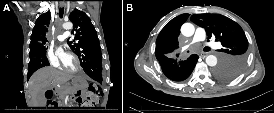
Figure 1. A: CT of the chest (coronal image) demonstrating large right hilar and mediastinal adenopathy, leading to moderate to severe narrowing of the superior vena cava (SVC). B: CT of the chest (axial image) demonstrating moderate to severe narrowing of the pulmonary artery trunk due to compression from mediastinal adenopathy. A left pleural effusion is noted.


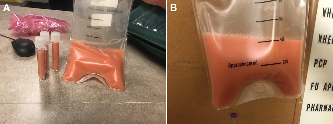
Figure 2. Pleural fluid sample demonstrating milky, pink fluid. The triglyceride level was 532 mg/dl and cholesterol level 63 mg/dl.


A 73-year-old man with untreated stage IV adenocarcinoma of the lung was admitted to the hospital with several days of progressively worsening dyspnea on exertion. The chest CT showed a large left pleural effusion with enlarging bilateral hilar and mediastinal lymphadenopathy, compression of the superior vena cava and right main pulmonary artery consistent with progressive lung cancer (Figure 1). Therapeutic and diagnostic left sided thoracentesis was performed, removing approximately 450 ml of milky, pink fluid suggestive of hemochylothorax (Figure 2). Analysis of the fluid was significant for 27,720 red blood cells, 476 total nucleated cells with lymphocyte predominance (87%), glucose 158 mg/dl, cholesterol 63 mg/dl, and amylase 28 U/L. The pleural fluid was exudative (protein 4.4 g/dl) with a significantly elevated triglyceride level of 532 mg/dl. No malignant cells were identified in the fluid.

This case illustrates a nontraumatic chylothorax secondary to metastatic adenocarcinoma of the lung. The leading cause of non-traumatic chylothorax is malignancy by compression and/or lymphangitic invasion (1). Thoracic duct invasion or leak can only be seen with nuclear medicine scintigraphy; however, this test was not performed on this patient. The appearance of the pleural fluid in chylothorax can be deceiving as less than half of pleural fluid samples will be milky in appearance (2). In addition, milky appearing pleural fluid is not specific for a chylothorax, as milky fluid can be seen in a cholesterol pleural effusion (pseudochylothorax) or an empyema. The detection of chylomicrons on pleural fluid lipoprotein electrophoresis is the definitive diagnostic criterion for chylothorax, however it is not widely available and is costly (3). The classic diagnostic criterion is a pleural fluid triglyceride level of >110 mg/dl in an appropriate clinical setting of mediastinal malignancy, lymphoma, recent thoracic surgery or penetrating trauma to the neck or thorax (4). A pleural fluid triglyceride level between 50 and 110 mg/dl does not exclude the diagnosis of chylothorax and clinicians should perform lipoprotein electrophoresis of the pleural fluid to detect chylomicrons. To distinguish a chylothorax from a pseudochylothorax (both have milky appearance), clinicians should obtain a cholesterol level on the fluid. The cholesterol level in a chylothorax is usually less than 200 mg/dl while a pseudochylothorax will have high levels, typically greater than 200 mg/dl.

The patient chose to undergo palliative radiation of the chest and symptomatic treatment of his dyspnea.  

John Dicken MD1, Madhav Chopra MD2, Faraz Jaffer MD2 and Linda Snyder MD2

1Department of Internal Medicine and 2Division of Pulmonary, Allergy, Critical Care and Sleep

Banner University Medical Center-Tucson

Tucson, AZ USA

References

  1. McGrath EE, Blades Z, Anderson PB. Chylothorax: aetiology, diagnosis and therapeutic options. Respir Med. 2010 Jan;104(1):1-8. [CrossRef] [PubMed]
  2. Maldonado F, Hawkins FJ, Daniels CE, Doerr CH, Decker PA, Ryu JH. Pleural fluid characteristics of chylothorax. Mayo Clin Proc. 2009 Feb;84(2):129-33. [CrossRef] [PubMed]
  3. Hooper C, Lee YC, Maskell N; BTS Pleural Guideline Group. Investigation of a unilateral pleural effusion in adults: British Thoracic Society Pleural Disease Guideline 2010. Thorax. 2010 Aug;65 Suppl 2:ii4-17. [CrossRef] [PubMed]
  4. Staats BA, Ellefson RD, Budahn LL, Dines DE, Prakash UB, Offord K. The lipoprotein profile of chylous and nonchylous pleural effusions. Mayo Clin Proc. 1980 Nov;55(11):700-4. [PubMed]

Cite as: Dicken J, Chopra M, Jaffer F, Snyder L. Medical image of the week: Chylothorax. Southwest J Pulm Crit Care. 2018;17(2):70-1. doi: https://doi.org/10.13175/swjpcc100-18 PDF 

Wednesday
Nov022016

Medical Image of the Week: Lynch Syndrome

Figure 1. Coronal CT of the chest showing bilateral filling defects in pulmonary arteries representing pulmonary emobolism.


Figure 2. Coronal CT of the abdomen/pelvis showing periaortic lymphadenopathy suggestive for metastatic disease.


Figure 3. Coronal CT of the abdomen/ pelvis showing a 13 x 13.6 cm solid and cystic mass above and to the right of the uterus concerning for right ovarian neoplasm.


A 43-year-old woman with a history of anemia, thrombocytopenia, and recent treatment for pyelonephritis was transferred to our hospital for increasing shortness of breath. Four months prior to admission, she developed unprovoked bilateral deep vein thrombosis (DVT) and pulmonary emboli (PE) and was started on rivaroxaban at that time. At presentation, she was complaining of worsening shortness of breath, heavy menstrual bleeding and pain in her calves. CT angiography of chest showed multiple pulmonary emboli to the lower lobes and left upper lobe (Figure 1) and lower extremity venous Doppler showed extensive, acute deep vein thrombosis involving the femoral, popliteal and calf veins bilaterally.

Rivaroxaban was held due to anemia and thrombocytopenia and there was concern for respiratory failure since she developed new DVT and PE. She was transfused with 1 unit of packed red blood cells and started on a heparin drip. She continued to have significant menorrhagia, the heparin drip was discontinued, and subsequently, an inferior vena cava filter was placed.

On further questioning, the patient reported a 26 pound weight loss over the past three weeks. This combined with her menorrhagia requiring blood transfusion prompted further imaging. CT of the abdomen and pelvis showed a 13 x 13.6 cm solid and cystic mass representing a right ovarian neoplasm that was contiguous with the uterus as well as periaortic adenopathy suggestive of metastasis (Figures 2 and 3). Further investigation into the patient’s family history identified significant history for breast and ovarian cancers on her maternal side. Genetic testing of the patient showed a germline mutation in the MSH2 gene, consistent with Lynch syndrome.

Lynch syndrome, also known as hereditary non-polyposis colorectal cancer, is a hereditary cancer syndrome characterized by mutations in DNA mismatch repair genes. The majority of affected individuals will develop colorectal or endometrial malignancies; however these individuals are also at increased risk for developing ovarian neoplasms. The lifetime risk of developing ovarian cancer in women with Lynch syndrome is 7% (3-14%) compared to 1.4% in the general population (1). However, there is no survival difference between women with Lynch syndrome and the general population (1). If ovarian malignancy is present at diagnosis of Lynch syndrome, prophylactic hysterectomy and bilateral salpingo-oophorectomy is recommended (2). Otherwise, management can include prophylactic surgery or screening with annual pelvic exams and transvaginal ultrasounds. Persons with lynch syndrome should also receive surveillance for other associated malignancies such as colorectal or endometrial cancer (1,2).

Abha Athale MS1, Christopher Morrison MD2, Robert Betancourt MD3 and Jennifer Segar MD3

1 Midwestern University Arizona College of Osteopathic Medicine

2 Tucson Hospitals Medical Education Program

3 Department of Internal Medicine, Banner University Medical Center-Tucson Medical Center, Tucson, AZ

References

  1. Koornstra JJ, Mourits MJ, Sijmons RH, Leliveld AM, Hollema H, Kleibeuker JH. Management of extracolonic tumours in patients with Lynch syndrome. Lancet Oncol. 2009 Apr;10(4):400-8. [CrossRef] [PubMed]
  2. Lindor NM, Petersen GM, Hadley DW, Kinney AY, Miesfeldt S, Lu KH, Lynch P, Burke W, Press N. Recommendations for the care of individuals with an inherited predisposition to Lynch syndrome: a systematic review. JAMA. 2006 Sep 27;296(12):1507-17. [CrossRef] [PubMed]

Cite as: Athale A, Morrison C, Betancourt R, Segar J. Medical image of the week: Lynch syndrome. Southwest J Pulm Crit Care. 2016;13(5):202-4. doi: http://dx.doi.org/10.13175/swjpcc087-16 PDF