Search Journal-type in search term and press enter
Southwest Pulmonary and Critical Care Fellowships
Social Media

Imaging

Last 50 Imaging Postings

(Most recent listed first. Click on title to be directed to the manuscript.)

June 2025 Medical Image of the Month: Neurofibromatosis-Associated Diffuse
   Cystic Lung Disease
May 2025 Medical Image of the Month: Aspirated Dental Screw
April 2025 Medical Image of the Month: An Unfortunate Case of Mimicry
March 2025 Medical Image of the Month: An Unusual Case of Pulmonary
   Infarction
February 2025 Medical Image of the Month: Unexpected Complications of
   Transjugular Intrahepatic Portosystemic Shunt (TIPS) 
February 2025 Imaging Case of the Month: A Wolf in Sheep’s Clothing
January 2025 Medical Image of the Month: Psoriasis with Pulmonary
   Involvement
December 2024 Medical Image of the Month: An Endobronchial Tumor
November 2024 Medical Image of the Month: A Case of Short Telomeres
November 2024 Imaging Case of the Month: A Recurring Issue
October 2024 Medical Image of the Month: Lofgren syndrome with Erythema
   Nodosum
September 2024 Medical Image of the Month: A Curious Case of Nasal
   Congestion
August 2024 Image of the Month: Lymphomatoid Granulomatosis
August 2024 Imaging Case of the Month: An Unexplained Pleural Effusion
July 2024 Medical Image of the Month: Vocal Cord Paralysis on PET-CT 
June 2024 Medical Image of the Month: A 76-year-old Man Presenting with
   Acute Hoarseness
May 2024 Medical Image of the Month: Hereditary Hemorrhagic
   Telangiectasia in a Patient on Veno-Arterial Extra-Corporeal Membrane
   Oxygenation
May 2024 Imaging Case of the Month: Nothing Is Guaranteed
April 2024 Medical Image of the Month: Wind Instruments Player Exhibiting
   Exceptional Pulmonary Function
March 2024 Medical Image of the Month: Sputum Cytology in Patients with
   Suspected Lung Malignancy Presenting with Acute Hypoxic Respiratory
   Failure
February 2024 Medical Image of the Month: Pulmonary Alveolar Proteinosis
   in Myelodysplastic Syndrome
February 2024 Imaging Case of the Month: Connecting Some Unusual Dots
January 2024 Medical Image of the Month: Polyangiitis Overlap Syndrome
   (POS) Mimicking Fungal Pneumonia 
December 2023 Medical Image of the Month: Metastatic Pulmonary
   Calcifications in End-Stage Renal Disease 
November 2023 Medical Image of the Month: Obstructive Uropathy
   Extremis
November 2023 Imaging Case of the Month: A Crazy Association
October 2023 Medical Image of the Month: Swyer-James-MacLeod
   Syndrome
September 2023 Medical Image of the Month: Aspergillus Presenting as a
   Pulmonary Nodule in an Immunocompetent Patient
August 2023 Medical Image of the Month: Cannonball Metastases from
   Metastatic Melanoma
August 2023 Imaging Case of the Month: Chew Your Food Carefully
July 2023 Medical Image of the Month: Primary Tracheal Lymphoma
June 2023 Medical Image of the Month: Solitary Fibrous Tumor of the Pleura
May 2023 Medical Image of the Month: Methamphetamine Inhalation
   Leading to Cavitary Pneumonia and Pleural Complications
April 2023 Medical Image of the Month: Atrial Myxoma in the Setting of
   Raynaud’s Phenomenon: Early Echocardiography and Management of
   Thrombotic Disease
April 2023 Imaging Case of the Month: Large Impact from a Small Lesion
March 2023 Medical Image of the Month: Spontaneous Pneumomediastinum
   as a Complication of Marijuana Smoking Due to Müller's Maneuvers
February 2023 Medical Image of the Month: Reversed Halo Sign in the
   Setting of a Neutropenic Patient with Angioinvasive Pulmonary
   Zygomycosis
January 2023 Medical Image of the Month: Abnormal Sleep Study and PFT
   with Supine Challenge Related to Idiopathic Hemidiaphragmatic Paralysis
December 2022 Medical Image of the Month: Bronchoesophageal Fistula in
   the Setting of Pulmonary Actinomycosis
November 2022 Medical Image of the Month: COVID-19 Infection
   Presenting as Spontaneous Subcapsular Hematoma of the Kidney
November 2022 Imaging Case of the Month: Out of Place in the Thorax
October 2022 Medical Image of the Month: Infected Dasatinib Induced
   Chylothorax-The First Reported Case 
September 2022 Medical Image of the Month: Epiglottic Calcification
Medical Image of the Month: An Unexpected Cause of Chronic Cough
August 2022 Imaging Case of the Month: It’s All About Location
July 2022 Medical Image of the Month: Pulmonary Nodule in the
   Setting of Pyoderma Gangrenosum (PG) 
June 2022 Medical Image of the Month: A Hard Image to Swallow
May 2022 Medical Image of the Month: Pectus Excavatum
May 2022 Imaging Case of the Month: Asymmetric Apical Opacity–
   Diagnostic Considerations
April 2022 Medical Image of the Month: COVID Pericarditis
March 2022 Medical Image of the Month: Pulmonary Nodules in the
   Setting of Diffuse Idiopathic Pulmonary NeuroEndocrine Cell Hyperplasia
   (DIPNECH) 
February 2022 Medical Image of the Month: Multifocal Micronodular
   Pneumocyte Hyperplasia in the Setting of Tuberous Sclerosis
February 2022 Imaging Case of the Month: Between A Rock and a
   Hard Place
January 2022 Medical Image of the Month: Bronchial Obstruction
   Due to Pledget in Airway Following Foregut Cyst Resection
December 2021 Medical Image of the Month: Aspirated Dental Implant
   Medical Image of the Month: Cavitating Pseudomonas
   aeruginosa Pneumonia
November 2021 Imaging Case of the Month: Let’s Not Dance
   the Twist


For complete imaging listings click here

Those who care for patients with pulmonary, critical care or sleep disorders rely heavily on chest radiology and pathology to determine diagnoses. The Southwest Journal of Pulmonary, Critical Care & Sleep publishes case-based articles with characteristic chest imaging and related pathology. The editor of this section will oversee and coordinate the publication of a core of the most important chest imaging topics. In doing so, they encourage the submission of unsolicited manuscripts. It cannot be overemphasized that both radiologic and pathologic images must be of excellent quality. As a rule, 600 DPI is sufficient for radiographic and pathologic images. Taking pictures of plain chest radiographs and CT scans with a digital camera is strongly discouraged. The figures should be cited in the text and numbered consecutively. The stain used for pathology specimens and magnification should be mentioned in the figure legend. Those who care for patients with pulmonary, critical care or sleep disorders rely heavily on chest radiology and pathology to determine diagnoses. The Southwest Journal of Pulmonary, Critical Care & Sleep publishes case-based articles with characteristic chest imaging and related pathology. The editor of this section will oversee and coordinate the publication of a core of the most important chest imaging topics. In doing so, they encourage the submission of unsolicited manuscripts. It cannot be overemphasized that both radiologic and pathologic images must be of excellent quality. As a rule, 600 DPI is sufficient for radiographic and pathologic images. Taking pictures of plain chest radiographs and CT scans with a digital camera is strongly discouraged. The figures should be cited in the text and numbered consecutively. The stain used for pathology specimens and magnification should be mentioned in the figure legend.

-------------------------------------------------------------------------------------------  

Entries in hemoptysis (21)

Wednesday
Aug282013

Medical Image of the Week: Fontan Procedure

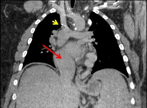
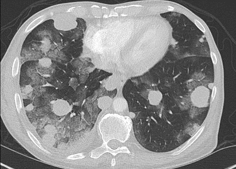
Figure 1. Thoracic CT scan showing Fontan anatomy, with the superior vena cava (SVC) connected to the pulmonary arteries (yellow arrow) and a single atrium and ventricle (red arrow).


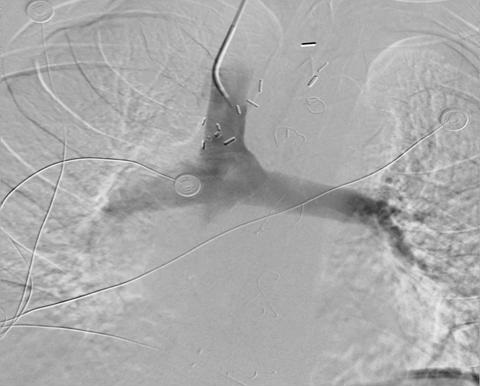
Figure 2. SVC venography shows SVC connected to the pulmonary artery.

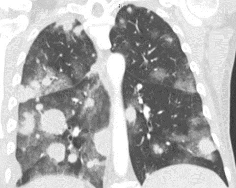
A 25-year-old man with a history of transposition of the great vessels (L-TGA) was admitted for persistent hemoptysis. He had a history of a double inlet left ventricle, pulmonary hypertension and was postoperative for a Fontan procedure completed at age of 2. No anatomical source for the hemoptysis was found.  A thoracic CT showed the Fontan anatomy: SVC connected to the pulmonary artery as per the Glenn connection (IVC drained to right pulmonary artery through the Fontan pathway) and a single ventricle and atrium (Figure 1). SVC venography showed the SVC connected to the pulmonary artery (Figure 2). The hemoptysis resolved after started sidenafil and bosentan for pulmonary hypertension.

Mohammed Alzoubaidi MD, Carmen Luraschi Monjagatta MD, Maria Tumanik DO, Naomi Jean Young MD

University of Arizona

Department of Pulmonary and Critical Care Medicine

Internal Medicine, South Campus.

Family Medicine, South Campus

Tucson, AZ

Reference as: Alzoubaidi M, Monjagatta CL, Tumanik M, Young NJ. Medical image of the week: Fontan procedure. Southwest J Pulm Crit Care. 2013;7(2):112-3. doi: http://dx.doi.org/10.13175/swjpcc114-13 PDF

Wednesday
Jun192013

Medical Image of the Week: Metastatic Melanoma with Hemorrhage

Figure 1. Axial image of CT Chest with contrast showing pulmonary metastatic masses and alveolar opacities consistent with pulmonary hemorrhage.


Figure 2. Coronal image of CT Chest with contrast showing innumerable pulmonary nodules and masses along with ground-glass alveolar opacities representing alveolar hemorrhage.

A 62 year-old gentleman presented with right leg swelling due to an extensive DVT in the right femoral vein.  He was found to have a right groin mass attributed to metastatic malignant melanoma.  Chest X-ray and CT revealed multiple bilateral pulmonary nodules.  He was started on warfarin 3mg daily for acute DVT and referred to Oncology.  2 weeks later he developed hemoptysis and was found to be hypoxemic.  He was admitted to our MICU.  His INR upon admission was 8.2 and hemoglobin 6.4.  CT Chest showed innumerable bilateral pulmonary nodules and ground-glass alveolar opacities with thickening and nodularity of intra-lobular septa adjacent to the nodules.  Warfarin was held and packed RBC and FFP transfusions were given with progressive improvement in hemoptysis and pulmonary status.

Tauseef Afaq Siddiqi, MD; Abdulmajid Eddib, MD; Phillip Factor, DO; and Steven Knoper, MD

Department of Medicine

Section of Pulmonary, Allergy, Critical Care and Sleep Medicine

The University of Arizona

Tucson, AZ 85724, USA

Reference as: Siddiqi TA, Eddib A, Factor P, Knoper S. Medical image of the week: metastatic melanoma with hemorrhage. Southwest J Pulm Crit Care. 2013;6(6):287-8. http://dx.doi.org/10.13175/swjpcc079-13 PDF

Page 1 ... 6 7 8 9 10 ... 11 Next 2 Entries »