Imaging

Last 50 Imaging Postings

(Most recent listed first. Click on title to be directed to the manuscript.)

August 2025 Medical Image of the Month: Crazy Paving in a Case of 
   Eosinophilic Granulomatosis with Polyangiitis
July 2025 Medical Image of the Month: A Case of Severe Hiatal Hernia
   Presenting as Atypical Chest Pain
July 2025 Imaging Case of the Month: A Growing Lung Nodule in a
   Patient with Heart Disease
June 2025 Medical Image of the Month: Neurofibromatosis-Associated Diffuse
   Cystic Lung Disease
May 2025 Medical Image of the Month: Aspirated Dental Screw
April 2025 Medical Image of the Month: An Unfortunate Case of Mimicry
March 2025 Medical Image of the Month: An Unusual Case of Pulmonary
   Infarction
February 2025 Medical Image of the Month: Unexpected Complications of
   Transjugular Intrahepatic Portosystemic Shunt (TIPS) 
February 2025 Imaging Case of the Month: A Wolf in Sheep’s Clothing
January 2025 Medical Image of the Month: Psoriasis with Pulmonary
   Involvement
December 2024 Medical Image of the Month: An Endobronchial Tumor
November 2024 Medical Image of the Month: A Case of Short Telomeres
November 2024 Imaging Case of the Month: A Recurring Issue
October 2024 Medical Image of the Month: Lofgren syndrome with Erythema
   Nodosum
September 2024 Medical Image of the Month: A Curious Case of Nasal
   Congestion
August 2024 Image of the Month: Lymphomatoid Granulomatosis
August 2024 Imaging Case of the Month: An Unexplained Pleural Effusion
July 2024 Medical Image of the Month: Vocal Cord Paralysis on PET-CT 
June 2024 Medical Image of the Month: A 76-year-old Man Presenting with
   Acute Hoarseness
May 2024 Medical Image of the Month: Hereditary Hemorrhagic
   Telangiectasia in a Patient on Veno-Arterial Extra-Corporeal Membrane
   Oxygenation
May 2024 Imaging Case of the Month: Nothing Is Guaranteed
April 2024 Medical Image of the Month: Wind Instruments Player Exhibiting
   Exceptional Pulmonary Function
March 2024 Medical Image of the Month: Sputum Cytology in Patients with
   Suspected Lung Malignancy Presenting with Acute Hypoxic Respiratory
   Failure
February 2024 Medical Image of the Month: Pulmonary Alveolar Proteinosis
   in Myelodysplastic Syndrome
February 2024 Imaging Case of the Month: Connecting Some Unusual Dots
January 2024 Medical Image of the Month: Polyangiitis Overlap Syndrome
   (POS) Mimicking Fungal Pneumonia 
December 2023 Medical Image of the Month: Metastatic Pulmonary
   Calcifications in End-Stage Renal Disease 
November 2023 Medical Image of the Month: Obstructive Uropathy
   Extremis
November 2023 Imaging Case of the Month: A Crazy Association
October 2023 Medical Image of the Month: Swyer-James-MacLeod
   Syndrome
September 2023 Medical Image of the Month: Aspergillus Presenting as a
   Pulmonary Nodule in an Immunocompetent Patient
August 2023 Medical Image of the Month: Cannonball Metastases from
   Metastatic Melanoma
August 2023 Imaging Case of the Month: Chew Your Food Carefully
July 2023 Medical Image of the Month: Primary Tracheal Lymphoma
June 2023 Medical Image of the Month: Solitary Fibrous Tumor of the Pleura
May 2023 Medical Image of the Month: Methamphetamine Inhalation
   Leading to Cavitary Pneumonia and Pleural Complications
April 2023 Medical Image of the Month: Atrial Myxoma in the Setting of
   Raynaud’s Phenomenon: Early Echocardiography and Management of
   Thrombotic Disease
April 2023 Imaging Case of the Month: Large Impact from a Small Lesion
March 2023 Medical Image of the Month: Spontaneous Pneumomediastinum
   as a Complication of Marijuana Smoking Due to Müller's Maneuvers
February 2023 Medical Image of the Month: Reversed Halo Sign in the
   Setting of a Neutropenic Patient with Angioinvasive Pulmonary
   Zygomycosis
January 2023 Medical Image of the Month: Abnormal Sleep Study and PFT
   with Supine Challenge Related to Idiopathic Hemidiaphragmatic Paralysis
December 2022 Medical Image of the Month: Bronchoesophageal Fistula in
   the Setting of Pulmonary Actinomycosis
November 2022 Medical Image of the Month: COVID-19 Infection
   Presenting as Spontaneous Subcapsular Hematoma of the Kidney
November 2022 Imaging Case of the Month: Out of Place in the Thorax
October 2022 Medical Image of the Month: Infected Dasatinib Induced
   Chylothorax-The First Reported Case 
September 2022 Medical Image of the Month: Epiglottic Calcification
Medical Image of the Month: An Unexpected Cause of Chronic Cough
August 2022 Imaging Case of the Month: It’s All About Location
July 2022 Medical Image of the Month: Pulmonary Nodule in the
   Setting of Pyoderma Gangrenosum (PG) 
June 2022 Medical Image of the Month: A Hard Image to Swallow
May 2022 Medical Image of the Month: Pectus Excavatum
May 2022 Imaging Case of the Month: Asymmetric Apical Opacity–
   Diagnostic Considerations
April 2022 Medical Image of the Month: COVID Pericarditis
March 2022 Medical Image of the Month: Pulmonary Nodules in the
   Setting of Diffuse Idiopathic Pulmonary NeuroEndocrine Cell Hyperplasia
   (DIPNECH) 
February 2022 Medical Image of the Month: Multifocal Micronodular
   Pneumocyte Hyperplasia in the Setting of Tuberous Sclerosis
February 2022 Imaging Case of the Month: Between A Rock and a
   Hard Place


For complete imaging listings click here

Those who care for patients with pulmonary, critical care or sleep disorders rely heavily on chest radiology and pathology to determine diagnoses. The Southwest Journal of Pulmonary, Critical Care & Sleep publishes case-based articles with characteristic chest imaging and related pathology. The editor of this section will oversee and coordinate the publication of a core of the most important chest imaging topics. In doing so, they encourage the submission of unsolicited manuscripts. It cannot be overemphasized that both radiologic and pathologic images must be of excellent quality. As a rule, 600 DPI is sufficient for radiographic and pathologic images. Taking pictures of plain chest radiographs and CT scans with a digital camera is strongly discouraged. The figures should be cited in the text and numbered consecutively. The stain used for pathology specimens and magnification should be mentioned in the figure legend. Those who care for patients with pulmonary, critical care or sleep disorders rely heavily on chest radiology and pathology to determine diagnoses. The Southwest Journal of Pulmonary, Critical Care & Sleep publishes case-based articles with characteristic chest imaging and related pathology. The editor of this section will oversee and coordinate the publication of a core of the most important chest imaging topics. In doing so, they encourage the submission of unsolicited manuscripts. It cannot be overemphasized that both radiologic and pathologic images must be of excellent quality. As a rule, 600 DPI is sufficient for radiographic and pathologic images. Taking pictures of plain chest radiographs and CT scans with a digital camera is strongly discouraged. The figures should be cited in the text and numbered consecutively. The stain used for pathology specimens and magnification should be mentioned in the figure legend.

-------------------------------------------------------------------------------------------  

Wednesday
Oct182017

Medical Image of the Week: Pulmonary Infarction- the “Reverse Halo Sign”

Figure 1. CT Chest with contrast. Two different levels in the same patient displayed on mediastinal windows. Several triangular shaped subpleural lesions with annular peripheral solid appearance are depicted, better characterized in the lung windows below (yellow arrows). Note the partial filling defect (red arrow on B), indicating a non-occlusive thrombus(arrow). Bilateral pleural effusions are also identified.


Figure 2. CT Chest with contrast, lung window corresponding to the levels in Figure 1 above. Note the triangular shaped subpleural lesions with peripheral solid appearance and ground glass center, characteristic of the atoll sign (arrows). As above, bilateral pleural effusions are present.


Pulmonary infarction is a known complication of pulmonary embolism (PE), a common disorder that results in 100,000-200,000 deaths annually in the United States. Computed tomography (CT) is the first-line modality to assess the pulmonary circulation with the ability to directly the visualize pulmonary emboli as well as pleuro-parenchymal abnormalities.

The appearance of a pulmonary infarct varies depending on the degree of ischemic injury in the setting of a dual blood supply to the lung. Infarcts occur more commonly in the periphery of the lung, given, the alternate blood supply by the bronchial arteries, is not as efficient as it is centrally. This location is also favored by the more common occurrence after occlusion of small peripheral arteries of 3 mm or less in caliber.

On CT lung infarcts can take the can take the “reverse halo” sign, also known as the “atoll” sign configuration, representing a focal area of decreased enhancement, and surrounding solid appearance. In the case of lung infarcts, the lesions typically have a broad pleural base triangular form with apex toward the hilum (1). Pathologically this corresponds to a hemorrhagic consolidation. The center of the lesion appears to correspond to aerated non-infarcted lung coexisting side by side with infarcted lung in the same lobule. The broad-based configuration is explained by the fan shaped distribution of the arteries as they extend out into the periphery. The convex border reflects the extravasated blood within the infarcted lung. Once the hemorrhage reabsorbs, the infarct heals completely or may leave behind a linear band of scarring.

From the imaging stand point, the reverse halo sign initially described in cryptogenic organizing pneumonia, has also been noted in patients with fungal disease, granulomatosis with polyangiitis, sarcoidosis and neoplastic disease among others (2).

George R Wu MS IV1, Berndt Schmit MD2, Veronica Arteaga MD2, and Diana Palacio MD2

1Arizona College of Osteopathic Medicine, Midwestern University, Glendale, AZ USA

2Division of Thoracic Imaging, University of Arizona, Tucson, AZ USA

References

  1. He H, Stein MW, Zalta B, Haramati LB. Pulmonary infarction: spectrum of findings on multidetector helical CT. J Thorac Imaging. 2006;21(1):1-7. [CrossRef] [PubMed]
  2. Godoy MC, Viswanathan C, Marchiori E, et al. The reversed halo sign: update and differential diagnosis. Br J Radiol. 2012;85(1017):1226-35. [CrossRef] [PubMed]

Cite as: Wu GR, Schmit B, Arteaga V, Palacio D. Medical image of the week: pulmonary infarction- the “reverse halo sign”. Southwest J Pulm Crit Care. 2017;15(4):162-3. doi: https://doi.org/10.13175/swjpcc124-17 PDF 

Wednesday
Oct112017

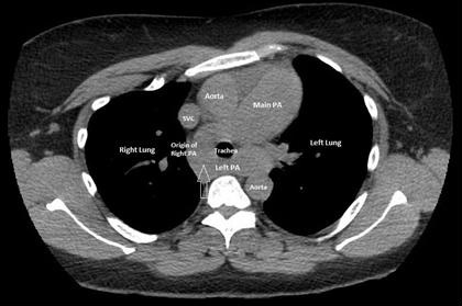
Medical Image of the Week: Pulmonary Artery Sling

Figure 1. Computerized tomography (CT) of the chest showing the aberrant origin of the left pulmonary artery from the right pulmonary artery creating a pulmonary artery sling with mild tracheal narrowing (arrow).


Figure 2. Cardiac magnetic resonance imaging (MRI) confirming the presence of a pulmonary artery sling with aberrant origin of the left pulmonary artery from the right pulmonary artery.


A 42-year-old year woman with asthma was admitted to the hospital with an asthma exacerbation. The patient complained of dyspnea on exertion, two-pillow orthopnea and bipedal edema. An echocardiogram showed a severely dilated right ventricle (RV) with elevated right ventricular systolic pressure of 71 mmHg. The systolic left ventricular (LV) function was also reduced with an ejection fraction of 45%. Computerized tomography (CT) of the chest showed an aberrant origin of the left pulmonary artery (PA) creating a pulmonary artery sling with mild tracheal narrowing (Figure 1, arrow). Cardiac magnetic resonance imaging (MRI) confirmed the presence of a pulmonary artery sling with the aberrant origin of the left PA from the right PA (Figure 2). Cardiac catheterization showed a mean PA pressure of 46mmHg with LV end diastolic pressure of 12mm Hg. The patient was diagnosed with WHO Group I pulmonary hypertension and started on treatment with sildenafil with a stable outpatient course.

Pulmonary artery sling is an uncommon form of vascular ring. The anomaly is a result of formation of the left PA from the right sixth vascular arch (rather than the left), leading to the left PA arising from the posterior aspect of the right PA (1). Pulmonary artery slings may produce symptoms of airway compression and esophageal compression and usually presents in childhood (2). In asymptomatic cases, a PA sling may mimic a mediastinal mass on chest radiographs and CT and MRI may be used to establish the diagnosis (3).

Abhinav Agrawal MD1, Stuart L Cohen MD2, Rakesh Shah MD2, Arunabh Talwar MD FCCP1

1Division of Pulmonary, Critical Care & Sleep Medicine, Department of Medicine

2Division of Thoracic Radiology, Department of Radiology

Hofstra-Northwell School of Medicine

New Hyde Park, NY USA

References

  1. Casta-er E, Gallardo X, Rimola J, Pallardó Y, Mata JM, Perendreu J, Martin C, Gil D. Congenital and acquired pulmonary artery anomalies in the adult: radiologic overview. Radiographics. 2006 Mar-Apr;26(2):349-71. [CrossRef] [PubMed]
  2. Odell DD, Gangadharan SP, Majid A. Pulmonary artery sling: a rare cause of tracheomalacia in the adult. J Bronchology Interv Pulmonol. 2011 Jul;18(3):278-80. [CrossRef] [PubMed]
  3. Ganesh V, Hoey ET, Gopalan D. Pulmonary artery sling: an unexpected finding on cardiac multidetector CT. Postgrad Med J. 2009 Mar;85(1001):128. [CrossRef] [PubMed] 

Cite as: Agrawal A, Cohen SL, Shah R, Talwar A. Medical image of the week: pulmonary artery sling. Southwest J Pulm Crit Care. 2017;15:160-61. doi: https://doi.org/10.13175/swjpcc116-17 PDF