Search Journal-type in search term and press enter
Southwest Pulmonary and Critical Care Fellowships
In Memoriam
Social Media

Imaging

Last 50 Imaging Postings

(Most recent listed first. Click on title to be directed to the manuscript.)

May 2025 Medical Image of the Month: Aspirated Dental Screw
April 2025 Medical Image of the Month: An Unfortunate Case of Mimicry
March 2025 Medical Image of the Month: An Unusual Case of Pulmonary
   Infarction
February 2025 Medical Image of the Month: Unexpected Complications of
   Transjugular Intrahepatic Portosystemic Shunt (TIPS) 
February 2025 Imaging Case of the Month: A Wolf in Sheep’s Clothing
January 2025 Medical Image of the Month: Psoriasis with Pulmonary
   Involvement
December 2024 Medical Image of the Month: An Endobronchial Tumor
November 2024 Medical Image of the Month: A Case of Short Telomeres
November 2024 Imaging Case of the Month: A Recurring Issue
October 2024 Medical Image of the Month: Lofgren syndrome with Erythema
   Nodosum
September 2024 Medical Image of the Month: A Curious Case of Nasal
   Congestion
August 2024 Image of the Month: Lymphomatoid Granulomatosis
August 2024 Imaging Case of the Month: An Unexplained Pleural Effusion
July 2024 Medical Image of the Month: Vocal Cord Paralysis on PET-CT 
June 2024 Medical Image of the Month: A 76-year-old Man Presenting with
   Acute Hoarseness
May 2024 Medical Image of the Month: Hereditary Hemorrhagic
   Telangiectasia in a Patient on Veno-Arterial Extra-Corporeal Membrane
   Oxygenation
May 2024 Imaging Case of the Month: Nothing Is Guaranteed
April 2024 Medical Image of the Month: Wind Instruments Player Exhibiting
   Exceptional Pulmonary Function
March 2024 Medical Image of the Month: Sputum Cytology in Patients with
   Suspected Lung Malignancy Presenting with Acute Hypoxic Respiratory
   Failure
February 2024 Medical Image of the Month: Pulmonary Alveolar Proteinosis
   in Myelodysplastic Syndrome
February 2024 Imaging Case of the Month: Connecting Some Unusual Dots
January 2024 Medical Image of the Month: Polyangiitis Overlap Syndrome
   (POS) Mimicking Fungal Pneumonia 
December 2023 Medical Image of the Month: Metastatic Pulmonary
   Calcifications in End-Stage Renal Disease 
November 2023 Medical Image of the Month: Obstructive Uropathy
   Extremis
November 2023 Imaging Case of the Month: A Crazy Association
October 2023 Medical Image of the Month: Swyer-James-MacLeod
   Syndrome
September 2023 Medical Image of the Month: Aspergillus Presenting as a
   Pulmonary Nodule in an Immunocompetent Patient
August 2023 Medical Image of the Month: Cannonball Metastases from
   Metastatic Melanoma
August 2023 Imaging Case of the Month: Chew Your Food Carefully
July 2023 Medical Image of the Month: Primary Tracheal Lymphoma
June 2023 Medical Image of the Month: Solitary Fibrous Tumor of the Pleura
May 2023 Medical Image of the Month: Methamphetamine Inhalation
   Leading to Cavitary Pneumonia and Pleural Complications
April 2023 Medical Image of the Month: Atrial Myxoma in the setting of
   Raynaud’s Phenomenon: Early Echocardiography and Management of
   Thrombotic Disease
April 2023 Imaging Case of the Month: Large Impact from a Small Lesion
March 2023 Medical Image of the Month: Spontaneous Pneumomediastinum
   as a Complication of Marijuana Smoking Due to Müller's Maneuvers
February 2023 Medical Image of the Month: Reversed Halo Sign in the
   Setting of a Neutropenic Patient with Angioinvasive Pulmonary
   Zygomycosis
January 2023 Medical Image of the Month: Abnormal Sleep Study and PFT
   with Supine Challenge Related to Idiopathic Hemidiaphragmatic Paralysis
December 2022 Medical Image of the Month: Bronchoesophageal Fistula in
   the Setting of Pulmonary Actinomycosis
November 2022 Medical Image of the Month: COVID-19 Infection
   Presenting as Spontaneous Subcapsular Hematoma of the Kidney
November 2022 Imaging Case of the Month: Out of Place in the Thorax
October 2022 Medical Image of the Month: Infected Dasatinib Induced
   Chylothorax-The First Reported Case 
September 2022 Medical Image of the Month: Epiglottic Calcification
Medical Image of the Month: An Unexpected Cause of Chronic Cough
August 2022 Imaging Case of the Month: It’s All About Location
July 2022 Medical Image of the Month: Pulmonary Nodule in the
   Setting of Pyoderma Gangrenosum (PG) 
June 2022 Medical Image of the Month: A Hard Image to Swallow
May 2022 Medical Image of the Month: Pectus Excavatum
May 2022 Imaging Case of the Month: Asymmetric Apical Opacity–
   Diagnostic Considerations
April 2022 Medical Image of the Month: COVID Pericarditis
March 2022 Medical Image of the Month: Pulmonary Nodules in the
   Setting of Diffuse Idiopathic Pulmonary NeuroEndocrine Cell Hyperplasia
   (DIPNECH) 
February 2022 Medical Image of the Month: Multifocal Micronodular
   Pneumocyte Hyperplasia in the Setting of Tuberous Sclerosis
February 2022 Imaging Case of the Month: Between A Rock and a
   Hard Place
January 2022 Medical Image of the Month: Bronchial Obstruction
   Due to Pledget in Airway Following Foregut Cyst Resection
December 2021 Medical Image of the Month: Aspirated Dental Implant
Medical Image of the Month: Cavitating Pseudomonas
   aeruginosa Pneumonia
November 2021 Imaging Case of the Month: Let’s Not Dance
   the Twist
Medical Image of the Month: COVID-19-Associated Pulmonary
   Aspergillosis in a Post-Liver Transplant Patient


For complete imaging listings click here

Those who care for patients with pulmonary, critical care or sleep disorders rely heavily on chest radiology and pathology to determine diagnoses. The Southwest Journal of Pulmonary, Critical Care & Sleep publishes case-based articles with characteristic chest imaging and related pathology. The editor of this section will oversee and coordinate the publication of a core of the most important chest imaging topics. In doing so, they encourage the submission of unsolicited manuscripts. It cannot be overemphasized that both radiologic and pathologic images must be of excellent quality. As a rule, 600 DPI is sufficient for radiographic and pathologic images. Taking pictures of plain chest radiographs and CT scans with a digital camera is strongly discouraged. The figures should be cited in the text and numbered consecutively. The stain used for pathology specimens and magnification should be mentioned in the figure legend. Those who care for patients with pulmonary, critical care or sleep disorders rely heavily on chest radiology and pathology to determine diagnoses. The Southwest Journal of Pulmonary, Critical Care & Sleep publishes case-based articles with characteristic chest imaging and related pathology. The editor of this section will oversee and coordinate the publication of a core of the most important chest imaging topics. In doing so, they encourage the submission of unsolicited manuscripts. It cannot be overemphasized that both radiologic and pathologic images must be of excellent quality. As a rule, 600 DPI is sufficient for radiographic and pathologic images. Taking pictures of plain chest radiographs and CT scans with a digital camera is strongly discouraged. The figures should be cited in the text and numbered consecutively. The stain used for pathology specimens and magnification should be mentioned in the figure legend.

-------------------------------------------------------------------------------------------  

Wednesday
Feb052014

Medical Image of the Week: Neurogenic Pulmonary Edema


Figure 1. CT scan showing intraparenchymal hemorrhage in the left frontal lobe, scattered subdural, subarachnoid and intraventricular hemorrhage with 1.1 cm of left to right midline shift.


Figure 2. Chest X-ray showing central vascular congestion with bilateral pulmonary edema throughout the lung fields


A 79 year old woman with a history of diabetes, hypertension and subarachnoid hemorrhage presented to the emergency department (ED) with altered mental status. The patient had a fall one day prior to admission, and hit her head on the ground. There was no loss of consciousness or seizure activity at that moment, however, she was found unresponsive in the bathroom the next day with brownish vomitus in the mouth and on her face. CT of the head without contrast showed a large intraparenchymal hemorrhage on the left frontal lobe with subdural hemorrhage on the right frontal and temporal lobe. Also, intraventricular blood with 1.1 cm left to right midline shift was observed (Figure 1). Although she had no history of left heart failure or pulmonary disease, physical exam showed coarse lung sound and chest X-ray showed acute change with prominence central vasculature with fluffy central airspace opacities, which were consistent with neurogenic pulmonary edema secondary to intracranial hemorrhage (Figure 2). An external ventricular drain was placed by neurosurgery and patient was intubated for airway protection, however she passed away the next day after her family decided comfort care.

The most common cause of neurogenic pulmonary edema is central nervous system injury including cerebral hemorrhage, head trauma and epileptic seizure (1). It usually develops several hours after an insult, although cases of immediate or delayed onset have been reported. The most common symptoms and signs include dyspnea, hemoptysis, tachypnea, tachycardia, which are not secondary to heart or lung parenchymal disease. Aspiration pneumonia is common presentation of patients with altered mental status, and it is hard to differentiate neurogenic pulmonary edema from aspiration pneumonia, however neurogenic pulmonary edema tends to develop and resolve more rapidly with no signs of infection such as fever and focal infiltration (2). The prognosis of neurogenic pulmonary edema mainly depends on the neurologic pathology rather than pulmonary edema itself, and the mainstream of treatment is supportive care, although medications including β-agonists, dobutamine or chlorpromazine can be tried.

Seongseok Yun, MD PhD; Tuan Phan, MD; Natasha Sharda, MD

Department of Medicine, University of Arizona, Tucson, AZ 85724, USA

References

  1. Neurogenic pulmonary oedema. Lancet. 1985;1(8443):1430-1. [PubMed]
  2. Colice GL, Matthay MA, Bass E, Matthay RA. Neurogenic pulmonary edema. Am Rev Respir Dis. 1984;130(5):941-8. [PubMed] 

Reference as: Yun S, Phan T, Sharda N. Medical image of the week: neurogenic pulmonary edema. Southwest J Pulm Crit Care. 2014;8(1): . doi: http://dx.doi.org/10.13175/swjpcc004-14 PDF

Monday
Feb032014

February 2014 Imaging Case of the Month

Michael B. Gotway, MD


Department of Radiology

Mayo Clinic Arizona

Scottsdale, AZ


Clinical History: A 60-year-old man presented with a history of weight loss and dysphagia for about 2 weeks duration. There was a possible history of asthma accompanied by ongoing shortness of breath first noticed nearly 2 years ago. Frontal chest radiography (Figure 1) was performed.
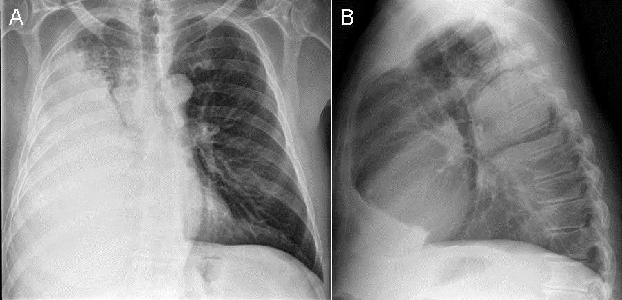
Figure 1.  Frontal chest radiography.

Which of the following statements regarding the chest radiograph is most accurate? (Choose the correct answer to move to the next panel)

  1. The chest radiograph shows a mass
  2. The chest radiograph shows hilar and mediastinal lymph node enlargement
  3. The chest radiograph shows multifocal consolidation
  4. The chest radiograph shows multifocal, somewhat basal predominant linear opacities suggesting fibrosis
  5. The chest radiograph shows multiple nodules

Reference as: Gotway MB. February 2014 imaging case of the month. Southwest J Pulm Crit Care. 2014;8(2):88-95. doi: http://dx.doi.org/10.13175/swjpcc010-14 PDF