
Medical Image of the Week: Constrictive Pericarditis
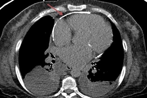
Figure 1. A computerized tomography of the chest revealed cardiomegaly, bilateral pleural effusions and pericardial calcification noted diffusely with focal regions of pericardial thickening greater than 4 mm.
A 62-year-old woman, with a past medical history significant for oxygen dependent COPD, paroxysmal atrial fibrillation, and obstructive sleep apnea, presented to the hospital with hypoxemic respiratory failure requiring intubation and mechanical ventilation. A computerized tomography of the chest revealed cardiomegaly, bilateral pleural effusions, and pericardial calcification that was noted diffusely with focal regions of pericardial thickening greater than 4 mm. A cardiac catheterization revealed elevated right-sided pressure; markedly elevated left ventricular end diastolic pressure; equalization of LV-RV diastolic pressures; and sharp Y descent on the right atrial pressure waveform; which is all suggestive of constrictive physiology. The patient was medically optimized and diuresed and eventually underwent a successful pericardiectomy.
Mohammed Alzoubaidi MD, John Bloom MD, Jarrod Mosier MD, Linda Snyder MD
Department of Pulmonary and Critical Care Medicine, University of Arizona,
Tucson, AZ
Reference as: Alzoubaidi M, Bloom J, Mosier J, Snyder L. Medical image of the week: constrictive pericaditis. Southwest J Pulm Crit Care. 2014;8(5):280. doi: http://dx.doi.org/10.13175/swjpcc042-14 PDF
 Post a Comment