Imaging

Last 50 Imaging Postings

(Most recent listed first. Click on title to be directed to the manuscript.)

August 2025 Medical Image of the Month: Crazy Paving in a Case of 
   Eosinophilic Granulomatosis with Polyangiitis
July 2025 Medical Image of the Month: A Case of Severe Hiatal Hernia
   Presenting as Atypical Chest Pain
July 2025 Imaging Case of the Month: A Growing Lung Nodule in a
   Patient with Heart Disease
June 2025 Medical Image of the Month: Neurofibromatosis-Associated Diffuse
   Cystic Lung Disease
May 2025 Medical Image of the Month: Aspirated Dental Screw
April 2025 Medical Image of the Month: An Unfortunate Case of Mimicry
March 2025 Medical Image of the Month: An Unusual Case of Pulmonary
   Infarction
February 2025 Medical Image of the Month: Unexpected Complications of
   Transjugular Intrahepatic Portosystemic Shunt (TIPS) 
February 2025 Imaging Case of the Month: A Wolf in Sheep’s Clothing
January 2025 Medical Image of the Month: Psoriasis with Pulmonary
   Involvement
December 2024 Medical Image of the Month: An Endobronchial Tumor
November 2024 Medical Image of the Month: A Case of Short Telomeres
November 2024 Imaging Case of the Month: A Recurring Issue
October 2024 Medical Image of the Month: Lofgren syndrome with Erythema
   Nodosum
September 2024 Medical Image of the Month: A Curious Case of Nasal
   Congestion
August 2024 Image of the Month: Lymphomatoid Granulomatosis
August 2024 Imaging Case of the Month: An Unexplained Pleural Effusion
July 2024 Medical Image of the Month: Vocal Cord Paralysis on PET-CT 
June 2024 Medical Image of the Month: A 76-year-old Man Presenting with
   Acute Hoarseness
May 2024 Medical Image of the Month: Hereditary Hemorrhagic
   Telangiectasia in a Patient on Veno-Arterial Extra-Corporeal Membrane
   Oxygenation
May 2024 Imaging Case of the Month: Nothing Is Guaranteed
April 2024 Medical Image of the Month: Wind Instruments Player Exhibiting
   Exceptional Pulmonary Function
March 2024 Medical Image of the Month: Sputum Cytology in Patients with
   Suspected Lung Malignancy Presenting with Acute Hypoxic Respiratory
   Failure
February 2024 Medical Image of the Month: Pulmonary Alveolar Proteinosis
   in Myelodysplastic Syndrome
February 2024 Imaging Case of the Month: Connecting Some Unusual Dots
January 2024 Medical Image of the Month: Polyangiitis Overlap Syndrome
   (POS) Mimicking Fungal Pneumonia 
December 2023 Medical Image of the Month: Metastatic Pulmonary
   Calcifications in End-Stage Renal Disease 
November 2023 Medical Image of the Month: Obstructive Uropathy
   Extremis
November 2023 Imaging Case of the Month: A Crazy Association
October 2023 Medical Image of the Month: Swyer-James-MacLeod
   Syndrome
September 2023 Medical Image of the Month: Aspergillus Presenting as a
   Pulmonary Nodule in an Immunocompetent Patient
August 2023 Medical Image of the Month: Cannonball Metastases from
   Metastatic Melanoma
August 2023 Imaging Case of the Month: Chew Your Food Carefully
July 2023 Medical Image of the Month: Primary Tracheal Lymphoma
June 2023 Medical Image of the Month: Solitary Fibrous Tumor of the Pleura
May 2023 Medical Image of the Month: Methamphetamine Inhalation
   Leading to Cavitary Pneumonia and Pleural Complications
April 2023 Medical Image of the Month: Atrial Myxoma in the Setting of
   Raynaud’s Phenomenon: Early Echocardiography and Management of
   Thrombotic Disease
April 2023 Imaging Case of the Month: Large Impact from a Small Lesion
March 2023 Medical Image of the Month: Spontaneous Pneumomediastinum
   as a Complication of Marijuana Smoking Due to Müller's Maneuvers
February 2023 Medical Image of the Month: Reversed Halo Sign in the
   Setting of a Neutropenic Patient with Angioinvasive Pulmonary
   Zygomycosis
January 2023 Medical Image of the Month: Abnormal Sleep Study and PFT
   with Supine Challenge Related to Idiopathic Hemidiaphragmatic Paralysis
December 2022 Medical Image of the Month: Bronchoesophageal Fistula in
   the Setting of Pulmonary Actinomycosis
November 2022 Medical Image of the Month: COVID-19 Infection
   Presenting as Spontaneous Subcapsular Hematoma of the Kidney
November 2022 Imaging Case of the Month: Out of Place in the Thorax
October 2022 Medical Image of the Month: Infected Dasatinib Induced
   Chylothorax-The First Reported Case 
September 2022 Medical Image of the Month: Epiglottic Calcification
Medical Image of the Month: An Unexpected Cause of Chronic Cough
August 2022 Imaging Case of the Month: It’s All About Location
July 2022 Medical Image of the Month: Pulmonary Nodule in the
   Setting of Pyoderma Gangrenosum (PG) 
June 2022 Medical Image of the Month: A Hard Image to Swallow
May 2022 Medical Image of the Month: Pectus Excavatum
May 2022 Imaging Case of the Month: Asymmetric Apical Opacity–
   Diagnostic Considerations
April 2022 Medical Image of the Month: COVID Pericarditis
March 2022 Medical Image of the Month: Pulmonary Nodules in the
   Setting of Diffuse Idiopathic Pulmonary NeuroEndocrine Cell Hyperplasia
   (DIPNECH) 
February 2022 Medical Image of the Month: Multifocal Micronodular
   Pneumocyte Hyperplasia in the Setting of Tuberous Sclerosis
February 2022 Imaging Case of the Month: Between A Rock and a
   Hard Place


For complete imaging listings click here

Those who care for patients with pulmonary, critical care or sleep disorders rely heavily on chest radiology and pathology to determine diagnoses. The Southwest Journal of Pulmonary, Critical Care & Sleep publishes case-based articles with characteristic chest imaging and related pathology. The editor of this section will oversee and coordinate the publication of a core of the most important chest imaging topics. In doing so, they encourage the submission of unsolicited manuscripts. It cannot be overemphasized that both radiologic and pathologic images must be of excellent quality. As a rule, 600 DPI is sufficient for radiographic and pathologic images. Taking pictures of plain chest radiographs and CT scans with a digital camera is strongly discouraged. The figures should be cited in the text and numbered consecutively. The stain used for pathology specimens and magnification should be mentioned in the figure legend. Those who care for patients with pulmonary, critical care or sleep disorders rely heavily on chest radiology and pathology to determine diagnoses. The Southwest Journal of Pulmonary, Critical Care & Sleep publishes case-based articles with characteristic chest imaging and related pathology. The editor of this section will oversee and coordinate the publication of a core of the most important chest imaging topics. In doing so, they encourage the submission of unsolicited manuscripts. It cannot be overemphasized that both radiologic and pathologic images must be of excellent quality. As a rule, 600 DPI is sufficient for radiographic and pathologic images. Taking pictures of plain chest radiographs and CT scans with a digital camera is strongly discouraged. The figures should be cited in the text and numbered consecutively. The stain used for pathology specimens and magnification should be mentioned in the figure legend.

-------------------------------------------------------------------------------------------  

Wednesday
Aug062014

Medical Image of the Week: Idiopathic Pulmonary Artery Hypertension

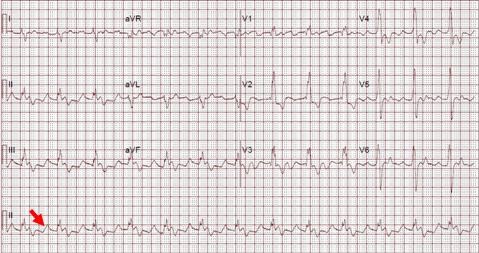
Figure 1. EKG showing sinus rhythm, right bundle branch block and peaked ('pulmonary') p waves (arrow).


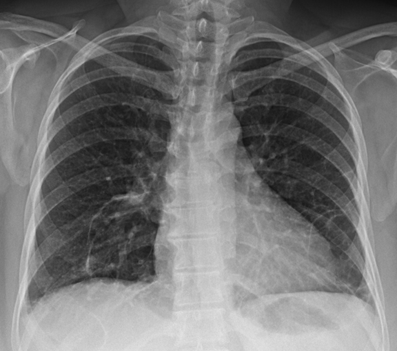
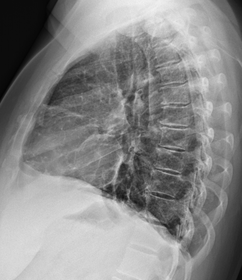
Figure 2. Two view chest X-ray showing right ventricular hypertrophy (arrows, note filling of the retrosternal space by an enlarged right ventricle in the lateral view) and enlarged central pulmonary arteries (arrowhead).


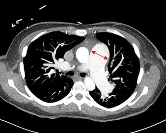
  Figure 3. Axial CT angiogram of the chest below the carina showing dilated pulmonary artery (diameter of pulmonary artery greater than aorta, arrow).


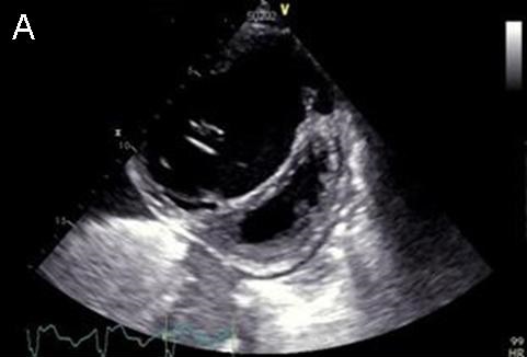
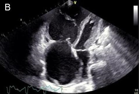
Figure 4. Panel A: Parasternal short axis view shows septal bowing to the left, a severely dilated right ventricle and a D-shaped left ventricle. Panel B: Four chamber view shows right atrial and ventricular dilatation.


A 39-year-old woman presented to the clinic with a history of progressive shortness of breath of 6-month duration associated with bilateral lower extremity edema, fatigue, lightheadedness, palpitations and occasional substernal chest pain. Her past medical history was unremarkable other than mild anemia. On physical exam her respiratory rate was 20 breaths per minute and O2 saturation 94% on room air by pulse oximetry. There was jugular venous distention at 12 cm, 2+ bilateral lower extremity edema, a 5/6 systolic murmur over the left sternal border with a sternal heave. Labwork was unremarkable except for an elevated BNP 657 (normal value < 100 pg/mL).

EKG (Figure 1) showed sinus rhythm with right bundle branch block. A 2-view chest X-ray (Figure 2) showed an enlarged right ventricle as well as dilated pulmonary arteries with no parenchymal infiltrates. CT angiography confirmed CXR findings (Figure 3) and was negative for pulmonary embolism. A 2D echocardiogram revealed a preserved left ventricle ejection fraction with right ventricular pressure of 80 mmHg + CVP, severe tricuspid regurgitation, decreased right ventricular function (as assessed by a Tricuspid annular plane systolic excursion of 10 mm) and flattened septum, suggestive of right ventricular overload (Figure 4). A right heart catheterization was performed revealing pulmonary pressures of 105/45 mmHg with a mean of 63 mmHg, a wedge pressure of 11 mmHg, a pulmonary vascular resistance of 13.19 Wood units and a cardiac output of 3.94 L/min.

The patient was admitted to the intensive care unit to start treatment with intravenous treprostinil and was eventually discharged home with subcutaneous treprostrinil.

Pulmonary arterial hypertension (PAH) is a disease of the pulmonary circulation characterized by a progressive elevation in pulmonary vascular resistance that leads to right ventricular failure and premature death. It is defined as a mean pulmonary artery pressure at rest of 25 mmHg or higher (1). Idiopathic (group 1) PAH requires the exclusion of parenchymal pathology or venous thromboembolic disease as well as a mean wedge pressure less than 15 mmHg. The initial symptoms of PH are the result of an inability to adequately increase cardiac output during exercise which eventually will progress to signs and symptoms of right ventricular failure such as lower extremity edema, syncope/presyncope and chest pain (2,3). Early recognition is of paramount importance to institute adequate treatment.

Roberto J. Bernardo, MD and Carlos Tafich Rios, MD

Internal Medicine Residency, Department of Medicine

University of Arizona, Tucson, AZ

References 

  1. McLaughlin VV, Archer SL, Badesch DB, et al. ACCF/AHA 2009 expert consensus document on pulmonary hypertension: a report of the American College of Cardiology Foundation Task Force on Expert Consensus Documents and the American Heart Association: developed in collaboration with the American College. Circulation. 2009. 119:2250-94. [CrossRef] [PubMed] 
  2. Runo JR, Loyd JE. Primary pulmonary hypertension. Lancet. 2003. 361:1533-44. [CrossRef] [PubMed]
  3. Peacock AJ. Primary pulmonary hypertension. Thorax. 1999;54:1107-18. [CrossRef] [PubMed]

Reference as: Bernardo RJ, Rios CT. Medical image of the week: idiopathic pulmonary artery hypertension. Southwest J Pulm Crit Care. 2014;9(2):101-3. doi: http://dx.doi.org/10.13175/swjpcc101-14   PDF

Sunday
Aug032014

August 2014 Imaging Case of the Month 

Michael B. Gotway, MD


Department of Radiology

Mayo Clinic Arizona

Scottsdale, AZ


Clinical History: A 42-year-old non-smoking woman presented with a history of relatively sudden onset left chest pain and shortness of breath. Her past medical history was remarkable for psoriasis, treated with Enbrel® (etanercept). She also had a history of partial hysterectomy for fibroids and right oophorectomy. Frontal and lateral chest radiography (Figure 1) were performed.

Figure 1. Frontal (A) and lateral (B) chest radiography.

Which of the following statements regarding the chest radiograph is most accurate? (Click on the correct answer to move to the next panel)

Reference as: Gotway MB. August 2014 imaging case of the month. Southwest J Pulm Crit Care. 2014;9(2):83-90. doi: http://dx.doi.org/10.13175/swjpcc104-14 PDF