Imaging

Last 50 Imaging Postings

(Most recent listed first. Click on title to be directed to the manuscript.)

August 2025 Medical Image of the Month: Crazy Paving in a Case of 
   Eosinophilic Granulomatosis with Polyangiitis
July 2025 Medical Image of the Month: A Case of Severe Hiatal Hernia
   Presenting as Atypical Chest Pain
July 2025 Imaging Case of the Month: A Growing Lung Nodule in a
   Patient with Heart Disease
June 2025 Medical Image of the Month: Neurofibromatosis-Associated Diffuse
   Cystic Lung Disease
May 2025 Medical Image of the Month: Aspirated Dental Screw
April 2025 Medical Image of the Month: An Unfortunate Case of Mimicry
March 2025 Medical Image of the Month: An Unusual Case of Pulmonary
   Infarction
February 2025 Medical Image of the Month: Unexpected Complications of
   Transjugular Intrahepatic Portosystemic Shunt (TIPS) 
February 2025 Imaging Case of the Month: A Wolf in Sheep’s Clothing
January 2025 Medical Image of the Month: Psoriasis with Pulmonary
   Involvement
December 2024 Medical Image of the Month: An Endobronchial Tumor
November 2024 Medical Image of the Month: A Case of Short Telomeres
November 2024 Imaging Case of the Month: A Recurring Issue
October 2024 Medical Image of the Month: Lofgren syndrome with Erythema
   Nodosum
September 2024 Medical Image of the Month: A Curious Case of Nasal
   Congestion
August 2024 Image of the Month: Lymphomatoid Granulomatosis
August 2024 Imaging Case of the Month: An Unexplained Pleural Effusion
July 2024 Medical Image of the Month: Vocal Cord Paralysis on PET-CT 
June 2024 Medical Image of the Month: A 76-year-old Man Presenting with
   Acute Hoarseness
May 2024 Medical Image of the Month: Hereditary Hemorrhagic
   Telangiectasia in a Patient on Veno-Arterial Extra-Corporeal Membrane
   Oxygenation
May 2024 Imaging Case of the Month: Nothing Is Guaranteed
April 2024 Medical Image of the Month: Wind Instruments Player Exhibiting
   Exceptional Pulmonary Function
March 2024 Medical Image of the Month: Sputum Cytology in Patients with
   Suspected Lung Malignancy Presenting with Acute Hypoxic Respiratory
   Failure
February 2024 Medical Image of the Month: Pulmonary Alveolar Proteinosis
   in Myelodysplastic Syndrome
February 2024 Imaging Case of the Month: Connecting Some Unusual Dots
January 2024 Medical Image of the Month: Polyangiitis Overlap Syndrome
   (POS) Mimicking Fungal Pneumonia 
December 2023 Medical Image of the Month: Metastatic Pulmonary
   Calcifications in End-Stage Renal Disease 
November 2023 Medical Image of the Month: Obstructive Uropathy
   Extremis
November 2023 Imaging Case of the Month: A Crazy Association
October 2023 Medical Image of the Month: Swyer-James-MacLeod
   Syndrome
September 2023 Medical Image of the Month: Aspergillus Presenting as a
   Pulmonary Nodule in an Immunocompetent Patient
August 2023 Medical Image of the Month: Cannonball Metastases from
   Metastatic Melanoma
August 2023 Imaging Case of the Month: Chew Your Food Carefully
July 2023 Medical Image of the Month: Primary Tracheal Lymphoma
June 2023 Medical Image of the Month: Solitary Fibrous Tumor of the Pleura
May 2023 Medical Image of the Month: Methamphetamine Inhalation
   Leading to Cavitary Pneumonia and Pleural Complications
April 2023 Medical Image of the Month: Atrial Myxoma in the Setting of
   Raynaud’s Phenomenon: Early Echocardiography and Management of
   Thrombotic Disease
April 2023 Imaging Case of the Month: Large Impact from a Small Lesion
March 2023 Medical Image of the Month: Spontaneous Pneumomediastinum
   as a Complication of Marijuana Smoking Due to Müller's Maneuvers
February 2023 Medical Image of the Month: Reversed Halo Sign in the
   Setting of a Neutropenic Patient with Angioinvasive Pulmonary
   Zygomycosis
January 2023 Medical Image of the Month: Abnormal Sleep Study and PFT
   with Supine Challenge Related to Idiopathic Hemidiaphragmatic Paralysis
December 2022 Medical Image of the Month: Bronchoesophageal Fistula in
   the Setting of Pulmonary Actinomycosis
November 2022 Medical Image of the Month: COVID-19 Infection
   Presenting as Spontaneous Subcapsular Hematoma of the Kidney
November 2022 Imaging Case of the Month: Out of Place in the Thorax
October 2022 Medical Image of the Month: Infected Dasatinib Induced
   Chylothorax-The First Reported Case 
September 2022 Medical Image of the Month: Epiglottic Calcification
Medical Image of the Month: An Unexpected Cause of Chronic Cough
August 2022 Imaging Case of the Month: It’s All About Location
July 2022 Medical Image of the Month: Pulmonary Nodule in the
   Setting of Pyoderma Gangrenosum (PG) 
June 2022 Medical Image of the Month: A Hard Image to Swallow
May 2022 Medical Image of the Month: Pectus Excavatum
May 2022 Imaging Case of the Month: Asymmetric Apical Opacity–
   Diagnostic Considerations
April 2022 Medical Image of the Month: COVID Pericarditis
March 2022 Medical Image of the Month: Pulmonary Nodules in the
   Setting of Diffuse Idiopathic Pulmonary NeuroEndocrine Cell Hyperplasia
   (DIPNECH) 
February 2022 Medical Image of the Month: Multifocal Micronodular
   Pneumocyte Hyperplasia in the Setting of Tuberous Sclerosis
February 2022 Imaging Case of the Month: Between A Rock and a
   Hard Place


For complete imaging listings click here

Those who care for patients with pulmonary, critical care or sleep disorders rely heavily on chest radiology and pathology to determine diagnoses. The Southwest Journal of Pulmonary, Critical Care & Sleep publishes case-based articles with characteristic chest imaging and related pathology. The editor of this section will oversee and coordinate the publication of a core of the most important chest imaging topics. In doing so, they encourage the submission of unsolicited manuscripts. It cannot be overemphasized that both radiologic and pathologic images must be of excellent quality. As a rule, 600 DPI is sufficient for radiographic and pathologic images. Taking pictures of plain chest radiographs and CT scans with a digital camera is strongly discouraged. The figures should be cited in the text and numbered consecutively. The stain used for pathology specimens and magnification should be mentioned in the figure legend. Those who care for patients with pulmonary, critical care or sleep disorders rely heavily on chest radiology and pathology to determine diagnoses. The Southwest Journal of Pulmonary, Critical Care & Sleep publishes case-based articles with characteristic chest imaging and related pathology. The editor of this section will oversee and coordinate the publication of a core of the most important chest imaging topics. In doing so, they encourage the submission of unsolicited manuscripts. It cannot be overemphasized that both radiologic and pathologic images must be of excellent quality. As a rule, 600 DPI is sufficient for radiographic and pathologic images. Taking pictures of plain chest radiographs and CT scans with a digital camera is strongly discouraged. The figures should be cited in the text and numbered consecutively. The stain used for pathology specimens and magnification should be mentioned in the figure legend.

-------------------------------------------------------------------------------------------  

Wednesday
Jul292015

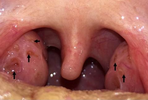
Medical Image of the Week: Teenage Tonsils

Figure 1. Tonsils showing shallow ulcers (arrows) secondary to coxsakievirus.


Figure 2. Enlarged tonsils R>L secondary to infectious mononucleosis.


An 18 year old woman complained of gradual onset throat pain and symptoms of a viral URI with nasal congestion, conjunctivitis and coryza. Later, faint macular rash appeared on her hands. Shallow ulcers developed on her tonsils (Figure 1). She was diagnosed with coxsakie viral infection and treated conservatively with ibuprofen.

A 19 year old man was seen for fatigue, malaise and odynophagia. Because of concern for peritonsilar abscess due to R>L tonsil enlargement (Figure 2) he was given intravenous steroids and antibiotics. His pain improved dramatically and he tested positive for infectious mononucleosis.

Adam M. Knox and Alexander G. Chiu, MD

Department of Otolaryngology

University of Arizona, Tucson

Reference as: Knox AM, Chiu AG. Medical image of the week: teenage tonsils. Southwest J Pulm Crit Care. 2015;11(1):51-2. doi: http://dx.doi.org/10.13175/swjpcc071-15 PDF

Wednesday
Jul222015

Medical Image of the Week: Focal Myopericarditis

Figure 1. Transverse view (panel A) and longitudinal view (panel B) of MRI with gadolinium enhanced contrast of chest showing abnormal enhancement at the level of the cardiac apex and also at the mid aspect of the infero-lateral wall and near the cardiac base within the lateral wall, consistent with the clinically suspected diagnosis of focal myopericarditis.

A 44-year-old man with no significant past medical history was admitted with a history of two episodes of substernal chest pain unrelated to exertion which had resolved spontaneously. Admission vital signs were within normal limits and physical examination was unremarkable. Basic lab tests were normal and urine toxicology was negative. Electrocardiogram was unremarkable with no ST/T changes. Troponin I was elevated at 4.19 which trended up to 6.57. An urgent cardiac angiogram was done which revealed normal patent coronaries. His transthoracic echocardiogram was also reported to be normal. He continued to have intermittent episodes of chest pain that was partially relieved by morphine. Erythrocyte sedimentation rate and C-reactive protein were elevated. Work up for autoimmune diseases, vasculitis, myocarditis panel were insignificant. Later, magnetic resonance imaging (MRI) with gadolinium enhanced contrast (Figure 1) was obtained which showed abnormal epicardial/subepicardial myocardial enhancement within the inferolateral wall and cardiac apex consistent with focal myopericarditis. He was started on Ibuprofen and colchicine. His chest pain significantly improved and he is currently following up as an outpatient.

Acute myo-pericarditis is primarily a pericarditic syndrome with variable myocardial involvement which is usually seen in male adolescents (1,2). There are 3 main etiologic categories: idiopathic, infectious or immune mediated. Patients present with chest pain that is sharp in nature with gradual onset radiating to the interscapular region, increasing with inspiration and easing with leaning forward. Pericardial friction rub on physical exam is considered pathognomonic. A typical pattern of ECG evolution includes diffuse ST segment elevation and PR depression, followed by normalization of ST and PR segments and then diffuse T wave inversion. Troponin I levels may be elevated, provides a rough estimate of the extent of myocardial inflammation. Cardiac MRI with gadolinium contrast is the best imaging modality to define the extent of myocardial involvement and patency of coronary system which is not always readily available. Therapy of choice is aspirin (1-1.5 g/day as mean dose) or nonsteroidal anti-inflammatory drugs such as ibuprofen for 7-10 days until symptom resolution. Colchicine should be the initial therapy in all refractory cases and in recurrent pericarditis. Physical exercise is contraindicated for at least 6 months from the onset of illness.

Chandramohan Meenakshisundaram MD, Nanditha Malakkla MD, and Venu Ganipisetti MD

Department of Internal Medicine,

Presence Saint Francis Hospital

Evanston, IL USA

References

  1. Imazio M, Cooper LT. Management of myopericarditis. Expert Rev Cardiovasc Ther. 2013;11(2):193-201. [CrossRef] [PubMed]
  2. Sharma J, Fernandes N, Alvarez D, Khanna S.Acute myopericarditis in an adolescent mimicking acute myocardial infarction. Pediatr Emerg Care. 2015;31(6):427-30. [CrossRef] [PubMed]

Reference as: Meenakshisundaram C, Malakkla N, Ganipisetti V. Medical image of the week: focal myopericaditis. Southwest J Pulm Crit Care. 2015;11(1):47-8. doi: http://dx.doi.org/10.13175/swjpcc064-15 PDF