
November 2016 Imaging Case of the Month
Michael B. Gotway, MD
Department of Radiology
Mayo Clinic Arizona
Scottsdale, Arizona USA
Imaging Case of the Month CME Information
Members of the Arizona, New Mexico, Colorado and California Thoracic Societies and the Mayo Clinic are able to receive 0.25 AMA PRA Category 1 Credits™. Completion of an evaluation form is required to receive credit and a link is provided on the last panel of the activity.
0.25 AMA PRA Category 1 Credit(s)™
Estimated time to complete this activity: 0.25 hours
Lead Author(s): Michael B. Gotway, MD. All Faculty, CME Planning Committee Members, and the CME Office Reviewers have disclosed that they do not have any relevant financial relationships with commercial interests that would constitute a conflict of interest concerning this CME activity.
Learning Objectives:
As a result of this activity I will be better able to:
- Correctly interpret and identify clinical practices supported by the highest quality available evidence.
- Will be better able to establsh the optimal evaluation leading to a correct diagnosis for patients with pulmonary, critical care and sleep disorders.
- Will improve the translation of the most current clinical information into the delivery of high quality care for patients.
- Will integrate new treatment options in discussing available treatment alternatives for patients with pulmonary, critical care and sleep related disorders.
Learning Format: Case-based, interactive online course, including mandatory assessment questions (number of questions varies by case). Please also read the Technical Requirements.
CME Sponsor: University of Arizona College of Medicine at the Arizona Health Sciences Center.
Current Approval Period: January 1, 2015-December 31, 2016
Clinical History: A 38-year-old man presented to his primary care physician with complaints of pruritus, jaundice, and poor appetite. The patient had been diagnosed with hypertension one year earlier and was treated with hydrochlorothiazide and an angiotensin-converting enzyme inhibitor, but evidently did not tolerate the regimen well, and developed “tea-colored” urine following initiation of this therapy. He was also recently diagnosed with diabetes mellitus and also complained of intermittent right upper quadrant pain.
Laboratory data, including white blood cell count and serum chemistries were within normal limits. Oxygen saturation on room air was 99%.
Frontal and lateral chest radiographs (Figure 1) were performed.
Figure 1. Frontal (A) and lateral (B) chest radiography.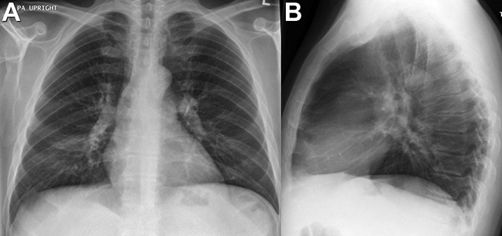
Which of the following statements regarding the chest radiograph is most accurate? (Click on the correct answer to proceed to the second of seven pages)
Cite as: Gotway MB. November 2016 imaging case of the month. Southwest J Pulm Crit Care. 2016;13(5):207-15. doi: http://dx.doi.org/10.13175/swjpcc112-16 PDF
 Post a Comment

