Search Journal-type in search term and press enter
Southwest Pulmonary and Critical Care Fellowships
Social Media

Imaging

Last 50 Imaging Postings

(Most recent listed first. Click on title to be directed to the manuscript.)

May 2025 Medical Image of the Month: Aspirated Dental Screw
April 2025 Medical Image of the Month: An Unfortunate Case of Mimicry
March 2025 Medical Image of the Month: An Unusual Case of Pulmonary
   Infarction
February 2025 Medical Image of the Month: Unexpected Complications of
   Transjugular Intrahepatic Portosystemic Shunt (TIPS) 
February 2025 Imaging Case of the Month: A Wolf in Sheep’s Clothing
January 2025 Medical Image of the Month: Psoriasis with Pulmonary
   Involvement
December 2024 Medical Image of the Month: An Endobronchial Tumor
November 2024 Medical Image of the Month: A Case of Short Telomeres
November 2024 Imaging Case of the Month: A Recurring Issue
October 2024 Medical Image of the Month: Lofgren syndrome with Erythema
   Nodosum
September 2024 Medical Image of the Month: A Curious Case of Nasal
   Congestion
August 2024 Image of the Month: Lymphomatoid Granulomatosis
August 2024 Imaging Case of the Month: An Unexplained Pleural Effusion
July 2024 Medical Image of the Month: Vocal Cord Paralysis on PET-CT 
June 2024 Medical Image of the Month: A 76-year-old Man Presenting with
   Acute Hoarseness
May 2024 Medical Image of the Month: Hereditary Hemorrhagic
   Telangiectasia in a Patient on Veno-Arterial Extra-Corporeal Membrane
   Oxygenation
May 2024 Imaging Case of the Month: Nothing Is Guaranteed
April 2024 Medical Image of the Month: Wind Instruments Player Exhibiting
   Exceptional Pulmonary Function
March 2024 Medical Image of the Month: Sputum Cytology in Patients with
   Suspected Lung Malignancy Presenting with Acute Hypoxic Respiratory
   Failure
February 2024 Medical Image of the Month: Pulmonary Alveolar Proteinosis
   in Myelodysplastic Syndrome
February 2024 Imaging Case of the Month: Connecting Some Unusual Dots
January 2024 Medical Image of the Month: Polyangiitis Overlap Syndrome
   (POS) Mimicking Fungal Pneumonia 
December 2023 Medical Image of the Month: Metastatic Pulmonary
   Calcifications in End-Stage Renal Disease 
November 2023 Medical Image of the Month: Obstructive Uropathy
   Extremis
November 2023 Imaging Case of the Month: A Crazy Association
October 2023 Medical Image of the Month: Swyer-James-MacLeod
   Syndrome
September 2023 Medical Image of the Month: Aspergillus Presenting as a
   Pulmonary Nodule in an Immunocompetent Patient
August 2023 Medical Image of the Month: Cannonball Metastases from
   Metastatic Melanoma
August 2023 Imaging Case of the Month: Chew Your Food Carefully
July 2023 Medical Image of the Month: Primary Tracheal Lymphoma
June 2023 Medical Image of the Month: Solitary Fibrous Tumor of the Pleura
May 2023 Medical Image of the Month: Methamphetamine Inhalation
   Leading to Cavitary Pneumonia and Pleural Complications
April 2023 Medical Image of the Month: Atrial Myxoma in the setting of
   Raynaud’s Phenomenon: Early Echocardiography and Management of
   Thrombotic Disease
April 2023 Imaging Case of the Month: Large Impact from a Small Lesion
March 2023 Medical Image of the Month: Spontaneous Pneumomediastinum
   as a Complication of Marijuana Smoking Due to Müller's Maneuvers
February 2023 Medical Image of the Month: Reversed Halo Sign in the
   Setting of a Neutropenic Patient with Angioinvasive Pulmonary
   Zygomycosis
January 2023 Medical Image of the Month: Abnormal Sleep Study and PFT
   with Supine Challenge Related to Idiopathic Hemidiaphragmatic Paralysis
December 2022 Medical Image of the Month: Bronchoesophageal Fistula in
   the Setting of Pulmonary Actinomycosis
November 2022 Medical Image of the Month: COVID-19 Infection
   Presenting as Spontaneous Subcapsular Hematoma of the Kidney
November 2022 Imaging Case of the Month: Out of Place in the Thorax
October 2022 Medical Image of the Month: Infected Dasatinib Induced
   Chylothorax-The First Reported Case 
September 2022 Medical Image of the Month: Epiglottic Calcification
Medical Image of the Month: An Unexpected Cause of Chronic Cough
August 2022 Imaging Case of the Month: It’s All About Location
July 2022 Medical Image of the Month: Pulmonary Nodule in the
   Setting of Pyoderma Gangrenosum (PG) 
June 2022 Medical Image of the Month: A Hard Image to Swallow
May 2022 Medical Image of the Month: Pectus Excavatum
May 2022 Imaging Case of the Month: Asymmetric Apical Opacity–
   Diagnostic Considerations
April 2022 Medical Image of the Month: COVID Pericarditis
March 2022 Medical Image of the Month: Pulmonary Nodules in the
   Setting of Diffuse Idiopathic Pulmonary NeuroEndocrine Cell Hyperplasia
   (DIPNECH) 
February 2022 Medical Image of the Month: Multifocal Micronodular
   Pneumocyte Hyperplasia in the Setting of Tuberous Sclerosis
February 2022 Imaging Case of the Month: Between A Rock and a
   Hard Place
January 2022 Medical Image of the Month: Bronchial Obstruction
   Due to Pledget in Airway Following Foregut Cyst Resection
December 2021 Medical Image of the Month: Aspirated Dental Implant
Medical Image of the Month: Cavitating Pseudomonas
   aeruginosa Pneumonia
November 2021 Imaging Case of the Month: Let’s Not Dance
   the Twist
Medical Image of the Month: COVID-19-Associated Pulmonary
   Aspergillosis in a Post-Liver Transplant Patient


For complete imaging listings click here

Those who care for patients with pulmonary, critical care or sleep disorders rely heavily on chest radiology and pathology to determine diagnoses. The Southwest Journal of Pulmonary, Critical Care & Sleep publishes case-based articles with characteristic chest imaging and related pathology. The editor of this section will oversee and coordinate the publication of a core of the most important chest imaging topics. In doing so, they encourage the submission of unsolicited manuscripts. It cannot be overemphasized that both radiologic and pathologic images must be of excellent quality. As a rule, 600 DPI is sufficient for radiographic and pathologic images. Taking pictures of plain chest radiographs and CT scans with a digital camera is strongly discouraged. The figures should be cited in the text and numbered consecutively. The stain used for pathology specimens and magnification should be mentioned in the figure legend. Those who care for patients with pulmonary, critical care or sleep disorders rely heavily on chest radiology and pathology to determine diagnoses. The Southwest Journal of Pulmonary, Critical Care & Sleep publishes case-based articles with characteristic chest imaging and related pathology. The editor of this section will oversee and coordinate the publication of a core of the most important chest imaging topics. In doing so, they encourage the submission of unsolicited manuscripts. It cannot be overemphasized that both radiologic and pathologic images must be of excellent quality. As a rule, 600 DPI is sufficient for radiographic and pathologic images. Taking pictures of plain chest radiographs and CT scans with a digital camera is strongly discouraged. The figures should be cited in the text and numbered consecutively. The stain used for pathology specimens and magnification should be mentioned in the figure legend.

-------------------------------------------------------------------------------------------  

Entries in emphysema (7)

Thursday
Aug152019

Medical Image of the Month: Mounier-Kuhn Syndrome

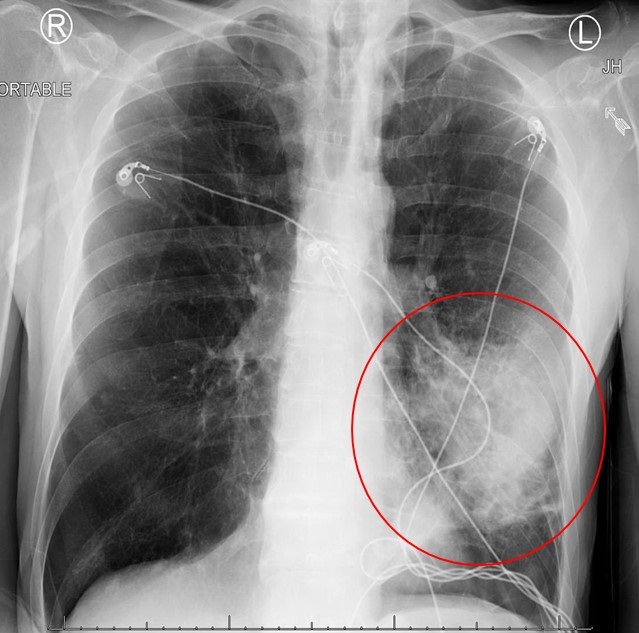
Figure 1. AP chest x-ray showing significant tracheomegaly (diameter 30.8 mm), bilateral interstitial infiltrates with dense consolidation more at the lower lobes (left>right).


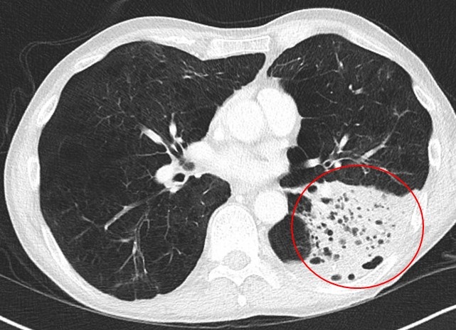
Figure 2. Axial thoracic CT in lung windows (A-D) and soft tissue windows (E-F). Sagittal CT in soft tissue windows (G-H). A: tracheal diameters in 2 dimensions (coronal 30.4 mm, sagittal 37.6 mm), para-septal emphysema (yellow arrows). B: showing tracheomegaly (23.2 x 34.3 mm) and para-septal emphysema changes (yellow arrows. C: enlarged mainstem bronchi diameters (right mainstem 22.3 x 30.6 mm, left mainstem 24.4 x 16.0 mm). In addition to central bronchiectatic changes (red arrows), left lower lobe consolidative changes (blue arrow). D: dense left lower lobe consolidation and para-septal emphysema. E: Significant tracheomegaly (31.5 x 41.a mm) and dilated esophagus (orange arrow). F: Significant tracheomegaly and dilated esophagus.


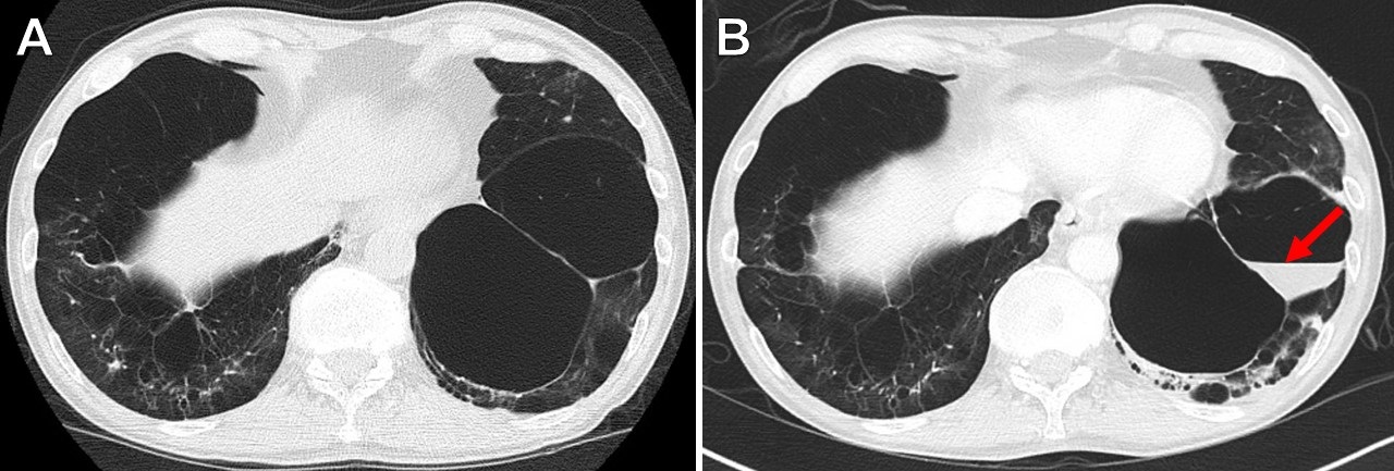
Figure 3. A: Sagittal CT scan (soft tissue window) showing significant tracheomegaly (sagittal diameter 35.8 mm). B: Sagittal CT chest (lung window) showing significant tracheomegaly, multiple tracheal diverticuli (green arrows) on the upper posterior tracheal wall.


Figure 4. Pulmonary function testing.


A 52-year-old non-smoking, Caucasian male patient with a past medical history of reported chronic obstructive pulmonary disease (COPD), recurrent lower respiratory tract infections, prior history of pneumothorax, and dysphagia presented with fevers and shortness of breathing associated with a productive cough for one week. Clinically, he was mildly tachypneic and chest auscultation revealed crackles bilaterally - more prominent at the left base. A chest radiograph (Figure 1) showed bilateral lower lobe pulmonary opacities (left more than right). Computed tomography (CT) of the chest demonstrated airspace disease in the lower lobes in addition to significant tracheobronchomegaly along with paraseptal emphysema and central bronchiectatic changes (Figures 2 and 3). Upper posterior tracheal wall diverticulae were also noted (Figure 3). Serum α1-antitrypsin level and serum immunoglobulins, including IgE levels, were normal. Our patient declined performing diagnostic bronchoscopy. He had a pulmonary function test performed few months prior to his hospital admission which showed combined mild obstructive/restrictive pattern (Figure 4). He responded well to empiric antibiotics and chest percussion therapy. He was discharged in stable condition.

Discussion

On the basis of above findings, a diagnosis of Mounier-Kuhn syndrome complicated by pneumonia was made. The syndrome was first described by P. Mounier-Kuhn in 1932 (1). The diagnosis is usually made when the tracheal diameter is greater than 3 cm on a CT chest (measured 2 cm above the aortic arch) (2). Other diagnostic criteria include a mainstem bronchial diameter of 20-24 mm (right) and 15-23 mm (left) (3). Our patient’s tracheal diameter was around 37 mm. Both mainstem bronchi were dilated.

The abnormal tracheobronchial dilatation in this syndrome is attributed to atrophy of the muscular and elastic tissues in the tracheal and the bronchial walls (3). Hence, in addition to tracheobronchomegaly, these patients can also develop tracheal diverticulosis along with varicose and cystic bronchiectasis (3). These patients usually present in the 3rd or 4th decade of life with nonspecific respiratory symptoms including recurrent bronchitis and subsequently end up being misdiagnosed with COPD (3).

Three subtypes of this syndrome had been described. Subtype 1 has symmetric dilation of the trachea and mainstem bronchi. Subtype 2 demonstrates tracheal dilation and tracheal diverticula. Subtype 3 has diverticular and saccular structures extending to the level of the distal bronchi (3). Our patient likely fits subtype 3 of this syndrome. Overall, treatment is supportive - usually with antibiotics, physiotherapy and postural drainage. In rare instances, tracheal stenting has been used (4). Special consideration should be taken post intubation as achieving good cuff seal can be potentially challenging.

Dysphagia has not been well documented in this syndrome and could be a coincidental finding in our case. However, theoretically, the etiology of this patient’s dysphagia could be secondary to extrinsic compression of the anterior esophageal wall by his markedly dilated trachea. Historically, he underwent multiple esophageal dilatations and at least one Botox injection over the last 5 years without any significant improvement.

Abdulmonam Ali MD and Naga S. Sirikonda MD

Pulmonary and Critical Care

Good Samaritan Hospital

Mount Vernon, Illinois

References

  1. Mounier-Kuhn P. "Dilatation de la trachee: constatations, radiographiques et bronchoscopies." Lyon Med. 1932;150:106-9.
  2. Menon B, Aggarwal B, Iqbal A. Mounier-Kuhn syndrome: report of 8 cases of tracheobronchomegaly with associated complications. South Med J. 2008;101(1):83-7. [CrossRef] [PubMed]
  3. Falconer M, Collins DR, Feeney J, Torreggiani WC. Mounier-Kuhn syndrome in an older patient. Age Ageing. 2008;37(1):115-6. [CrossRef] [PubMed]
  4. Schwartz M, Rossoff L. Tracheobronchomegaly. Chest 1994;106(5):1589-90. [CrossRef] [PubMed]

Cite as: Ali A, Sirikonda NS. Medical image of the month: Mounier-Kuhn syndrome. Southwest J Pulm Crit Care. 2019;19(2):73-5. doi: https://doi.org/10.13175/swjpcc044-19 PDF 

Wednesday
Jan182017

Medical Image of the Week: Infected Emphysematous Bulla

Figure 1. Portable AP chest X-ray revealing dense opacity within the lingula of left lung.


Figure 2. Thoracic CT with contrast showing lobar consolidation with increased lucency compatible with emphysema.


Figure 3. (A) Chest CT one year prior demonstrating severe emphysema. (B) Chest CT on admission showing new fluid-filled bulla (red arrow) in the setting pneumococcal pneumonia.


A 65 year-old man with chronic obstructive lung disease (COPD), hypertension and alcohol abuse presented to the emergency department with complaints of feeling unwell and shortness of breath. He was tachycardic but otherwise hemodynamically stable, afebrile, and requiring 3 liters/min supplemental oxygen. Pertinent initial laboratory findings revealed a neutrophilic predominant leukocytosis (WBC 37.8 x 10³ micro/L) with lactic acidosis (2.7 mMol/L). Chest radiograph showed a dense opacity within the region of the lingula (Figure 1). Follow-up CT chest confirmed a consolidation likely representing lobar pneumonia in the setting of severe bullous emphysema (Figure 2). A large fluid-containing emphysematous bulla (Figure 3) was present which was not visualized one year prior. 

He was started on broad spectrum antibiotics after peripheral blood cultures were drawn which revealed Streptococcus pneumoniae. Broad spectrum antibiotics were discontinued and patient was started on intravenous ceftriaxone 2g every 24 hours. He improved clinically and was discharged home after 4 days.

Pneumococcal pneumonia remains the most common cause of community-acquired pneumonia and accounts for nearly 66% of all bacteremic pneumonias (1,2). Our patient had multiple risk factors for developing pneumococcal pneumonia including alcohol abuse, COPD, and history of cigarette smoking. Pneumococcal pneumonia often causes dense consolidation within the lung in a well-defined lobar or segmental distribution. In emphysema areas of lucency may be seen within the consolidation which may mimic other processes such as necrosis. The pathogenesis of fluid accumulation in an emphysematous bulla is not well understood but can be associated with severe lung infection (3). Percutaneous drainage is not recommended and bronchoscopy is not usually required unless there is another indication (3). Antibiotic therapy in those who are asymptomatic has not shown to add any benefit in resolution or preventing infection (3).

Norman Beatty MD1, Kyle McKeown MPH2, Kelly M. Hager MPH2, and Stephen J. Scholand MD3

1 Department of Medicine, Banner-University Medical Center South, Tucson, AZ USA

2 University of Arizona College of Medicine, Tucson, AZ USA

3 Division of Infectious Diseases, Department of Medicine, MidState Medical Center, Meriden, CT USA

References

  1. Torres A, Peetermans WE, Viegi G, Blasi F. Risk factors for community-acquired pneumonia in adults in Europe: a literature review. Thorax. 2013 Nov;68(11):1057-65. [CrossRef] [PubMed]
  2. Fine MJ, Smith MA, Carson CA, Mutha SS, Sankey SS, Weissfeld LA, Kapoor WN. Prognosis and outcomes of patients with community-acquired pneumonia. A meta-analysis. JAMA. 1996 Jan 10;275(2):134-41. [CrossRef] [PubMed]
  3. Chandra D, Rose SR, Carter RB, Musher DM, Hamill RJ. Fluid-containing emphysematous bullae: a spectrum of illness. Eur Respir J. 2008 Aug;32(2):303-6. [CrossRef] [PubMed]

Cite as: Beatty N, McKeown K, Hager KM, Scholand SJ. Medical image of the week: infected emphysematous bulla. Southwest J Pulm Crit Care. 2016;14(1):37-8. doi: https://doi.org/10.13175/swjpcc006-17 PDF