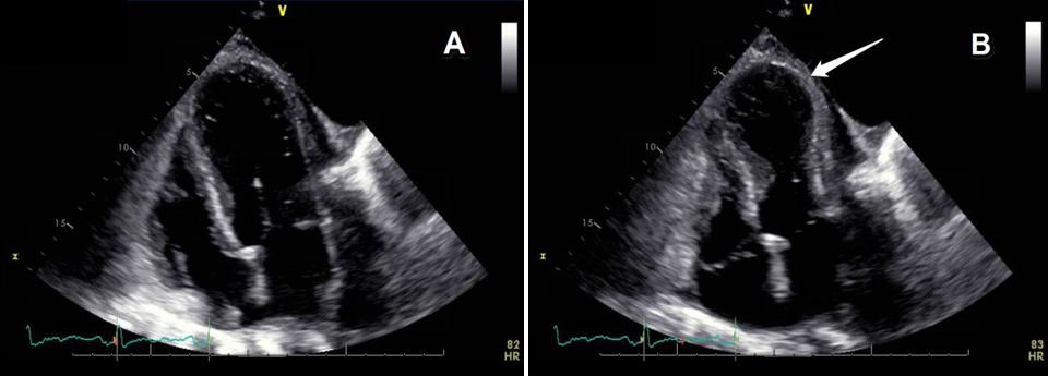
Imaging

Last 50 Imaging Postings

(Most recent listed first. Click on title to be directed to the manuscript.)

July 2025 Imaging Case of the Month: A Growing Lung Nodule in a 
   Patient with Heart Disease
June 2025 Medical Image of the Month: Neurofibromatosis-Associated Diffuse
   Cystic Lung Disease
May 2025 Medical Image of the Month: Aspirated Dental Screw
April 2025 Medical Image of the Month: An Unfortunate Case of Mimicry
March 2025 Medical Image of the Month: An Unusual Case of Pulmonary
   Infarction
February 2025 Medical Image of the Month: Unexpected Complications of
   Transjugular Intrahepatic Portosystemic Shunt (TIPS) 
February 2025 Imaging Case of the Month: A Wolf in Sheep’s Clothing
January 2025 Medical Image of the Month: Psoriasis with Pulmonary
   Involvement
December 2024 Medical Image of the Month: An Endobronchial Tumor
November 2024 Medical Image of the Month: A Case of Short Telomeres
November 2024 Imaging Case of the Month: A Recurring Issue
October 2024 Medical Image of the Month: Lofgren syndrome with Erythema
   Nodosum
September 2024 Medical Image of the Month: A Curious Case of Nasal
   Congestion
August 2024 Image of the Month: Lymphomatoid Granulomatosis
August 2024 Imaging Case of the Month: An Unexplained Pleural Effusion
July 2024 Medical Image of the Month: Vocal Cord Paralysis on PET-CT 
June 2024 Medical Image of the Month: A 76-year-old Man Presenting with
   Acute Hoarseness
May 2024 Medical Image of the Month: Hereditary Hemorrhagic
   Telangiectasia in a Patient on Veno-Arterial Extra-Corporeal Membrane
   Oxygenation
May 2024 Imaging Case of the Month: Nothing Is Guaranteed
April 2024 Medical Image of the Month: Wind Instruments Player Exhibiting
   Exceptional Pulmonary Function
March 2024 Medical Image of the Month: Sputum Cytology in Patients with
   Suspected Lung Malignancy Presenting with Acute Hypoxic Respiratory
   Failure
February 2024 Medical Image of the Month: Pulmonary Alveolar Proteinosis
   in Myelodysplastic Syndrome
February 2024 Imaging Case of the Month: Connecting Some Unusual Dots
January 2024 Medical Image of the Month: Polyangiitis Overlap Syndrome
   (POS) Mimicking Fungal Pneumonia 
December 2023 Medical Image of the Month: Metastatic Pulmonary
   Calcifications in End-Stage Renal Disease 
November 2023 Medical Image of the Month: Obstructive Uropathy
   Extremis
November 2023 Imaging Case of the Month: A Crazy Association
October 2023 Medical Image of the Month: Swyer-James-MacLeod
   Syndrome
September 2023 Medical Image of the Month: Aspergillus Presenting as a
   Pulmonary Nodule in an Immunocompetent Patient
August 2023 Medical Image of the Month: Cannonball Metastases from
   Metastatic Melanoma
August 2023 Imaging Case of the Month: Chew Your Food Carefully
July 2023 Medical Image of the Month: Primary Tracheal Lymphoma
June 2023 Medical Image of the Month: Solitary Fibrous Tumor of the Pleura
May 2023 Medical Image of the Month: Methamphetamine Inhalation
   Leading to Cavitary Pneumonia and Pleural Complications
April 2023 Medical Image of the Month: Atrial Myxoma in the Setting of
   Raynaud’s Phenomenon: Early Echocardiography and Management of
   Thrombotic Disease
April 2023 Imaging Case of the Month: Large Impact from a Small Lesion
March 2023 Medical Image of the Month: Spontaneous Pneumomediastinum
   as a Complication of Marijuana Smoking Due to Müller's Maneuvers
February 2023 Medical Image of the Month: Reversed Halo Sign in the
   Setting of a Neutropenic Patient with Angioinvasive Pulmonary
   Zygomycosis
January 2023 Medical Image of the Month: Abnormal Sleep Study and PFT
   with Supine Challenge Related to Idiopathic Hemidiaphragmatic Paralysis
December 2022 Medical Image of the Month: Bronchoesophageal Fistula in
   the Setting of Pulmonary Actinomycosis
November 2022 Medical Image of the Month: COVID-19 Infection
   Presenting as Spontaneous Subcapsular Hematoma of the Kidney
November 2022 Imaging Case of the Month: Out of Place in the Thorax
October 2022 Medical Image of the Month: Infected Dasatinib Induced
   Chylothorax-The First Reported Case 
September 2022 Medical Image of the Month: Epiglottic Calcification
Medical Image of the Month: An Unexpected Cause of Chronic Cough
August 2022 Imaging Case of the Month: It’s All About Location
July 2022 Medical Image of the Month: Pulmonary Nodule in the
   Setting of Pyoderma Gangrenosum (PG) 
June 2022 Medical Image of the Month: A Hard Image to Swallow
May 2022 Medical Image of the Month: Pectus Excavatum
May 2022 Imaging Case of the Month: Asymmetric Apical Opacity–
   Diagnostic Considerations
April 2022 Medical Image of the Month: COVID Pericarditis
March 2022 Medical Image of the Month: Pulmonary Nodules in the
   Setting of Diffuse Idiopathic Pulmonary NeuroEndocrine Cell Hyperplasia
   (DIPNECH) 
February 2022 Medical Image of the Month: Multifocal Micronodular
   Pneumocyte Hyperplasia in the Setting of Tuberous Sclerosis
February 2022 Imaging Case of the Month: Between A Rock and a
   Hard Place
January 2022 Medical Image of the Month: Bronchial Obstruction
   Due to Pledget in Airway Following Foregut Cyst Resection
December 2021 Medical Image of the Month: Aspirated Dental Implant


For complete imaging listings click here

Those who care for patients with pulmonary, critical care or sleep disorders rely heavily on chest radiology and pathology to determine diagnoses. The Southwest Journal of Pulmonary, Critical Care & Sleep publishes case-based articles with characteristic chest imaging and related pathology. The editor of this section will oversee and coordinate the publication of a core of the most important chest imaging topics. In doing so, they encourage the submission of unsolicited manuscripts. It cannot be overemphasized that both radiologic and pathologic images must be of excellent quality. As a rule, 600 DPI is sufficient for radiographic and pathologic images. Taking pictures of plain chest radiographs and CT scans with a digital camera is strongly discouraged. The figures should be cited in the text and numbered consecutively. The stain used for pathology specimens and magnification should be mentioned in the figure legend. Those who care for patients with pulmonary, critical care or sleep disorders rely heavily on chest radiology and pathology to determine diagnoses. The Southwest Journal of Pulmonary, Critical Care & Sleep publishes case-based articles with characteristic chest imaging and related pathology. The editor of this section will oversee and coordinate the publication of a core of the most important chest imaging topics. In doing so, they encourage the submission of unsolicited manuscripts. It cannot be overemphasized that both radiologic and pathologic images must be of excellent quality. As a rule, 600 DPI is sufficient for radiographic and pathologic images. Taking pictures of plain chest radiographs and CT scans with a digital camera is strongly discouraged. The figures should be cited in the text and numbered consecutively. The stain used for pathology specimens and magnification should be mentioned in the figure legend.

-------------------------------------------------------------------------------------------  

Wednesday
Mar252015

Medical Image of the Week: REM without Atonia

Figure 1. Polysomnogram with thirty-second epoch showing leg movements and relative increase in chin tone during REM sleep.


Figure 2. Polysomnogram with thirty-second epoch demonstrating similar interval of REM without atonia with the addition of sleep-talking.


A 78 year-old man with a past medical history of Parkinson’s disease (PD) presented to the sleep medicine clinic for evaluation of obstructive sleep apnea (OSA). An overnight polysomnogram (PSG) study was consistent with sleep apnea and revealed frequent leg and arm movements and sleep-talking during rapid eye movement (REM) sleep.

REM sleep behavior disorder (RBD) is a parasomnia characterized by repeated episodes of abnormal behavior occurring during REM sleep (1,2). On PSG, REM sleep without atonia is seen while features of “normal REM” such as number of REM periods and REM cycling remain intact (2). RBD emerges most often in the context of alpha-synucleinopathies, and occurs in up to 60% of PD patients (3). The presence of RBD may be an important preclinical symptom prior to the onset of PD. Cases of PD with RBD are associated with a unique phenotype with an older age of onset, longer disease duration, more profound motor disability, and greater degrees of hallucinations and cognitive dysfunction (3). Establishing a safe sleep environment can be of primary importance in patients with RBD as REM without atonia is associated with injurious behavior. Melatonin is effective as a first-line agent in patients with dementia. In non-demented patients without OSA, low-dose clonazepam is the first line intervention and is rarely associated with withdrawal or need for dose escalation (1). Treating concomitant OSA is important adjunctive therapy.

Jared Bartell, Safal Shetty MD, and Kenneth S. Knox MD

University of Arizona Medical Center

Tucson, AZ

References

  1. Aurora RN, Zak RS, Maganti RK, Auerbach SH, Casey KR, Chowdhuri S, Karippot A, Ramar K, Kristo DA, Morgenthaler TI; Standards of Practice Committee; American Academy of Sleep Medicine. Best practice guide for the treatment of REM sleep behavior disorder (RBD). J Clin Sleep Med. 2010;6(1):85-95. [PubMed]
  2. Schenck CH, Mahowald MW. REM sleep behavior disorder: clinical, developmental, and neuroscience perspectives 16 years after its formal identification in SLEEP. Sleep. 2002;25(2):120-38. [PubMed]
  3. Kim YE, Jeon BS. Clinical implication of REM sleep behavior disorder in Parkinson's disease. Parkinsons Dis. 2014;4(2):237-44. [CrossRef] [PubMed]

Reference as: Bartell J, Shetty S, Knox KS. Medical image of the week: REM without atonia. Southwest J Pulm Crit Care. 2015;10(3):147-8. doi: http://dx.doi.org/10.13175/swjpcc022-15 PDF

Wednesday
Mar182015

Medical Image of the Week: Cheyne-Stokes Respiration

Figure 1. Cheyne-Stokes Breathing pattern seen. The red arrow indicates the cycle time which is defined as the duration of the central apnea (or hypopnea) + the duration of a respiratory phase.

A 62 year-old male with a past medical history congestive heart failure, chronic obstructive pulmonary disease, and obesity with a body mass index of 38.02 kg/m2 underwent an overnight polysomnogram for clinical suspicion for obstructive sleep apnea. He was found to have a periodic breathing as seen in the image above.

Cheyne-stokes respiration (CSR) is a type of periodic breathing characterized by crescendo-decrescendo pattern of respiration separated by central sleep apneas (CSA) or hypopneas (1). CSR-CSA may be seen in up to 15-37% of systolic heart failure patients (2,3). A longer cycle length, usually between 45-90 seconds, as well as the waxing and waning breathing pattern differentiate CSR from other forms of cyclic central apnea. CSA leads to chronically increased sympathetic activity and exerts multiple deleterious effects on the failing heart (2). The presence of CSR has been associated with higher mortality and rapid deterioration in cardiac function (4).

Jared Bartell and Safal Shetty, MD

University of Arizona Medical Center

Tucson, AZ

References

  1. Berry RB, Budhiraja R, Gottlieb DJ, Gozal D, Iber C, Kapur VK, Marcus CL, Mehra R, Parthasarathy S, Quan SF, Redline S, Strohl KP, Davidson Ward SL, Tangredi MM; American Academy of Sleep Medicine. Rules for scoring respiratory events in sleep: update of the 2007 AASM Manual for the Scoring of Sleep and Associated Events. Deliberations of the Sleep Apnea Definitions Task Force of the American Academy of Sleep Medicine. J Clin Sleep Med. 2012;8(5):597-619. [CrossRef]  [PubMed]
  2. Yumino D, Bradley TD. Central sleep apnea and Cheyne-Stokes respiration. Proc Am Thorac Soc. 2008;5(2):226-36. [CrossRef] [PubMed]
  3. Garcia-Touchard A, Somers VK, Olson LJ, Caples SM. Central sleep apnea: implications for congestive heart failure. Chest. 2008;133(6):1495-504. [CrossRef] [PubMed]
  4. Hanly PJ, Zuberi-Khokhar NS. Increased mortality associated with Cheyne-Stokes respiration in patients with congestive heart failure. Am J Respir Crit Care Med. 1996;153(1):272-6. [CrossRef] [PubMed] 

Reference as: Bartell J, Shetty S. Medical image of the week: Cheyne-Stokes respiration. Southwest J Pulm Crit Care. 2015;10(3):145-6. doi: http://dx.doi.org/10.13175/swjpcc017-15 PDF