Search Journal-type in search term and press enter
Southwest Pulmonary and Critical Care Fellowships
In Memoriam

Sleep

(Click on title to be directed to posting , most recent listed first)

July 2023 Sleep Case of the Month: Fighting for a Good Night’s Sleep
Associations Between Insomnia and Obstructive Sleep Apnea with
   Nutritional Intake After Involuntary Job Loss
January 2023 Sleep Case of the Month: An Unexpected EEG Abnormality
July 2022 Sleep Case of the Month: A Sleepy Scout
Assessing Depression and Suicidality Among Recently Unemployed
   Persons with Obstructive Sleep Apnea and Socioeconomic
   Inequality
Impact of Recent Job Loss on Sleep, Energy Consumption and Diet
Long-term All-Cause Mortality Risk in Obstructive Sleep Apnea Using
   Hypopneas Defined by a ≥3 Percent Oxygen Desaturation or Arousal
The Association Between Obstructive Sleep Apnea Defined by 3 Percent
   Oxygen Desaturation or Arousal Definition and Self-Reported
Cardiovascular Disease in the Sleep Heart Health Study
Informe de políticas: Fatiga, sueño y salud del personal de enfermería, y 
   cómo garantizar la seguridad de los pacientes y el público
Sleep Tips for Shift Workers in the Time of Pandemic
Tips for Circadian Sleep Health While Working from Home
Impacto del Sueño y la Modalidad de Diálisis sobre la Calidad de Vida en
   una Población
The Effect of CPAP on HRQOL as Measured by the Quality of Well-Being
   Self-Administered Questionnaire (QWB-SA)
Declaración de posición: Reducir la fatiga asociada con la deficiencia de 
   sueño y las horas de trabajo en enfermeras
Impact of Sleep and Dialysis Mode on Quality of Life in a Mexican Population
Out of Center Sleep Testing in Ostensibly Healthy Middle Aged to Older
   Adults
Sleep Related Breathing Disorders and Neurally Mediated Syncope (SRBD
   and NMS)
Sleep Board Review Question: Restless Legs
Impact of Sleep Duration and Weekend Oversleep on Body Weight
   and Blood Pressure in Adolescents
Role of Spousal Involvement in Continuous Positive Airway Pressure
   (CPAP) Adherence in Patients with Obstructive Sleep Apnea (OSA)
The Impact of an Online Prematriculation Sleep Course (Sleep 101) on
   Sleep Knowledge and Behaviors in College Freshmen: A Pilot Study
Obstructive Sleep Apnea and Quality of Life: Comparison of the SAQLI,
   FOSQ, and SF-36 Questionnaires
Gender Differences in Real-Home Sleep of Young and Older Couples
Brief Review: Sleep Health and Safety for Transportation Workers
Lack of Impact of Mild Obstructive Sleep Apnea on Sleepiness, Mood and
   Quality of Life
Alpha Intrusion on Overnight Polysomnogram
Sleep Board Review Question: Insomnia in Obstructive Sleep Apnea
Long-Term Neurophysiologic Impact of Childhood Sleep Disordered 
   Breathing on Neurocognitive Performance
Sleep Board Review Question: Hyperarousal in Insomnia
Sleep Board Review Question: Epilepsy or Parasomnia?
Sleep Board Review Question: Nocturnal Hypoxemia in COPD
Sleep Board Review Questions: Medications and Their
   Adverse Effects
Sleep Board Review Questions: The Restless Sleeper
Obstructive Sleep Apnea and Cardiovascular Disease:
Back and Forward in Time Over the Last 25 Years
Sleep Board Review Questions: The Late Riser
Sleep Board Review Questions: CPAP Adherence in OSA
Sleep Board Review Questions: Sleep Disordered Breathing 
That Improves in REM
The Impact Of Sleep-Disordered Breathing On Body
   Mass Index (BMI): The Sleep Heart Health Study (SHHS)
Incidence and Remission of Parasomnias among Adolescent Children in the 
   Tucson Children’s Assessment of Sleep Apnea (TuCASA) Study 
A 45-Year Old Man with Excessive Daytime Somnolence, 
   and Witnessed Apnea at Altitude


The Southwest Journal of Pulmonary and Critical Care and Sleep publishes articles related to those who treat sleep disorders in sleep medicine from a variety of primary backgrounds, including pulmonology, neurology, psychiatry, psychology, otolaryngology, and dentistry. Manuscripts may be either basic or clinical original investigations or review articles. Potential authors of review articles are encouraged to contact the editors before submission, however, unsolicited review articles will be considered.

-------------------------------------------------------------------------------------

Friday
Apr192019

Out of Center Sleep Testing in Ostensibly Healthy Middle Aged to Older Adults

Stuart F. Quan, M.D.1,2

Brandon J. Lockyer1

Salma Batool-Anwar, M.D., M.P.H.1

Daniel Aeschbach, Ph.D.1,3

1Division of Sleep and Circadian Disorders, Department of Medicine and Department of Neurology; Brigham and Women's Hospital and Division of Sleep Medicine, Harvard Medical School, Boston, MA

2Asthma and Airways Research Center, University of Arizona College of Medicine, Tucson, AZ

3Department of Sleep and Human Factors Research; Institute of Aerospace Medicine; German Aerospace Center; Cologne, 51147; Germany

Abstract

Background: Out of Center Sleep Testing (OCST) is used increasingly to diagnose obstructive sleep apnea (OSA). However, there are few data using OCST that quantify the amount of intrinsic apneic and hypopneic events among asymptomatic healthy persons, especially those who are elderly. This analysis reports the results of OCST in a small group of ostensibly healthy asymptomatic individuals.

Methods: The study population was comprised of ostensibly healthy middle-aged to elderly volunteers for studies of circadian physiology. Before undergoing an OCST, they were found to be free of any chronic medical or psychiatry condition by history, physical and psychologic examination and by a variety of questionnaires and laboratory tests.

Results: There were 24 subjects ranging in age from 55-70 years who had an OCST performed. Repeat studies were required in only 3 subjects. Over half the study population was over the age of 60 years (54.2% vs 45.8%); the majority were men (70.8%). The mean apnea hypopnea index (AHI) was 9.2 /hour with no difference between younger and older subjects. However, 11 had an AHI > 5 /hour. Five had an AHI >15 /hour and 2 had an AHI >40 /hour. Those with an AHI <15 /hour had a mean AHI of 4.4 /hour (95% CI:2.8-6.0 /hour).

Conclusions: Although OCST has a low failure rate, there is a high prevalence of intrinsic obstructive apnea and hypopnea in ostensibly healthy asymptomatic persons.

Introduction

Out of center sleep testing (OCST) is increasingly used instead of laboratory polysomnography (PSG) for the identification of persons with obstructive sleep apnea (OSA) (1). There have been a number of studies validating the use of OCST for this purpose (1). Consequently, large research studies now are using OCST to identify subjects with OSA for clinical trials or observational studies (2-4). Out of center sleep testing also may have a role in identifying the presence of unrecognized OSA in studies of asymptomatic normal individuals. Studies reporting on the frequency of apneic and hypopneic events in healthy individuals using current PSG monitoring techniques (5, 6) found surprisingly high prevalence rates of OSA. Similar reports using OCST also noted that OSA was common in asymptomatic volunteers, but many of these subjects still had chronic medical conditions (7, 8). Thus, there is little information on the prevalence of OSA using OCST, especially in those who are older without any co-morbidities.

In this report, we describe the results of OCST in a small group of middle-aged to older adults who were screened intensively to exclude the presence of chronic sleep and medical conditions. We hypothesized that in this group of ostensibly healthy adults the prevalence of OSA would be less than previously observed.

Methods

Potential subjects between 55 and 70 years of age were identified through public advertisements for volunteers to participate in a circadian physiology research study. The study was approved by the Partners Health Care Human Research Committee, and subjects gave written informed consent prior to their participation. Initially, 5,225 individuals were screened by telephone for medical and psychiatric exclusion criteria. Those who passed (n=90) were invited for an interview and more intensive screening which included a medical history, the Pittsburgh Sleep Quality Index (PSQI) (9), Epworth Sleepiness Scale (ESS) (10) the Berlin Questionnaire (11), blood and urine tests, an electrocardiogram, and a complete physical examination by a physician. In addition, a structured psychologic examination was conducted by a clinical psychologist along with completion of several psychological screening tests including the Geriatric Depression Scale (12), Mattis Dementia Rating Scale (13) and the Mini Mental Status Examination (14). Potential subjects also could not have worked at night for the previous 3 years or recently traveled across time zones. Those who were determined to be free from any acute or chronic medical or psychiatric condition including but not limited to obesity (body mass index, BMI < 30 kg/m2), medication use, hypertension, cardiovascular or pulmonary disease, neoplasia, and disorders of the gastrointestinal, renal, endocrine, metabolic or neurologic systems were asked to undergo an OCST.

The OCST was performed using the Embletta Gold (Embla Systems, Broomfield, CO), a Type III OCST device. The testing montage included nasal pressure, pulse oximetry, bilateral leg electromyography, and chest and abdominal inductance plethysmography. Subjects were instructed to sleep at their habitual hours during OCST. Procedure for determining lights out and lights on, sleep onset and sleep offset, and wake periods during OCST in the sleep, aging and circadian rhythm disorders is given in Appendix 1. Estimated total sleep time was ascertained by the scoring technologist on the basis of changes in the recorded signals indicative of sleep onset or offset and a questionnaire administered to the subject on the morning after the study (see online supplement for the protocol). Studies were scored for apneas, hypopneas and periodic limb movements according to the following criteria. An apnea was defined as > 90% amplitude decrease from baseline of the nasal pressure signal lasting ≥ 10 s. Hypopneas were scored if an event was at least 10 s in duration and if there was a clear amplitude reduction of the nasal pressure signal that was associated with an oxygen desaturation > 4%. Obstructive or central apneas were identified by the presence or absence of respiratory effort, respectively. The apnea hypopnea index (AHI) was calculated as the sum of all apneas and hypopneas divided by the estimated total sleep time. Periodic limb movements were identified using American Academy of Sleep Medicine criteria (15). The periodic limb movement index (PLMI) was computed as the sum of all periodic limb movements divided by the estimated total sleep time. All OCST recordings were scored by a registered polysomnographic technologist and reviewed by a board-certified sleep physician.

Six subjects who had successfully completed the circadian physiology protocol were invited again to enroll in a related protocol approximately 1-3 years later. They repeated the aforementioned screening procedures including a second OCST.

Paired and unpaired Student’s t-tests were used to compare means. Comparisons between proportions were performed using c2. Data are expressed as means ± SD or number and % of cases. Data were analyzed using IBM SPSS Statistics V24 (Armonk, NY).

Results

As shown in Table 1, there were 24 subjects who successfully underwent a screening OCST.

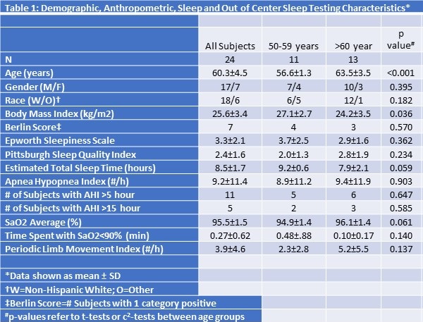
Their ages ranged from 55 to 70 years. Over half the study population was over the age of 60 years [13/24 (54.2 %) vs 11/24 (45.8%)], and the majority were men (70.8%). In 3 subjects, the initial OCST provided insufficient data and a repeat study was required. The mean AHI was 9.2 /hour overall and there were no differences between younger and older subjects. Notably, 11 subjects (46%) had an AHI greater than 5 /hour. Five (21%) had an AHI greater than 15 /hour and 2 subjects had an AHI greater than 40 /hour. Excluding subjects with an AHI ≥15 /hour yielded a mean AHI of 4.4 (95% CI: 2.8-6.0) /hour. Younger subjects had a higher BMI (27.1 ± 2.7 vs. 24.2 ± 3.5 kg/m2, p=0.036), tended to have a longer estimated total sleep time (9.2 ± 0.6 vs. 7.9 ± 2.1 hours, p=0.059), and a slightly lower average nocturnal oxygen saturation (94.9 ± 1.4 vs. 96.1 ± 1.4%, p=.061). No other differences were observed between young and older subjects.

In Table 2 are shown the sleep and anthropometric findings for the 6 subjects who had repeat testing.

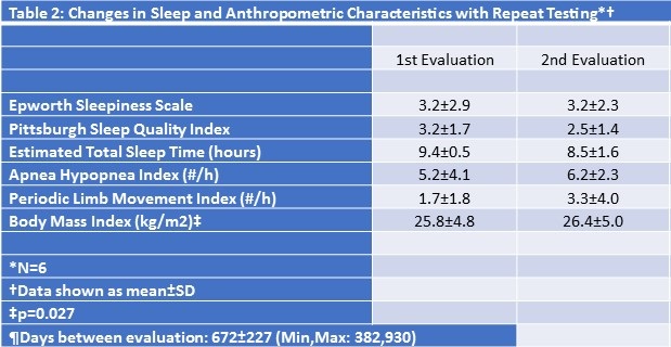
The interval between tests ranged from 382 to 930 days (mean: 672 ± 227 days). Although there was a slight increase in BMI over this interval, no changes were noted in their sleep quality, AHI or PLMI.

Discussion

In this study, we found that it was feasible to screen an ostensibly healthy asymptomatic group of middle to older aged adults for the presence of OSA using a Level III OCST device. The results of the OCST were replicable over an interval of several years and there were few technical failures. Furthermore, evidence of OSA was observed in a substantial proportion of these individuals underscoring that the diagnosis of OSA remains unrecognized in many elderly persons.

Over the past 20 years, there have been numerous clinical trials and cohort studies that have recorded data related to the presence of OSA (2-4, 8, 16, 17). Most have used PSG recorded in the laboratory or at home (16-18). However, the use of PSG is logistically complex and expensive. Additionally, the equipment required for data acquisition may artifactually disrupt the subject’s sleep. In contrast, OCST is less intrusive and expensive, but actual sleep is not recorded. However, to our knowledge there have not been previous studies that have determined the normative values for the AHI using OCST in a group of ostensibly healthy middle-aged to older adults. In this small cohort, the mean AHI was 9.2 /hour. According to the 3rd International Classification of Sleep Disorders (19), an AHI ≥15 /hour is diagnostic of OSA even in the absence of symptoms or associated medical conditions. Therefore, after excluding those who met criteria for at least moderate OSA (i.e., AHI ≥15 hour), our findings indicate an average value of 4.4 /hour in those individuals with a “normal” AHI using OCST. In contrast, a previous study of healthy volunteers for circadian physiology research using similar inclusion and exclusion criteria found the overall mean AHI of all subjects using PSG was substantially higher; hypopneas were identified if reductions in airflow were associated with a minimum 3% oxygen desaturation or a cortical arousal (6). However, a more recent study also using PSG found AHI values more similar to our observations (5). Although it would be expected that OCST may underreport the presence of hypopneas, the explanation for the differences between the two PSG studies is unclear given that the recording montages and respiratory scoring algorithms appeared to be similar.

A striking finding from this study is the high prevalence of OSA among these carefully screened, ostensibly healthy volunteers who had no evidence of a sleep disorder based on their Berlin Questionnaire, PSQI and ESS. The AHI of 2 subjects documented the presence of severe OSA which is usually an indication for treatment. The absence of hypersomnia in a substantial proportion of persons with PSG evidence of OSA has been clearly documented (20). Our data extend these observations by demonstrating the existence of severe OSA in ostensibly healthy middle-aged to older adults. Although some data suggest that such individuals are not at risk for cardiovascular disease or other sequelae of OSA (8), whether this prognosis is correct is still not settled.

Some, but not all epidemiologic data suggest that OSA slowly worsens with age. In contrast, we observed no progression of OSA over an interval of approximately 1-2.5 years (8, 21). However, it is likely that our follow up interval was insufficient to detect a change given the small number of subjects.

In conclusion, use of OCST to screen for OSA is feasible in research studies of normal individuals. Importantly, a relatively high proportion of even ostensibly healthy individuals can be expected to show evidence of OSA.

Acknowledgements

This study was supported by P01 AG009975 from the National Institute of Aging. We would like to express our gratitude to Alec Rader and Jacob Medina for their help with subject recruitment and Stephanie Marvin for scoring support.

References

  1. Kapur VK, Auckley DH, Chowdhuri S, et al. Clinical Practice Guideline for Diagnostic Testing for Adult Obstructive Sleep Apnea: An American Academy of Sleep Medicine Clinical Practice Guideline. J Clin Sleep Med. 2017;13(3):479-504. [CrossRef] [PubMed]
  2. Gottlieb DJ, Punjabi NM, Mehra R, et al. CPAP versus oxygen in obstructive sleep apnea. N Engl J Med. 2014;370(24):2276-85. [CrossRef] [PubMed]
  3. Zhao YY, Wang R, Gleason KJ, et al. Effect of Continuous Positive Airway Pressure Treatment on Health-Related Quality of Life and Sleepiness in High Cardiovascular Risk Individuals with Sleep Apnea: Best Apnea Interventions for Research (BestAIR) Trial. Sleep. 2017;40(4):10.1093/sleep/zsx040. [CrossRef] [PubMed]
  4. Redline S, Sotres-Alvarez D, Loredo J, et al. Sleep-disordered breathing in Hispanic/Latino individuals of diverse backgrounds. The Hispanic Community Health Study/Study of Latinos. Am J Respir Crit Care Med. 2014;189(3):335-44. [CrossRef] [PubMed]
  5. Mitterling T, Hogl B, Schonwald SV, et al. Sleep and Respiration in 100 Healthy Caucasian Sleepers--A Polysomnographic Study According to American Academy of Sleep Medicine Standards. Sleep. 2015;38(6):867-75. [CrossRef] [PubMed]
  6. Pavlova MK, Duffy JF, Shea SA. Polysomnographic respiratory abnormalities in asymptomatic individuals. Sleep. 2008;31(2):241-8. [CrossRef] [PubMed]
  7. Sforza E, Pichot V, Martin MS, Barthelemy JC, Roche F. Prevalence and determinants of subjective sleepiness in healthy elderly with unrecognized obstructive sleep apnea. Sleep Med. 2015;16(8):981-6.[CrossRef] [PubMed]
  8. Sforza E, Hupin D, Pichot V, Barthelemy JC, Roche F. A 7-year follow-up study of obstructive sleep apnoea in healthy elderly: The PROOF cohort study. Respirology. 2017;22(5):1007-14. [CrossRef] [PubMed]
  9. Buysse DJ, Reynolds CF,3rd, Monk TH, Berman SR, Kupfer DJ. The Pittsburgh Sleep Quality Index: a new instrument for psychiatric practice and research. Psychiatry Res. 1989;28(2):193-213. [CrossRef] [PubMed]
  10. Johns MW. A new method for measuring daytime sleepiness: the Epworth sleepiness scale. Sleep. 1991;14(6):540-5. [CrossRef] [PubMed]
  11. Netzer NC, Stoohs RA, Netzer CM, Clark K, Strohl KP. Using the Berlin Questionnaire to identify patients at risk for the sleep apnea syndrome. Ann Intern Med. 1999;131(7):485-91.[CrossRef] [PubMed]
  12. Yesavage JA, Brink TL, Rose TL, et al. Development and validation of a geriatric depression screening scale: a preliminary report. J Psychiatr Res. 1982;17(1):37-49. [CrossRef] [PubMed]
  13. Mattis S. Mental status examination for organic mental syndrome in the elderly patient. In: Geriatric psychiatry. Bellak L and Karasu TE, eds. New York: Grune and Stratton, 1976; 77-121
  14. Pangman VC, Sloan J, Guse L. An examination of psychometric properties of the mini-mental state examination and the standardized mini-mental state examination: implications for clinical practice. Appl Nurs Res. 2000;13(4):209-213. [CrossRef] [PubMed]
  15. Iber C, Ancoli-Israel S, Chesson A, Quan SF. The AASM Manual for the Scoring of Sleep and Associated Events: Rules, Terminology and Technical Specifications. 1st ed. Westchester, IL: American Academy of Sleep Medicine, 2007; 57.
  16. Quan SF, Howard BV, Iber C, et al. The Sleep Heart Health Study: design, rationale, and methods. Sleep. 1997;20(12):1077-85. [PubMed]
  17. Kushida CA, Nichols DA, Quan SF, et al. The Apnea Positive Pressure Long-term Efficacy Study (APPLES): rationale, design, methods, and procedures. J Clin Sleep Med. 2006;2(3):288-300. [PubMed]
  18. Chen X, Wang R, Zee P, et al. Racial/Ethnic Differences in Sleep Disturbances: The Multi-Ethnic Study of Atherosclerosis (MESA). Sleep. 2015;38(6):877-88. [CrossRef] [PubMed]
  19. Sateia MJ. International Classification of Sleep Disorders. 3rd ed. Darien, IL: American Academy of Sleep Medicine, 2014.
  20. He K, Kapur VK. Sleep-Disordered Breathing and Excessive Daytime Sleepiness. Sleep Med Clin. 2017;12(3):369-82.[CrossRef] [PubMed]
  21. Redline S, Schluchter MD, Larkin EK, Tishler PV. Predictors of longitudinal change in sleep-disordered breathing in a nonclinic population. Sleep. 2003;26(6):703-9. [CrossRef] [PubMed]

Cite as: Quan SF, Lockyer BJ, Batool-Anwar S, Aeschbach D. Out of center sleep testing in ostensibly healthy middle aged to older adults. Southwest J Pulm Crit Care. 2019;18:87-93. doi: https://doi.org/10.13175/swjpcc016-19 PDF 

Friday
Apr052019

Sleep Related Breathing Disorders and Neurally Mediated Syncope (SRBD and NMS)

Damian Valencia, MD1 

Stella Pak, MD1

Juan Linares, MD1

Victor Valencia, BS2

Christopher Lee, MD1

John-Philip Markovic, MD1

Hemant Shah, MD1 


1Department of Medicine, Kettering Medical Center, Kettering, Ohio USA

2Department of Medicine, Northwestern University Feinberg School of Medicine, Chicago, Illinois USA


Abstract

Introduction: Individuals with severe sleep related breathing disorders (SRBD) tend to experience intermittent hypoxia, sleep fragmentation and highly fluctuating intrathoracic pressures. Chronic exposure to these stressors sensitizes the parasympathetic system while suppressing the sympathetic system. Parasympathetic over-reactivity among patients with severe sleep related breathing disorders has been proposed as a predisposing factor for neurally mediated syncope.

Goal: We sought to determine the relative risk for neurally mediated syncope in patients with severe SRBD compared to the general population.

Methods: This is a retrospective cohort study of 228 cases selected from 2,598 patients who were referred for polysomnography on discharge from hospitalization. Incidence of neurally mediated syncope (NMS) was compared between patients with apnea-hypopnea-index (AHI) scores of 30 or greater and those with an AHI score below 5.

Results: Approximately 32% of patients with severe SRBD had a history of neurally mediated syncope compared to only 14% in patients with normal sleep breathing patterns (OR = 3.09, 95% CI: 1.25 - 7.62, p = 0.015).

Conclusion: Our multi-center retrospective study supports an association between SRBD and NMS.

Brief Summary

Current Knowledge/Study Rational. There are multiple reports that highlight a possible connection between sleep related breathing disorders and neurally mediated syncope. Deleterious effects on the autonomic and peripheral nervous system by severe sleep related breathing disorders have also been demonstrated. We sought to determine the association and relative risk of neurally mediated syncope in patients with severe sleep related breathing disorders.

Study Impact. Patients with severe sleep related breathing disorders are at increased risk for neurally mediated syncope. Early identification and appropriate treatment in this patient population may reduce rates of syncope, improve quality of life and clinical outcomes.

Introduction

Sleep related breathing disorders (SRBD), comprise a spectrum of disorders characterized by chronic intermittent apnea and hypopnea, which includes obstructive sleep apnea (OSA), central sleep apnea, sleep-related hypoventilation, and nocturnal hypoxemia (1). Neurally mediated syncope (NMS), also known as reflex syncope, is defined as a transient loss of consciousness secondary to decreased cerebral blood supply, typically as a result of reflexive cardiac inhibition and decreased vascular tone. NMS includes vasovagal syncope, situational syncope and carotid sinus syncope (2). Autonomic dysfunction may also play a role in cases of NMS (3). Researchers have previously documented the deleterious effects of SRBD on the autonomic and peripheral nervous system (4-7). A connection between SRBD and NMS has been proposed by some, detailing cases of patients suffering from incapacitating recurrent syncope which demonstrates dramatic improvement or resolution after diagnosis and treatment of OSA (8-10). In this study, we sought to determine the association and relative risk of neurally mediated syncope in patients with severe sleep related breathing disorders.

Methods

This retrospective cohort analysis was performed using electronic medical record data collection from hospitals within the Kettering Health Network, including Fort Hamilton Hospital (Hamilton, Ohio), Grandview Medical Center (Dayton, Ohio), Greene Memorial Hospital (Xenia, Ohio), Kettering Medical Center (Kettering, Ohio), Soin Medical Center (Beavercreek, Ohio), Southview Medical Center (Centerville, Ohio) and Sycamore Medical Center (Miamisburg, Ohio). Individuals who underwent and completed in-facility polysomnography were selected for study review. Patients under the age of 18 years old and those with a pacemaker or implantable cardiac defibrillator were excluded from the study. Patients were divided into two groups; those with severe SRBD, defined as having an Apnea-Hypopnea Index (AHI) score of/ or greater than 30, and a control group, defined as patients having an AHI score of/ or less than 5. This study was approved by the institutional review board (IRB) at Kettering Health Network.

Statistical Methods: The Kolmogorov-Smirnov and Shapiro-Wilk tests were utilized to compare baseline patient demographics between each group; control and severe SRBD group. These tests were selected to better represent the data, with median interquartile range (IQR), as outliers were included in the analysis. Categorical variables were compared using Pearson’s Chi-squared test. Continuous variables were compared using the Student’s t-test or Wilcoxon rank sum test (Mann-Whitney U test). All estimates were reported as 95% confidence intervals with p-values. Two-sided p-values less than 0.05 were considered statistically significant. Multivariate logistic regression modeling was used to determine the effects of each variable while controlling for confounding variables. Odds ratios (OR) were calculated for each type of syncope in both the severe SRBD group and control group. All statistical analyses were performed using IBM SPSS Statistics for Windows version 20.0 (IBM Corp., Armonk, NY, USA).

Results

A total of 2,598 patients were identified from the electronic medical record database, of which, only 228 patients fulfilled our inclusion criteria for severe SRBD (AHI score of/ or greater than 30), with 80 patients meeting criteria for the control group (AHI score of/ or less than 5). Among the 228 patients with severe SRBD, the most common subtype was obstructive sleep apnea (204 of 228 patients, 89.5%), followed by central sleep apnea (13 of 228 patients, 4.2%) and mixed type (11 of 228 patients, 3.6%). 

Initial comparison of demographic characteristics (Table 1) was done using univariate analysis.

Table 1: Baseline Characteristics of the Individuals with and without SRBD.

SRBD: sleep-related breathing disorder, IQR: interquartile range; BMI: body-mass index; AHI: apnea-hypopnea index; LEVF: left ventricular ejection fraction; COPD: chronic obstructive pulmonary disease; N: number; N/A: not applicable.

The SRBD group and control did not statistically significance differ in age (p = 0.79). Although gender differences were noted, 62.3% male in the SRBD group compared to 47.5% in the control group (p = 0.042), there were no statistically significant differences on multivariate logistic regression (p = 0.854). Differences in body mass index (BMI) between groups were noted on univariate (p = 0.042) and multivariate models (p = 0.041), with 36.5 (IQR 31.3 – 43.8) mean BMI of the SRBD group compared to 34.4 (IQR 27.3 – 40.4) in the control group.

The incidence of pre-existing comorbidities between groups was also compared. (Table 1) There were no statistically significant differences between the groups in terms of pulmonary artery pressure (p = 0.226), diabetes (p = 0.902) and coronary artery disease (p = 0.065). Univariate analysis did reveal differences amongst left ventricular ejection fraction (LVEF), 31.1% of patients with SRBD had LVEF <55% compared to only 17.5% in the control group (p = 0.19), hypertension (HTN), 75.4% of patients with SRBD had HTN compared to only 53.8% in the control group (p < 0.001), and chronic obstructive pulmonary disease (COPD), 29.8% of patients with SRBD compared to 43.8% in the control group (p = 0.023). These findings were not statistically significant on multivariate logistic regression; LVEF<55% (p = 0.326), HTN (p = 0.585), COPD (p = 0.576). 

The mean apnea hypopnea index (AHI) for the severe SRBD group was 53.2 (38.7-80.2), compared to 1.6 (0-2.6) in the control group (p < 0.001). The prevalence of NMS was higher in the SRBD group compared to the control group, 32% (73 of 228 patients) and 14% (11 of 80 patients), respectively; χ2 (2, N=308) = 9.96, p = 0.001. Prevalence of non-neurally mediated syncope did not differ significantly between the SRBD group and control group, 1% (2 of 228 patients) and 0% (no patients), respectively; Pearson’s Chi-squared test p = 0.571. Approximately 32% of patients with severe SRBD had a history of neurally mediated syncope compared to only 14% in patients with normal sleep breathing patterns (OR = 3.09, 95% CI: 1.25 - 7.62, p = 0.015). (Table 2).

Table 2: Multivariate Logistic Regression Modeling, Odds Ratio (OR) for Neurally Mediated Syncope.

CI: confidence interval; BMI: body mass index; AHI: apnea-hypopnea index; LEVF: left ventricular ejection fraction; COPD: chronic obstructive pulmonary disease.

Situational syncope has not been consistently recorded, OR and CI were not calculated.

Discussion

This study suggests that individuals with severe sleep related breathing disorders are at increased risk for developing neurally mediated syncope. Chrysostomakis et al. (11) showed that parasympathetic activity is increased during the night in patients with obstructive sleep apnea and that continuous positive airway pressure (CPAP) treatment may restore autonomic balance. Puel et al. (8) suggested that intermittent hypoxia, sleep fragmentation and variations of intra-thoracic pressures may result in chronic adaptations to the autonomic nervous system, which may predispose patients to vasovagal syncope. Cintra et al. (12) noted that patients with vasovagal syncope exhibited sympathetic suppression during rapid eye movement (REM) sleep. Previous studies indicate that chronic intermittent hypoxia can also increase activation of free-radical oxidation, which in turn can elicit rapid and sustained expression of pro-inflammatory cytokines (12). This oxidative stress and inflammatory response can induce tissue damage and intermittent academia, eventually leading to up-regulation of pH sensitive ion channels on chemo-afferent neurons at the carotid bodies. Overexpression of these channels potentiates the carotid body response to changes in arterial oxygen saturation (12). It is possible that this mechanism also contributes to the higher prevalence of carotid sinus hypersensitivity and syncope among patients with severe SRBD. In accordance with our findings, there have been reported cases of recurrent syncope, which have resolved with correction of underlying SRBD (8-10). Appropriate management of SRBD in this patient population may reduce rates of NMS. Given the high prevalence of SRBD and neurally mediated syncope in the United States, further investigation is warranted to delineate the association between the two disease processes and the mechanisms which are involved.

Study Limitations

Our retrospective study design has limited control over consistency and accuracy. This study did not use any matching algorithm to match the control group to the individuals with severe SRBD for baseline characteristics. We did not utilize a caliper matching process to identify controls, and as a result, the control cohort was smaller than the study cohort. The diagnosis of vasovagal syncope was made based on clinical presentation. All patients who were diagnosed with vasovagal syncope did not have confirmatory tilt-table testing, limiting diagnostic accuracy and consistency.

Conclusion

Our study suggests that individuals with severe sleep-related breathing disorders (SRBD) are approximately 3 times (OR = 3.09, 95% CI: 1.25 - 7.62, p = 0.015) more likely to have experienced neurally mediated syncope (NMS) compared to case matched controls.

Acknowledgement

Rosaria Jordan (table/figure formatting)

References

  1. Tsara V, Amfilochiou A, Papagrigorakis M, Georgopoulos D, Liolios E. Guidelines for diagnosis and treatment of sleep-related breathing disorders in adults and children. Definition and classification of sleep related breathing disorders in adults: Different types and indications for sleep studies (part 1). Hippokratia. 2009;13:187-91. [PubMed]
  2. Alboni P, Brignole M, Menozzi C, et al. Diagnostic value of history in patients with syncope with or without heart disease. J Am Coll Cardiol. 2001;37(7):1921-8. [CrossRef] [PubMed]
  3. Mosqueda-Garcia R, Furlan R, Tank J, Fernandez-Violante R. The elusive pathophysiology of neurally mediated syncope. Circulation. 2000;102(23):2898-2906. [CrossRef] [PubMed]
  4. Li Y, Veasey SC. Neurobiology and neuropathophysiology of obstructive sleep apnea. NeuroMolecular Med. 2012;14(3):168-179. doi:10.1007/s12017-011-8165-7. [CrossRef] [PubMed]
  5. Gozal D, Daniel JM, Dohanich GP. Behavioral and anatomical correlates of chronic episodic hypoxia during sleep in the rat. J Neurosci. 2001;21(7):2442-50. [PubMed]
  6. Mayer P, Dematteis M, Pépin JL, et al. Peripheral neuropathy in sleep apnea: A tissue marker of the severity of nocturnal desaturation. Am J Respir Crit Care Med. 1999;159(1):213-9. [CrossRef] [PubMed]
  7. Dziewas R, Schilling M, Engel P, et al. Treatment for obstructive sleep apnoea: effect on peripheral nerve function. J Neurol Neurosurg Psychiatry. 2007;78(3):295-7. [CrossRef] [PubMed]
  8. Puel V, Pepin JL, Gosse P. Sleep related breathing disorders and vasovagal syncope, a possible causal link? Int J Cardiol. 2013;168(2):1666-7. [CrossRef] [PubMed]
  9. Barone D, Fine L, Bhandari N, Ana C. Syncope in hypoxemic respiratory arrest. J Sleep Med Disord. 2015;2(2):2-4. [CrossRef].
  10. Willis FB, Isley AL, Geda YE, Shaygan A, Quarles L, Fredrickson P a. Resolution of syncope with treatment of sleep apnea. J Am Board Fam Med. 2008;21(5):466-8. [CrossRef] [PubMed]
  11. Chrysostomakis SI, Simantirakis EN, Schiza SE, et al. Continuous positive airway pressure therapy lowers vagal tone in patients with obstructive sleep apnoea-hypopnoea syndrome. Hell J Cardiol. 2006;47(1):13-20. [PubMed]
  12. Cintra F, Poyares D, DO Amaral A, et al. Heart rate variability during sleep in patients with vasovagal syncope. Pacing Clin Electrophysiol. 2005;28(12):1310-6. [CrossRef] [PubMed]

Cite as: Valencia D, Pak S, Linares J, Valencia V, Lee C, Markovic J-P, Shah H. Sleep related breathing disorders and neurally mediated syncope (SRBD and NMS). Southwest J Pulm Crit Care. 2019;18(4):76-81. doi: https://doi.org/10.13175/swjpcc015-19 PDF 

Monday
Feb052018

Sleep Board Review Question: Restless Legs

Olabimpe Omobomi, MD MPH

Rohit Budhiraja, MD

Click to read more ...

Friday
Jan122018

Impact of Sleep Duration and Weekend Oversleep on Body Weight and Blood Pressure in Adolescents

*Division of Sleep and Circadian Disorders, Brigham and Women’s Hospital, Harvard Medical School, Boston, MA USA

Asthma and Airways Research Center, University of Arizona College of Medicine, Tucson, AZ USA

Department of Pediatrics, University of Arizona College of Medicine, Tucson, AZ USA

§Department of Medicine, University of Arizona College of Medicine, Tucson, AZ USA

Center for Sleep and Circadian Sciences, University of Arizona Health Sciences Center, Tucson, AZ USA

Click to read more ...

Monday
May082017

Role of Spousal Involvement in Continuous Positive Airway Pressure (CPAP) Adherence in Patients with Obstructive Sleep Apnea (OSA)

Salma Batool-Anwar, MD, MPH 2

Carol M. Baldwin, PhD, MSN 3

Shira Fass, PhD4

Stuart F. Quan, MD 1,2


1University of Arizona College of Medicine, Tucson, AZ USA

2Brigham and Women’s Hospital, Boston, MA USA

3Arizona State University College of Nursing and Health Innovation and College of Health Solutions, Phoenix, AZ USA

4Case Western Reserve University, Cleveland, Ohio USA


Abstract

Introduction: Little is known about the impact of spousal involvement on continuous positive airway pressure (CPAP) adherence. The aim of this study was to determine whether spouse involvement affects adherence with CPAP therapy, and how this association varies with gender.  

Methods: 194 subjects recruited from Apnea Positive Pressure Long Term Efficacy Study (APPLES) completed the Dyadic Adjustment Scale (DAS). The majority of participants were Caucasian (83%), and males (73%), with mean age of 56 years, mean BMI of 31 kg/m2. & 62% had severe OSA. The DAS is a validated 32-item self-report instrument measuring dyadic consensus, satisfaction, cohesion, and affectional expression. A high score in the DAS is indicative of a person’s adjustment to the marriage. Additionally, questions related to spouse involvement with general health and CPAP use were asked. CPAP use was downloaded from the device and self-report, and compliance was defined as usage > 4 h per night.

Results: There were no significant differences in overall marital quality between the compliant and noncompliant subjects. However, level of spousal involvement was associated with increased CPAP adherence at 6 months (p=0.01). After stratifying for gender these results were significant only among males (p=0.03). Three years after completing APPLES, level of spousal involvement was not associated with CPAP compliance even after gender stratification.

Conclusion: Spousal involvement is important in determining CPAP compliance in males in the 1st 6 months after initiation of therapy but is not predictive of longer-term adherence. Involvement of the spouse should be considered an integral part of CPAP initiation procedures.


Abbreviations List

AASM: American Academy of Sleep Medicine

AHI: Apnena Hyponea Index

APPLES: Apnea Positive Pressure Long Term Efficacy Study

BMI: Body Mass Index

CPAP: Continuous positive airway pressure

DAS: Dyadic Adjustment Scale

EEG: Electroencephalogram

EMG: Electromyograms

EOG: Electroocculogram

OSA: Obstructive Sleep Apnea

PSG: polysomnography


Introduction

Obstructive Sleep apnea (OSA) is characterized by repetitive episodes of upper airway closure during sleep resulting in oxygen desaturation and frequent arousals. In addition to cardiovascular comorbidities, OSA has been linked to poor quality of life, depression and motor vehicle accidents. Recent data suggest an increase in the prevalence of OSA for both men and women (34% and 17.4% respectively) (1).

Continuous positive airway pressure (CPAP) is the treatment of choice for OSA. Poor adherence, however, remains a widely recognized problem limiting overall effectiveness of CPAP therapy. Prior studies have identified various factors and strategies to promote CPAP adherence (2). In addition to disease, educational, and technology-specific considerations that can affect CPAP adherence, social and psychological dynamics are important components of adherence as well.

Several studies have suggested that partner/spousal dyadic support can play a positive role in the patient’s overall health and health behaviors (3,4) . For example, higher CPAP adherence was reported among patients with bed partners (5), as well as persons who were married versus single (6). Little is known about the influence of spousal involvement on CPAP adherence. One study indicated that perceived spousal support predicted greater CPAP adherence among men with high disease severity; however, pressure to adhere to treatment by the wife was not of benefit and predicted poorer CPAP adherence (7). Another study indicated reduced marital conflict by OSA patients following 3 months of CPAP, suggesting that marital conflict resolution might serve as an intervention for CPAP adherence (8). Despite these hints that dyadic support may play a role in CPAP adherence, participants in both studies by Baron et al. (7.8) consisted primarily of men, and the studies focusing on CPAP adherence by Lewis et al. (5) and Gagnadoux et al. (6) included only men. Thus, the aim of the current study was to determine whether spouse involvement affects CPAP adherence and how this association differs by gender using data from a large randomized trial of CPAP versus sham CPAP to treat OSA. 


Methods

Study Population and Protocol

The Apnea Positive Pressure Long-term Efficacy Study (APPLES) was a 6-month multicenter, randomized, double-blinded, 2-arm, sham-controlled, intention-to-treat study of CPAP efficacy on three domains of neurocognitive function in OSA. Three of the 5 APPLES Clinical Centers, the University of Arizona, Stanford University and St. Luke’s Hospital (Chesterfield, MO) participated in this ancillary study. A detailed description of the protocol has previously been published (9). Briefly, participants were either recruited through local advertisement, or from attending sleep clinics for evaluation of possible OSA. Symptoms indicative of OSA were used to prescreen potential participants. The initial clinical evaluation included administering informed consent, screening questionnaires, a history and physical examination, and a medical assessment by a study physician. Participants subsequently returned 2-4 weeks later for a 24-h sleep laboratory visit, during which polysomnography (PSG) was performed to confirm the diagnosis, followed by a day of neurocognitive, mood, sleepiness, and quality of life survey testing. Inclusion and exclusion criteria have been published previously and included age ≥ 18 years and a clinical diagnosis of OSA as defined by American Academy of Sleep Medicine (AASM) criteria. Only participants with an apnea hypopnea index (AHI) ≥ 10 by PSG were randomized to continue in the APPLES study. Exclusion criteria were previous treatment for OSA with CPAP or surgery, oxygen saturation on baseline PSG <75% for >10% of the recording time, history of motor vehicle accident-related to sleepiness within the past 12 months, presence of chronic medical conditions, use of various medications known to affect sleep or neurocognitive function, and various health and social factors that may impact standardized testing procedures (e.g., shift work).

Following the PSG, participants with an AHI ≥ 10 who met other enrollment criteria were randomized to CPAP or sham CPAP for continued participation in APPLES. After randomization, participants returned to the sleep laboratory for a CPAP or sham CPAP titration PSG. Subsequent assessments were made at 2, and 6 months post-randomization at which time a test battery was re-administered. At the conclusion of their 6-month post-randomization evaluations, each participant was informed of their treatment group assignment and offered CPAP treatment going forward. Approximately 36 months after the conclusion of APPLES, participants were sent the Dyadic Adjustment Scale (DAS) questionnaire with the addition of several additional questions related to health.

Assessment of Spouse involvement

Inclusion in the current analysis required that subjects were married during the APPLES study and remained married at the time of questionnaire administration. The DAS (10), a quality of marriage questionnaire, was utilized to assess marital relationship. It is a 32-item self-report instrument that incorporates four dimensions, including a 13 item dyadic consensus, 10 item dyadic satisfaction, 5 item dyadic cohesion, and 4 item affectional expression. A high DAS score is indicative of a person’s positive adjustment to the marriage. Additionally, questions related to spouse involvement with general health and CPAP use were asked (See Appendix for full questionnaire).

Polysomnography

The PSG montage included monitoring of the electroencephalogram (EEG, C3-A2 or C4-A1, O2-A1 or O1-A2), electro-oculogram (EOG, ROC-A1, LOC-A2), chin and anterior tibialis electromyograms (EMG), heart rate by 2-lead electrocardiogram, snoring intensity (anterior neck microphone), nasal pressure (nasal cannula), nasal/oral thermistor, thoracic and abdominal movement (inductance plethysmography bands), and oxygen saturation (pulse oximetry). All PSG records were electronically transmitted to a centralized data coordinating and PSG reading center. Sleep and wakefulness were scored using Rechtschaffen and Kales criteria (11). Apneas and hypopneas were scored using American Academy of Sleep Medicine Task Force (1999) diagnostic criteria (12, 13). Briefly, an apnea was defined by a clear decrease (> 90%) from baseline in the amplitude of the nasal pressure or thermistor signal lasting ≥ 10 sec. Hypopneas were identified if there was a clear decrease (> 50% but ≤ 90%) from baseline in the amplitude of the nasal pressure or thermistor signal, or if there was a clear amplitude reduction of the nasal pressure signal ≥ 10 sec that did not reach the above criterion, but was associated with either an oxygen desaturation > 3% or an arousal.

Obstructive events were scored if there was persistence of chest or abdominal respiratory effort. Central events were noted if no displacement occurred on either the chest or abdominal channels. Sleep apnea was classified as mild (AHI 10.0 to 15.0 events per hour), moderate (AHI 15.1 to 30.0 events per hour), and severe (AHI more than 30 events per hour) (12).

CPAP adherence

The primary dependent variable of interest was CPAP adherence and was assessed by nightly use of CPAP at the 6-months follow up visit. CPAP use was downloaded from the device and the participants were considered to be adherent if the mean CPAP use was > 4 hours per night at 6-months. Long-term CPAP adherence was measured as self-reported adherence (hours per night) at the time of the DAS administration.

Statistical Analysis 

Statistical analyses were performed using STATA (Version 11, StataCorp TX USA). Univariate and multivariate logistic regression models were used to estimate the degree to which variables correlated with CPAP adherence. We examined the association between CPAP adherence and following variables: OSA severity as measured by the AHI, age, baseline body mass index (BMI, kg/m2), spousal involvement and the DAS. For these models, dichotomous variables were created for OSA severity (AHI < 15 vs. ≥ 15), obesity (BMI <30 kg/m2 vs. ≥30 kg/m2) and CPAP adherence (< 4 hours/night vs. ≥4 hours/night). Spousal involvement was ascertained using a 5 point Lickert scale and analyzed as a continuous variable.

To assess predictors of CPAP adherence we used multiple regression models. Unpaired t-tests were used to assess the effect of gender, age, OSA severity, BMI, and CPAP adherence in both the CPAP and Sham CPAP groups. Data for continuous and interval variables were expressed as mean ± SD, and as a percentage for categorical variables. Statistical significance was set at a P value <0.05, two-tailed. The variables that produced P value of < 0.05 were included in the final model.


Results

Baseline demographic data on participants (N=194) who completed the DAS are outlined in Table 1.


Table1. Baseline Characteristics of APPLES Participants Who Completed Dyadic Data.


The majority of the participants were Caucasian (83%) and males (73%), with mean age of 56 years and a mean BMI of 31 kg/m2. Over half of the participants had severe OSA (62%). Table 2a demonstrates CPAP adherence at 6 months using multivariate analysis.


Table 2A. Multivariate Analysis of Adherence to CPAP or Sham CPAP at 6 Months.


The CPAP adherence was independently associated with advanced age (p < 0.01) and increasing spousal involvement (p < 0.01). After stratifying by treatment group, the association between CPAP adherence and spousal involvement was seen only amongst the CPAP group (Table 2b).


Table 2B. Multivariate Analysis of Adherence to CPAP at 6 Months.


Adjustment to marriage as reflected by items on the DAS questionnaire, however, was not associated with CPAP adherence.

Notably, after gender stratification, significant association between spousal involvement and CPAP adherence was limited to men alone (p=0.03). Three years after completing APPLES, 82 participants were still adherent by self-report (Table 3).


Table 3. Multivariate Analysis CPAP Adherence 3 years After Completing APPLES Study (based on subjective adherence).


At this time point, spousal involvement was not associated with CPAP adherence even after gender stratification.


Discussion

This multicenter double blind study demonstrates that spousal involvement is important in determining CPAP adherence during the initial treatment period, but has no effect on long-term adherence. Notably, the positive results for adherence were seen only among husbands using CPAP, but there was no effect on wives using CPAP. In line with previous research, we also found that increase in age was associated with greater CPAP adherence among both men and women.

Prior studies have indicated that married versus single, CPAP patients with bed partners, perceived spousal support, and quality of marital relationship all play a role in promoting CPAP adherence (5-8). Although these studies support the idea of social support as a conduit to CPAP adherence, the role of spousal involvement was not clear, sample sizes in the spousal role studies were small, and CPAP users were men, which reduces generalizability.

Baron et al. (3) used a spousal involvement measure, including positive and negative collaboration and one-sided items one week after beginning CPAP treatment (N=23 married men on CPAP), in addition to an interpersonal measure of supportive behaviors at 3 months to evaluate interpersonal qualities (n=16/23 responded). These investigators found that perceived collaborative involvement was related to greater CPAP adherence at 3 months (p=0.002). These findings are similar to our study in that spousal support, at least for husbands on CPAP, fostered greater adherence during the initial period of treatment.

Our observations and those of Baron et al. (14) fit well with the theories of motivation. The fundamental fact of motivation and adherence in healthcare is that individuals cannot be forced to change their behaviors. The behavior change, in this case the CPAP adherence, may be initiated by extrinsic motivation. External motivation may be rewards, punishments, or pressure from other people, such as family members or healthcare providers. However, extrinsic motivation, such as spousal pressure, is less effective in the long-term. In order to sustain long term behavioral change for CPAP adherence one needs to rely on intrinsic motivation which can be strengthened by examining the decisional balance of the ratio between a patient’s perceived pros and cons for engaging in a health behavior. The decisional balance has been found to be predictive of adherence to treatment in a variety of healthcare settings.

Our study also found increased age as an independent predictor of CPAP adherence at 6-months, yet the results were not significant for long-term adherence. Previous studies have also demonstrated conflicting results on the association between age and CPAP adherence. Sin et al. (15) found that a 10 year increment in age resulted in 0.24 ± 0.11-h increase in CPAP use. Alternatively, McArdle and colleagues (16) found that older patients were less likely to use their CPAP machines. Similarly, Janson et al. (17) found older age to be an independent risk factor for discontinuing CPAP treatment, and this finding was thought to be secondary to nasal, or pharyngeal problems. In another study, Russo-Magno et al. (18) found that adherent patients were younger in age compared to non-adherents, and increasing age made CPAP adherence difficult. Cognitive and physical impairments were thought to be contributing to difficulty with CPAP adherence. Mean age in this cohort was 73 years, which was higher than the mean age in our study. It is possible that these inconsistent associations of age on CPAP adherence may be related to the length of follow-up as well. With longer durations, the effect of time on comorbidities in the elderly may make adherence more difficult.

To our knowledge, this is the first study to demonstrate a gender bias in support for CPAP adherence. While men on CPAP were significantly more likely to adhere with support from their wives, there was no such effect for married women on CPAP, suggesting little to no support from their husbands. Although the effect of gender on CPAP adherence and spousal involvement has not been studied, previous research suggests that women are more likely to be the health caregivers in families, and thus exercise more social control (19). It is the social norm and expectation that women are often involved in their husbands’ health. As indicated in the literature regarding type 2 diabetes (20), male patients and their wives shared an expectation that the wives will be involved in their care while female patients and their husbands did not have similar expectations. We can support this finding in relationship to CPAP adherence.

Not surprisingly, spousal support for adherence did not apply to sham CPAP. This suggests that if an intervention is not having any perceived benefit, spousal involvement will have little impact on adherence. 

There are several limitations to this study. A major limitation is self-reported long term CPAP adherence. Additionally, our study was limited to husbands and wives on CPAP completing the DAS; their respective spouses were not asked about their degree of involvement. Moreover, it is unclear which components of spouse involvement played a role in CPAP adherence. We cannot assume that patients welcome all types of spouse involvement. Spouse involvement may be perceived by patients as control and nagging and may not be advantageous for all patients (21). In the context of chronic illness significant differences are demonstrated across couples in expectations for spouse involvement (20).

Despite these limitations, to our knowledge this is the first study of its type that examined spousal support for both men and women on CPAP supporting generalizability of our findings. Other strengths of this study include a large number of participants across multiple sites, randomized CPAP and Sham CPAP control groups, and objective documentation of CPAP adherence at 6 months.

Dyadic coping has been utilized in other health related interventions and can also be used to improve CPAP adherence. Ye et al. (4) has provided a comprehensive review of dyadic support in CPAP adherence, including methodological considerations, recommendations for future research, and implications for interventions. In tandem with the Ye et al. (4) review, our findings, particularly with respect to the need for spousal support of wives on CPAP, can provide a springboard for future clinical/intervention studies to promote CPAP adherence for men and women, to develop gender-relevant training needs to support their spouse on CPAP, and to determine spousal support activities that are the most efficient at promoting CPAP adherence.


Acknowledgments

APPLES was funded by contract 5UO1-HL-068060 from the National Heart, Lung and Blood Institute. The APPLES pilot studies were supported by grants from the American Academy of Sleep Medicine and the Sleep Medicine Education and Research Foundation to Stanford University and by the National Institute of Neurological Disorders and Stroke (N44-NS-002394) to SAM Technology. In addition, APPLES investigators gratefully recognize the vital input and support of Dr. Sylvan Green who died before the results of this trial were analyzed, but was instrumental in its design and conduct.

Administrative Core

Clete A. Kushida, MD, PhD; Deborah A. Nichols, MS; Eileen B. Leary, BA, RPSGT; Pamela R. Hyde, MA; Tyson H. Holmes, PhD; Daniel A. Bloch, PhD; William C. Dement, MD, PhD

Data Coordinating Center

Daniel A. Bloch, PhD; Tyson H. Holmes, PhD; Deborah A. Nichols, MS; Rik Jadrnicek, Microflow, Ric Miller, Microflow Usman Aijaz, MS; Aamir Farooq, PhD; Darryl Thomander, PhD; Chia-Yu Cardell, RPSGT; Emily Kees, Michael E. Sorel, MPH; Oscar Carrillo, RPSGT; Tami Crabtree, MS; Booil Jo, PhD; Ray Balise, PhD; Tracy Kuo, PhD

Clinical Coordinating Center

Clete A. Kushida, MD, PhD, William C. Dement, MD, PhD, Pamela R. Hyde, MA, Rhonda M. Wong, BA, Pete Silva, Max Hirshkowitz, PhD, Alan Gevins, DSc, Gary Kay, PhD, Linda K. McEvoy, PhD, Cynthia S. Chan, BS, Sylvan Green, MD

Clinical Centers

Stanford University 

Christian Guilleminault, MD; Eileen B. Leary, BA, RPSGT; David Claman, MD; Stephen Brooks, MD; Julianne Blythe, PA-C, RPSGT; Jennifer Blair, BA; Pam Simi, Ronelle Broussard, BA; Emily Greenberg, MPH; Bethany Franklin, MS; Amirah Khouzam, MA; Sanjana Behari Black, BS, RPSGT; Viola Arias, RPSGT; Romelyn Delos Santos, BS; Tara Tanaka, PhD

University of Arizona 

Stuart F. Quan, MD; James L. Goodwin, PhD; Wei Shen, MD; Phillip Eichling, MD; Rohit Budhiraja, MD; Charles Wynstra, MBA; Cathy Ward, Colleen Dunn, BS; Terry Smith, BS; Dane Holderman, Michael Robinson, BS; Osmara Molina, BS; Aaron Ostrovsky, Jesus Wences, Sean Priefert, Julia Rogers, BS; Megan Ruiter, BS; Leslie Crosby, BS, RN

St. Mary Medical Center

Richard D. Simon Jr., MD; Kevin Hurlburt, RPSGT; Michael Bernstein, MD; Timothy Davidson, MD; Jeannine Orock-Takele, RPSGT; Shelly Rubin, MA; Phillip Smith, RPSGT; Erica Roth, RPSGT; Julie Flaa, RPSGT; Jennifer Blair, BA; Jennifer Schwartz, BA; Anna Simon, BA; Amber Randall, BA

St. Luke’s Hospital

James K. Walsh, PhD, Paula K. Schweitzer, PhD, Anup Katyal, MD, Rhody Eisenstein, MD, Stephen Feren, MD, Nancy Cline, Dena Robertson, RN, Sheri Compton, RN, Susan Greene, Kara Griffin, MS, Janine Hall, PhD

Brigham and Women’s Hospital

Daniel J. Gottlieb, MD, MPH, David P. White, MD, Denise Clarke, BSc, RPSGT, Kevin Moore, BA, Grace Brown, BA, Paige Hardy, MS, Kerry Eudy, PhD, Lawrence Epstein, MD, Sanjay Patel, MD

*Sleep HealthCenters for the use of their clinical facilities to conduct this research

Consultant Teams

Methodology Team: Daniel A. Bloch, PhD, Sylvan Green, MD, Tyson H. Holmes, PhD, Maurice M. Ohayon, MD, DSc, David White, MD, Terry Young, PhD

Sleep-Disordered Breathing Protocol Team: Christian Guilleminault, MD, Stuart Quan, MD, David White, MD

EEG/Neurocognitive Function Team: Jed Black, MD, Alan Gevins, DSc, Max Hirshkowitz, PhD, Gary Kay, PhD, Tracy Kuo, PhD

Mood and Sleepiness Assessment Team: Ruth Benca, MD, PhD, William C. Dement, MD, PhD, Karl Doghramji, MD, Tracy Kuo, PhD, James K. Walsh, PhD

Quality of Life Assessment Team: W. Ward Flemons, MD, Robert M. Kaplan, PhD

APPLES Secondary Analysis-Neurocognitive (ASA-NC) Team: Dean Beebe, PhD, Robert Heaton, PhD, Joel Kramer, PsyD, Ronald Lazar, PhD, David Loewenstein, PhD, Frederick Schmitt, PhD

National Heart, Lung, and Blood Institute (NHLBI)

Michael J. Twery, PhD, Gail G. Weinmann, MD, Colin O. Wu, PhD

Data and Safety Monitoring Board (DSMB)

Seven year term: Richard J. Martin, MD (Chair), David F. Dinges, PhD, Charles F. Emery, PhD, Susan M. Harding MD, John M. Lachin, ScD, Phyllis C. Zee, MD, PhD

Other term: Xihong Lin, PhD (2 yrs), Thomas H. Murray, PhD (1 yr)


References

  1. Peppard PE, Young T, Barnet JH, Palta M, Hagen EW, Hla KM. Increased prevalence of sleep-disordered breathing in adults. Am J Epidemiol. 2013;177(9):1006-14. [CrossRef] [PubMed]
  2. Sawyer AM, Gooneratne NS, Marcus CL, Ofer D, Richards KC, Weaver TE. A systematic review of CPAP adherence across age groups: clinical and empiric insights for developing CPAP adherence interventions. Sleep Med Rev. 2011;15(6):343-56. [CrossRef] [PubMed]
  3. Baron KG, Gunn HE, Czajkowski LA, Smith TW, Jones CR. Spousal involvement in CPAP: does pressure help? J Clin Sleep Med. 2012;8(2):147-53. [CrossRef] [PubMed]
  4. Ye L, Malhotra A, Kayser K, et al. Spousal involvement and CPAP adherence: A dyadic perspective. Sleep Med Rev. 2015;19:67-74. [CrossRef] [PubMed]
  5. Lewis KE, Seale L, Bartle IE, Watkins AJ, Ebden P. Early predictors of CPAP use for the treatment of obstructive sleep apnea. Sleep. 2004; 27(1):134-8. [CrossRef] [PubMed]
  6. Gagnadoux F, Le Vaillant M, Goupil F, et al. Influence of marital status and employment status on long-term adherence with continuous positive airway pressure in sleep apnea patients. PLoS One.6(8):e22503. [CrossRef] [PubMed]
  7. Baron KG, Smith TW, Berg CA, Czajkowski LA, Gunn H, Jones CR. Spousal involvement in CPAP adherence among patients with obstructive sleep apnea. Sleep Breath. 2011;15(3):525-34. [CrossRef] [PubMed]
  8. Baron KG, Smith TW, Czajkowski LA, Gunn HE, Jones CR. Relationship quality and CPAP adherence in patients with obstructive sleep apnea. Behav Sleep Med. 2009;7(1):22-36. [CrossRef] [PubMed]
  9. Kushida CA, Nichols DA, Quan SF, et al. The apnea positive pressure long-term efficacy study (APPLES): rationale, design, methods, and procedures. J Clin Sleep Med. 2006; 2(3):288-300. [PubMed]
  10. Carey MP, Spector IP, Lantinga LJ, Krauss DJ. Reliability of the dyadic adjustment scale. Psychol Assessment. 1993;5(2):238. [CrossRef] [PubMed]
  11. Rechtschaffen A, Kales A. A manual of standardized terminology, techniques and scoring system for sleep stages of human subjects. Bethesda, Md., U. S. National Institute of Neurological Diseases and Blindness, Neurological Information Network, 1968.
  12. Flemons WW, Buysse D, Redline S, Pack A. The report of American academy of sleep medicine task force. Sleep related breathing disorders in adults: recommendations for syndrome definition and measurement techniques in clinical research. Sleep. 1999;22(5):667-89. [CrossRef] [PubMed]
  13. Quan SF, Gillin JC, Littner MR, Shepard JW. Sleep-related breathing disorders in adults: Recommendations for syndrome definition and measurement techniques in clinical research. editorials. Sleep. 1999;22(5):662-89. [CrossRef] [PubMed]
  14. Prochaska JO, DiClemente CC. Transtheoretical therapy: Toward a more integrative model of change. Psychother-Theor Res. 1982;19(3):276. [CrossRef]
  15. Sin DD, Mayers I, Man GCW, Pawluk L. Long-term compliance rates to continuous positive airway pressure in obstructive sleep apnea: a population-based study. CHEST. 2002;121(2):430-5. [PubMed]
  16. McArdle N, Kingshott R, Engleman HM, Mackay TW, Douglas NJ. Partners of patients with sleep apnoea/hypopnoea syndrome: effect of CPAP treatment on sleep quality and quality of life. Thorax. 2001;56(7):513-8. [CrossRef] [PubMed]
  17. Janson C, Nöges E, Svedberg-Brandt S, Lindberg E. What characterizes patients who are unable to tolerate continuous positive airway pressure (CPAP) treatment? Respir Med. 2000; 94(2): 145-9. [CrossRef] [PubMed]
  18. Russo‐Magno P, O'Brien A, Panciera T, Rounds S. Compliance with CPAP therapy in older men with obstructive sleep apnea. J Am Geriatr Soc. 2001;49(9):1205-11. [CrossRef] [PubMed]
  19. Umberson D. Gender, marital status and the social control of health behavior. Soc Sci Med. 1992;34(8):907-17. [CrossRef] [PubMed]
  20. Seidel AJ, Franks MM, Stephens MAP, Rook KS. Spouse control and type 2 diabetes management: moderating effects of dyadic expectations for spouse involvement. Fam Relat. 2012; 61(4):698-709. [CrossRef] [PubMed]
  21. Tucker JS. Health-related social control within older adults' relationships. J of Gerontol B: Psychol Sci Soc Sci. 2002;57(5):P387-P395. [PubMed]

Cite as: Batool-Anwar S, Baldwin CM, Fass S, Quan SP. Role of spousal involvement in continuous positive airway pressure (CPAP) adherence in patients with obstructive sleep apnea (OSA). Southwest J Pulm Crit Care. 2017;14(5):213-27. doi: https://doi.org/10.13175/swjpcc034-17 PDF

Page 1 ... 2 3 4 5 6 ... 8 Next 5 Entries »