Respiratory Papillomatosis with Small Cell Carcinoma: Case Report and Brief Review
Priya Sharma
Anish Kumar
Bharath Janapati
Anil Kumar Jain
Department of Respiratory Medicine
National Institute of Tuberculosis and Respiratory Diseases
New Delhi 110030, India
Abstract
Respiratory Papillomatosis is a rare disease in which multiple exophytic squamous wart-like lesions occur within the respiratory tract. Recurrent Respiratory Papillomatosis (RRP) has the potential for malignant transformation to squamous lung cell carcinoma with a dismal prognosis. Most of the prior literature has shown malignant transformation of respiratory papillomatosis into squamous cell carcinoma. Here, we report a rare presentation of respiratory papillomatosis coexisting with small cell carcinoma and a review of relevant literature.
Introduction
RRP is a rare disease in which multiple exophytic squamous wart-like lesions occur within the respiratory tract. RRP has the potential for malignant transformation to squamous lung cell carcinoma with a dismal prognosis. The cases of squamous cell carcinomas developing within lung papillomas have been reported and these are usually associated with HPV 11 DNA (1,2). Here we present a rare case of respiratory papillomatosis coexisting with small cell carcinoma.
Case Report
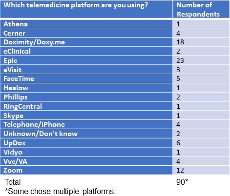
A 47-year-old woman presented with right sided chest pain and cough for 8 months. She had history of two episodes of blood streaked sputum four months ago. She also complained of loss of appetite and weight loss. She was a former smoker (1-2 cigarettes per day for 2-3 years quitting 5 years ago) and had a history of exposure to biomass fuel while working as a farmer. On examination pallor and clubbing was noted. Chest x-ray was suggestive of hilar enlargement (Figure 1).

Figure 1. Initial chest radiography.
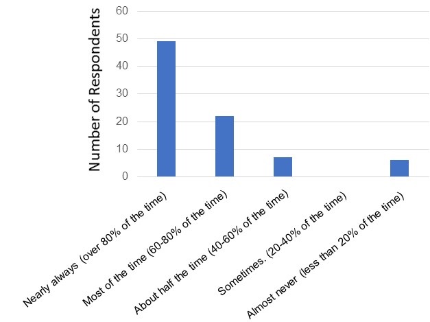
Contrast-enhanced CT of the chest showed homogeneously enhancing soft tissue density central lung mass that is narrowing and circumferentially encasing right main bronchus. The mass was abutting arch of aorta and the ascending aorta and circumferentially encasing and narrowing the superior vena cava and right main pulmonary artery. Subsegmental collapse of superior segment of right lower lobe was also seen with right paratracheal and pretracheal lymph node enlargement (Figure 2).

Figure 2. Representative axial image from thoracic CT scan in soft tissue windows showing the right lung mass.
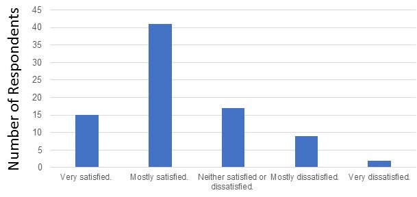
Flexible optic bronchoscopy showed an endoluminal irregular mass invading distal end of trachea along with carina and right main bronchus (Figure 3).

Figure 3. Photograph taken at bronchoscopy of the endobronchial mass in the distal trachea and right main bronchus.
Endobronchial biopsy showed papillary structures with fibro vascular cores lined with cell with moderate amount of cytoplasm with enlarged nuclei with granular chromatin and inconspicuous nuclei suggestive of RPR on histopathological examination. The patient was lost to follow up. Two months later she presented with increased breathlessness. Chest x-ray showed unilateral opaque right hemithorax with mediastinum slightly shifted to right (Figure 4).

Figure 4. Repeat chest radiography taken 2 months after initial presentation.
Hyponatremia was seen on routine blood investigation. CECT chest showed a well-defined heterogeneously enhancing soft tissue mass lesion with irregular margins involving the upper and middle lobe of right lung (Figure 5).

Figure 5. Coronal view of repeat thoracic CT in soft tissue windows.
The mass was encasing the right main bronchus and distal trachea, abutting large vessel, shifting trachea towards right side with moderate pleural effusion. Sputum analysis for acid-fast bacteria and malignant cells was negative. Ultrasound of abdomen showed no abnormality. Pleural fluid analysis showed paucicellular smear on cytology with ADA 20.5U/l, Protein 2.4 mg/dl and glucose 95.1 mg/dl. The patient refused bronchoscopy but consented to an ultrasound guided trans-thoracic biopsy. Histopathology showed pulmonary tissue with infiltrating tumor and the tumor was made up of sheets of small round cells with irregular contours suggestive of small cell carcinoma. Patient refused further management and left against medical advice. She passed away 11 days later.
Discussion
The incidence of RRP is bimodal, with the juvenile-onset form typically first occurring in children aged 2 to 4 years and adult-onset RRP typically occurring in adults aged 20 to 40 years. Juvenile-onset RRP is thought to be caused from peripartum exposure through an infected birth canal (3). Risk factors for adult-onset RRP include multiple lifetime sexual partners as well as a high frequency of oral sex. There was no statistically significant difference in illicit drug use between patients with adult-onset RRP vs a control group in a study by Ruiz et al. (4). RRP affects, from the most common site to the least common site, the true vocal cord, oral cavity, trachea, bronchi, and esophagus. Only 5% of the patients had the distal involvement of the trachea, and the involvement of the lung parenchyma is very rare, which is seen in, 1% of all cases (5). Therefore, patients present most commonly with hoarseness followed by stridor, cough, and dyspnea. Risk factor for malignant conversion includes smoking, prior irradiation, HPV-6. A recent study showed the presence of E6 and E7 oncogenes and their transcripts in HPV-positive lung cancer cases that are prerequisite for cancer development, thus reinforcing further the hypothesis that HPV could be a co-factor in bronchial carcinogenesis (6).Our patient had history of smoking as the only risk factor for malignant conversion.
Progressively increased expression of p53 and pRb proteins along with a reduced expression of p21WAF1 protein appears to be significant subsequent events in the progression to carcinoma (7). Talierco et al. (8) reported 100% of patients with adult onset RRP had concurrent HPV infection of the oral cavity; however, our patient had no evidence of oral cavity HPV infection on physical examination. Bronchoscopic pictures were suggestive of papillary lesion although association with HPV can’t be commented upon as patient refused for further testing. A literature review of RRP case reports revealed that patients usually have the diagnosis of RRP many years before evidence of malignant transformation (9-12). In contrast, our patient had evidence of malignant transformation about six months after diagnosis of respiratory papillomatosis.
DiMarco et al. (13) were the first to report the presence of the multiple RRP of the tracheobronchial tree with malignant degeneration, in 1978. One other case report showing coexistence of multiple squamous cell papilloma and carcinoma in the upper trachea with severe airway obstruction has been reported (14). A case study done in Taiwan suggests that HPV infection is an important risk factor for lung cancer among women (15).
Surgical excision of RRP is the current standard of care with objective of preserving adequate voice quality and airway patency (16). Lasers can also be employed for surgical excision of RRP. Either cutting/ablating lasers (CO2 and thallium lasers), or photoangiolytic lasers such as pulsatile (PDL) and potassium- titanil-phosphate lasers (KTP) can be used. Both KTP and PDL lasers are safe and effective for in office treatment of RRP (17). Microdebriders have distinct advantages over lasers and cold instruments because of their shorter operating time and absence of thermal injury (18). Adjuvant therapies for RRP include the usage of immunomodulators such as IFN, antivirals such as Cidofovir, Angiogenesis inhibitor (Bevacizumab) and PDL-1 inhibitor (19). The development of HPV- 1 vaccination is perhaps the most important modality in the management of RRP, by preventing infection with papilloma virus.
Conclusion
To the best of our knowledge, this is the first case report of coexisting respiratory papillomatosis with small cell carcinoma lung. Thus, coexistence of malignancy or malignant degeneration of respiratory papillomatosis is although unusual but can still occur without the associative factors. Patients with RRP should be radiographically monitored at regular intervals for pulmonary involvement and further evaluation actively pursued if any suspicion of malignancy arises.
References
- Magid MS, Chen YT, Soslow RA, Boulad F, Kernan NA, Szabolcs P. Juvenile-onset recurrent respiratory papillomatosis involving the lung: A case report and review of the literature. Pediatr Dev Pathol. 1998 Mar-Apr;1(2):157-63. [CrossRef] [PubMed]
- Kramer SS, Wehunt WD, Stocker JT, Kashima H. Pulmonary manifestations of juvenile laryngotracheal papillomatosis. AJR Am J Roentgenol. 1985 Apr;144(4):687-94. [CrossRef] [PubMed]
- Kashima HK, Shah F, Lyles A, Glackin R, Muhammad N, Turner L, Van Zandt S, Whitt S, Shah K. A comparison of risk factors in juvenile-onset and adult-onset recurrent respiratory papillomatosis. Laryngoscope. 1992 Jan;102(1):9-13. [CrossRef] [PubMed]
- Ruiz R, Achlatis S, Verma A, Born H, Kapadia F, Fang Y, Pitman M, Sulica L, Branski RC, Amin MR. Risk factors for adult-onset recurrent respiratory papillomatosis. Laryngoscope. 2014 Oct;124(10):2338-44. Epub 2014 Jun 10. [CrossRef] [PubMed].
- Cook JR, Hill DA, Humphrey PA, Pfeifer JD, El-Mofty SK. Squamous cell carcinoma arising in recurrent respiratory papillomatosis with pulmonary involvement: emerging common pattern of clinical features and human papillomavirus serotype association. Mod Pathol. 2000 Aug;13(8):914-8. [CrossRef] [PubMed]
- Giuliani L, Favalli C, Syrjanen K, Ciotti M. Human papillomavirus infections in lung cancer. Detection of E6 and E7 transcripts and review of the literature. Anticancer Res. 2007 Jul-Aug;27(4C):2697-704. [PubMed]
- Lele SM, Pou AM, Ventura K, Gatalica Z, Payne D. Molecular events in the progression of recurrent respiratory papillomatosis to carcinoma. Arch Pathol Lab Med. 2002 Oct;126(10):1184-8. [CrossRef] [PubMed]
- Taliercio S, Cespedes M, Born H, Ruiz R, Roof S, Amin MR, Branski RC. Adult-onset recurrent respiratory papillomatosis: a review of disease pathogenesis and implications for patient counseling. JAMA Otolaryngol Head Neck Surg. 2015 Jan;141(1):78-83. [CrossRef] [PubMed]
- Martina D, Kurniawan A, Pitoyo CW. Pulmonary papillomatosis: a rare case of recurrent respiratory papillomatosis presenting with multiple nodular and cavitary lesions. Acta Med Indones. 2014 Jul;46(3):238-43. [PubMed]
- Azadarmaki R, Lango MN. Malignant transformation of respiratory papillomatosis in a solid-organ transplant patient: case report and literature review. Ann Otol Rhinol Laryngol. 2013 Jul;122(7):457-60. [CrossRef] [PubMed]
- Hasegawa Y, Sato N, Niikawa H, Kamata S, Sannohe S, Kurotaki H, Sasaki T, Ebina A. Lung squamous cell carcinoma arising in a patient with adult-onset recurrent respiratory papillomatosis. Jpn J Clin Oncol. 2013 Jan;43(1):78-82. Epub 2012 Oct 30. [CrossRef] [PubMed]
- Lin HW, Richmon JD, Emerick KS, de Venecia RK, Zeitels SM, Faquin WC, Lin DT. Malignant transformation of a highly aggressive human papillomavirus type 11-associated recurrent respiratory papillomatosis. Am J Otolaryngol. 2010 Jul-Aug;31(4):291-6. Epub 2009 Jul 10. [CrossRef] [PubMed].
- DiMarco AF, Montenegro H, Payne CB Jr, Kwon KH. Papillomas of the tracheobronchial tree with malignant degeneration. Chest. 1978 Oct;74(4):464-5. [CrossRef] [PubMed].
- Paliouras D, Gogakos A, Rallis T, Chatzinikolaou F, Asteriou C, Tagarakis G, Organtzis J, Tsakiridis K, Tsavlis D, Zissimopoulos A, Kioumis I, Hohenforst-Schmidt W, Zarogoulidis K, Zarogoulidis P, Barbetakis N. Coexistence of squamous cell tracheal papilloma and carcinoma treated with chemotherapy and radiotherapy: a case report. Ther Clin Risk Manag. 2015 Dec 21;12:1-4. [CrossRef] [PubMed]
- Lin FC, Huang JY, Tsai SC, Nfor ON, Chou MC, Wu MF, Lee CT, Jan CF, Liaw YP. The association between human papillomavirus infection and female lung cancer: A population-based cohort study. Medicine (Baltimore). 2016 Jun;95(23):e3856. Erratum in: Medicine (Baltimore). 2016 Jul 18;95(28):e0916. [CrossRef] [PubMed]
- Kim HT, Baizhumanova AS. Is recurrent respiratory papillomatosis a manageable or curable disease? Laryngoscope. 2016 Jun;126(6):1359-64. [CrossRef] [PubMed] Epub 2015 Nov 26.
- Yan Y, Olszewski AE, Hoffman MR, Zhuang P, Ford CN, Dailey SH, Jiang JJ. Use of lasers in laryngeal surgery. J Voice. 2010 Jan;24(1):102-9. Epub 2009 May 31. [CrossRef] [PubMed]
- Holler T, Allegro J, Chadha NK, Hawkes M, Harrison RV, Forte V, Campisi P. Voice outcomes following repeated surgical resection of laryngeal papillomata in children. Otolaryngol Head Neck Surg. 2009 Oct;141(4):522-6. [CrossRef] [PubMed]
- Ivancic R, Iqbal H, deSilva B, Pan Q, Matrka L. Current and future management of recurrent respiratory papillomatosis. Laryngoscope Investig Otolaryngol. 2018 Jan 14;3(1):22-34. [CrossRef] [PubMed]
Cite as: Sharma P, Kumar A, Janapati B, Jain AK. Respiratory papillomatosis with small cell carcinoma: case report and brief review. Southwest J Pulm Crit Care. 2020;21:141-6. doi: https://doi.org/10.13175/swjpcc064-20 PDF
 Post a Comment