Search Journal-type in search term and press enter
Southwest Pulmonary and Critical Care Fellowships
In Memoriam
Social Media

Critical Care

Last 50 Critical Care Postings

(Most recent listed first. Click on title to be directed to the manuscript.)

May 2025 Critical Care Case of the Month: Where’s the Rub?
April 2025 Critical Care Case of the Month: Being Decisive During a
   Difficult Treatment Dilemma 
January 2025 Critical Care Case of the Month: A 35-Year-Old Admitted After
   a Fall
October 2024 Critical Care Case of the Month: Respiratory Failure in a
   Patient with Ulcerative Colitis
July 2024 Critical Care Case of the Month: Community-Acquired
   Meningitis
April 2024 Critical Care Case of the Month: A 53-year-old Man Presenting
   with Fatal Acute Intracranial Hemorrhage and Cryptogenic Disseminated
   Intravascular Coagulopathy 
Delineating Gastrointestinal Dysfunction Variants in Severe Burn Injury
   Cases: A Retrospective Case Series with Literature Review
Doggonit! A Classic Case of Severe Capnocytophaga canimorsus Sepsis
January 2024 Critical Care Case of the Month: I See Tacoma
October 2023 Critical Care Case of the Month: Multi-Drug Resistant
   K. pneumoniae
May 2023 Critical Care Case of the Month: Not a Humerus Case
Essentials of Airway Management: The Best Tools and Positioning for 
   First-Attempt Intubation Success (Review)
March 2023 Critical Care Case of the Month: A Bad Egg
The Effect of Low Dose Dexamethasone on the Reduction of Hypoxaemia
   and Fat Embolism Syndrome After Long Bone Fractures
Unintended Consequence of Jesse’s Law in Arizona Critical Care Medicine
Impact of Cytomegalovirus DNAemia Below the Lower Limit of
   Quantification: Impact of Multistate Model in Lung Transplant Recipients
October 2022 Critical Care Case of the Month: A Middle-Aged Couple “Not
   Acting Right”
Point-of-Care Ultrasound and Right Ventricular Strain: Utility in the
   Diagnosis of Pulmonary Embolism
Point of Care Ultrasound Utility in the Setting of Chest Pain: A Case of
   Takotsubo Cardiomyopathy
A Case of Brugada Phenocopy in Adrenal Insufficiency-Related Pericarditis
Effect Of Exogenous Melatonin on the Incidence of Delirium and Its 
   Association with Severity of Illness in Postoperative Surgical ICU Patients
Pediculosis As a Possible Contributor to Community-Acquired MRSA
   Bacteremia and Native Mitral Valve Endocarditis
April 2022 Critical Care Case of the Month: Bullous Skin Lesions in
   the ICU
Leadership in Action: A Student-Run Designated Emphasis in
   Healthcare Leadership
MSSA Pericarditis in a Patient with Systemic Lupus
   Erythematosus Flare
January 2022 Critical Care Case of the Month: Ataque Isquémico
   Transitorio in Spanish 
Rapidly Fatal COVID-19-associated Acute Necrotizing
Encephalopathy in a Previously Healthy 26-year-old Man 
Utility of Endobronchial Valves in a Patient with Bronchopleural Fistula in
   the Setting of COVID-19 Infection: A Case Report and Brief Review
October 2021 Critical Care Case of the Month: Unexpected Post-
   Operative Shock 
Impact of In Situ Education on Management of Cardiac Arrest after
   Cardiac Surgery
A Case and Brief Review of Bilious Ascites and Abdominal Compartment
   Syndrome from Pancreatitis-Induced Post-Roux-En-Y Gastric Remnant
   Leak
Methylene Blue Treatment of Pediatric Patients in the Cardiovascular
   Intensive Care Unit
July 2021 Critical Care Case of the Month: When a Chronic Disease
   Becomes Acute
Arizona Hospitals and Health Systems’ Statewide Collaboration Producing a
   Triage Protocol During the COVID-19 Pandemic
Ultrasound for Critical Care Physicians: Sometimes It’s Better to Be Lucky
   than Smart
High Volume Plasma Exchange in Acute Liver Failure: A Brief Review
April 2021 Critical Care Case of the Month: Abnormal Acid-Base Balance
   in a Post-Partum Woman
First-Attempt Endotracheal Intubation Success Rate Using A Telescoping
   Steel Bougie
January 2021 Critical Care Case of the Month: A 35-Year-Old Man Found
   Down on the Street
A Case of Athabaskan Brainstem Dysgenesis Syndrome and RSV
   Respiratory Failure
October 2020 Critical Care Case of the Month: Unexplained
   Encephalopathy Following Elective Plastic Surgery
Acute Type A Aortic Dissection in a Young Weightlifter: A Case Study with
  an In-Depth Literature Review
July 2020 Critical Care Case of the Month: Not the Pearl You Were
   Looking For...
Choosing Among Unproven Therapies for the Treatment of Life-Threatening
   COVID-19 Infection: A Clinician’s Opinion from the Bedside
April 2020 Critical Care Case of the Month: Another Emerging Cause
   for Infiltrative Lung Abnormalities
Further COVID-19 Infection Control and Management Recommendations for
   the ICU
COVID-19 Prevention and Control Recommendations for the ICU
Loperamide Abuse: A Case Report and Brief Review
Single-Use Telescopic Bougie: Case Series


For complete critical care listings click here.

The Southwest Journal of Pulmonary and Critical Care publishes articles directed to those who treat patients in the ICU, CCU and SICU including chest physicians, surgeons, pediatricians, pharmacists/pharmacologists, anesthesiologists, critical care nurses, and other healthcare professionals. Manuscripts may be either basic or clinical original investigations or review articles. Potential authors of review articles are encouraged to contact the editors before submission, however, unsolicited review articles will be considered.

------------------------------------------------------------------------------------

Thursday
Jan142016

Ultrasound for Critical Care Physicians: Hungry Heart

A 31-year-old incarcerated man with a past medical history of intravenous drug use and hepatitis C, presented with a one week history of dry, non-productive cough, orthopnea and exertional dyspnea. He denied current intravenous drug use, and endorsed that the last time he used was before he was incarcerated over 3 years ago, his last tattoo was in prison, 6 months prior. He was found to have an oxygen saturation of 77% on room air, fever of 40º C, heart rate of 114 bpm, and blood pressure of 80/50 mmHg. The patient had a leukocytosis of 14 x109/L, and a chest x-ray demonstrating patchy airspace disease. Blood cultures were sent and he was treated with antibiotics and vasopressors for septic shock. The patient was intubated for acute hypoxemic respiratory failure secondary to multifocal pneumonia. A bedside transthoracic echocardiogram was performed. 
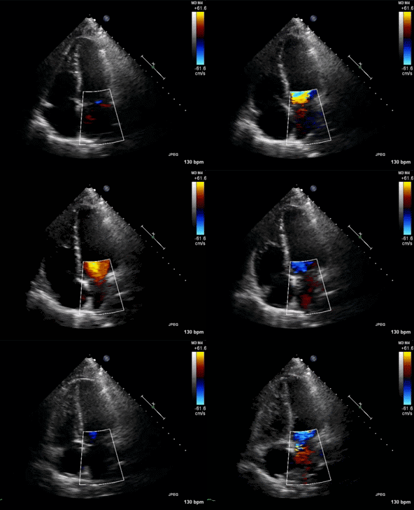
Figure 1. Apical four chamber view echocardiogram with color Doppler over the mitral valve.
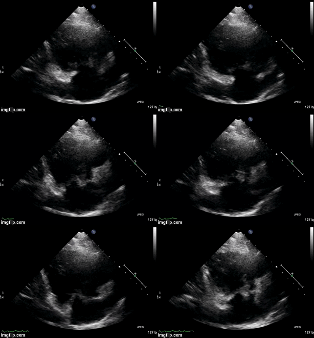
 

Figure 2. Right Ventricular (RV) inflow view echocardiogram from same patient


What is the likely diagnosis supported by the echocardiogram? (Click on the correct answer for an explanation)

Cite as: Villalobos N, Stoltze K, Azeem M. Ultrasound for critical care physicians: hungry heart. Southwest J Pulm Crit Care. 2016;12(1):24-7. doi: http://dx.doi.org/10.13175/swjpcc007-16 PDF

Saturday
Jan022016

January 2016 Critical Care Case of the Month

Sandra L. Till, DO

Banner University Medical Center Phoenix

Phoenix, AZ USA  

History of Present Illness

The patient is an 18-year-old woman who was driving to high school on a frontage road when she fell asleep at the wheel and her car rolled over. She was wearing her seatbelt but there was no airbag deployment. She did not lose consciousness and she was responsive and answering questions at the scene. She self-extricated from the vehicle. She had left arm pain with a boney deformity and she walked to the ambulance that transferred her to the hospital emergency department (ED).

Upon arrival in the ED she appeared pale and had difficulty breathing. In addition to her arm pain with an obvious left humeral fracture she also complained of upper abdominal and anterior chest pain. O2 saturation was initially 90% but declined to 70%.

Which of the following should be ordered immediately? (Click on the correct answer to proceed to the second of six panels)

  1. Begin intravenous lines with large bore needles
  2. X-ray of humerus
  3. Hemoglobin and hematocrit
  4. 1 and 3
  5. All of the above

Cite as: Till SL. January 2016 critical care case of the month. Southwest J Pulm Crit Care. 2016;12:6-12. doi: http://dx.doi.org/10.13175/swjpcc151-15 PDF 

Friday
Dec042015

Ultrasound for Critical Care Physicians: The Pleura and the Answers that Lie Within

Heidi L. Erickson MD 

Division of Pulmonary, Critical Care and Occupational Medicine

University of Iowa Hospitals and Clinics

Iowa City, IA

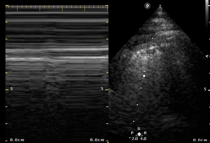
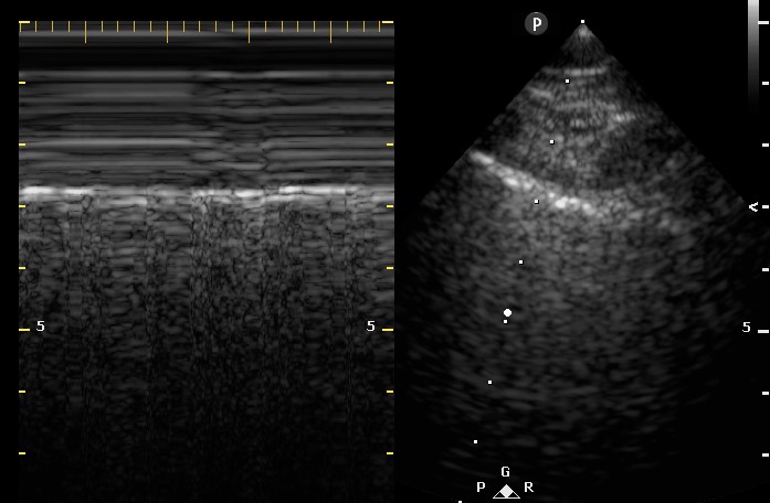
A 67-year-old woman with a 40-pack-year smoking history was admitted to the intensive care unit with acute respiratory failure secondary to adult respiratory distress syndrome (ARDS) in the setting of pneumococcal bacteremia. On admission, she required endotracheal intubation and vasopressor support.  She was ventilated using a low tidal volume strategy and was relatively easy to oxygenate with a PEEP of 5 and 40% FiO2. After 48 hours of clinical improvement, the patient developed sudden onset tachypnea and increased peak and plateau airway pressures. A bedside ultrasound was subsequently performed (Figures 1 and 2).


Figure 1. Two- dimensional ultrasound image of the right lung with associated M-mode image.


Figure 2. Two- dimensional ultrasound image of the left lung with associated M-mode image.

What is the cause of this patient’s acute respiratory decompensation and increased airway pressures? (Click on the correct answer for an explanation)

  1. Pericardial effusion
  2. Pneumothorax
  3. Pulmonary edema
  4. Pulmonary embolism

Cite as: Erickson HL. Ultrasound for critical care physicians: the pleura and the answers that lie within. Southwest J Pulm Crit Care. 2015;11(6):260-3. doi: http://dx.doi.org/10.13175/swjpcc149-15 PDF

Wednesday
Dec022015

December 2015 Critical Care Case of the Month

Samir Sultan, DO 

Banner University Medical Center Phoenix

Phoenix, AZ 

History of Present Illness

The patient is a 32-year-old woman who presented with flank pain for 3 days to an outside hospital. She was diagnosed with pyelonephritis and begun on ceftriaxone. She was discharged against medical advice on cephalexin.

She returned to the same hospital 3 days later by ambulance with labored breathing and weakness and was emergently intubated. She was transferred for ventilator management and respiratory failure.

Past Medical History

She has a long history of poorly controlled diabetes mellitus.

Physical Examination

She is orally intubated and sedated.

Vitals: Temperature - 100.9º F, Blood Pressure - 117/75 mm Hg, Heart Rate - 148 beats per minute,  Respiratory Rate - 31 breaths/min, SpO2 - 88 % on assist control of 30, tidal volume of 350 mL, PEEP 15, and an FiO2 100%.

There is scatted rhonchi and rales but the remainder of the physical examination is unremarkable.

Radiography

Her admission portable chest X-ray is shown in Figure 1.

Figure 1. Admission portable AP of the chest.

Which of the following should be ordered as part of her initial work-up? (Click on the correct answer to proceed to the second of five panels).

  1. Administer broad spectrum antibiotics
  2. Blood and urine cultures
  3. Rapid influenza test
  4. 1 and 3
  5. All of the above

Cite as: Sultan S. December critical care case of the month. Southwest J Pulm Crit Care. 2015;11(6):246-51. doi: http://dx.doi.org/10.13175/swjpcc147-15 PDF

Saturday
Nov212015

Ultrasound for Critical Care Physicians: 50 Ways to Line Your Liver

Seth Skiles ACNP

Theresa Heynekamp MD MPH


Division of Pulmonary, Critical care and Sleep Medicine,

University of New Mexico School of Medicine

Albuquerque, NM USA


A 54-year-old man with a past medical history significant for traumatic brain injury and aspiration pneumonia presented with hypoxic respiratory failure secondary to foreign body aspiration.

On presentation, the patient was found to be hypoxic and tachypneic, requiring endotracheal intubation and mechanical ventilation. Bronchoscopy was performed with removal of extensive food particles throughout both lungs. The patient subsequently developed sepsis secondary to aspiration pneumonia.  He became hypotensive, requiring central venous catheter placement for vasopressor therapy. A right subclavian central line was attempted under ultrasound guidance. A beside ultrasound was subsequently performed (Video 1).

Video 1. Sagittal view of the liver and heart at the level of the mid-abdomen and slightly right of mid-line.


What does the video obtained of a longitudinal view of the IVC at the level of the liver demonstrate? (Click on the correct answer for a discussion)

Cite as: Skiles S, Heynekamp T. Ultrasound for critical care physicians: 50 ways to line your liver. Southwest J Pulm Crit Care. 2015;11(5):235-7. doi: http://dx.doi.org/10.13175/swjpcc144-15 PDF