Search Journal-type in search term and press enter
Southwest Pulmonary and Critical Care Fellowships
In Memoriam
Social Media

Critical Care

Last 50 Critical Care Postings

(Most recent listed first. Click on title to be directed to the manuscript.)

May 2025 Critical Care Case of the Month: Where’s the Rub?
April 2025 Critical Care Case of the Month: Being Decisive During a
   Difficult Treatment Dilemma 
January 2025 Critical Care Case of the Month: A 35-Year-Old Admitted After
   a Fall
October 2024 Critical Care Case of the Month: Respiratory Failure in a
   Patient with Ulcerative Colitis
July 2024 Critical Care Case of the Month: Community-Acquired
   Meningitis
April 2024 Critical Care Case of the Month: A 53-year-old Man Presenting
   with Fatal Acute Intracranial Hemorrhage and Cryptogenic Disseminated
   Intravascular Coagulopathy 
Delineating Gastrointestinal Dysfunction Variants in Severe Burn Injury
   Cases: A Retrospective Case Series with Literature Review
Doggonit! A Classic Case of Severe Capnocytophaga canimorsus Sepsis
January 2024 Critical Care Case of the Month: I See Tacoma
October 2023 Critical Care Case of the Month: Multi-Drug Resistant
   K. pneumoniae
May 2023 Critical Care Case of the Month: Not a Humerus Case
Essentials of Airway Management: The Best Tools and Positioning for 
   First-Attempt Intubation Success (Review)
March 2023 Critical Care Case of the Month: A Bad Egg
The Effect of Low Dose Dexamethasone on the Reduction of Hypoxaemia
   and Fat Embolism Syndrome After Long Bone Fractures
Unintended Consequence of Jesse’s Law in Arizona Critical Care Medicine
Impact of Cytomegalovirus DNAemia Below the Lower Limit of
   Quantification: Impact of Multistate Model in Lung Transplant Recipients
October 2022 Critical Care Case of the Month: A Middle-Aged Couple “Not
   Acting Right”
Point-of-Care Ultrasound and Right Ventricular Strain: Utility in the
   Diagnosis of Pulmonary Embolism
Point of Care Ultrasound Utility in the Setting of Chest Pain: A Case of
   Takotsubo Cardiomyopathy
A Case of Brugada Phenocopy in Adrenal Insufficiency-Related Pericarditis
Effect Of Exogenous Melatonin on the Incidence of Delirium and Its 
   Association with Severity of Illness in Postoperative Surgical ICU Patients
Pediculosis As a Possible Contributor to Community-Acquired MRSA
   Bacteremia and Native Mitral Valve Endocarditis
April 2022 Critical Care Case of the Month: Bullous Skin Lesions in
   the ICU
Leadership in Action: A Student-Run Designated Emphasis in
   Healthcare Leadership
MSSA Pericarditis in a Patient with Systemic Lupus
   Erythematosus Flare
January 2022 Critical Care Case of the Month: Ataque Isquémico
   Transitorio in Spanish 
Rapidly Fatal COVID-19-associated Acute Necrotizing
Encephalopathy in a Previously Healthy 26-year-old Man 
Utility of Endobronchial Valves in a Patient with Bronchopleural Fistula in
   the Setting of COVID-19 Infection: A Case Report and Brief Review
October 2021 Critical Care Case of the Month: Unexpected Post-
   Operative Shock 
Impact of In Situ Education on Management of Cardiac Arrest after
   Cardiac Surgery
A Case and Brief Review of Bilious Ascites and Abdominal Compartment
   Syndrome from Pancreatitis-Induced Post-Roux-En-Y Gastric Remnant
   Leak
Methylene Blue Treatment of Pediatric Patients in the Cardiovascular
   Intensive Care Unit
July 2021 Critical Care Case of the Month: When a Chronic Disease
   Becomes Acute
Arizona Hospitals and Health Systems’ Statewide Collaboration Producing a
   Triage Protocol During the COVID-19 Pandemic
Ultrasound for Critical Care Physicians: Sometimes It’s Better to Be Lucky
   than Smart
High Volume Plasma Exchange in Acute Liver Failure: A Brief Review
April 2021 Critical Care Case of the Month: Abnormal Acid-Base Balance
   in a Post-Partum Woman
First-Attempt Endotracheal Intubation Success Rate Using A Telescoping
   Steel Bougie
January 2021 Critical Care Case of the Month: A 35-Year-Old Man Found
   Down on the Street
A Case of Athabaskan Brainstem Dysgenesis Syndrome and RSV
   Respiratory Failure
October 2020 Critical Care Case of the Month: Unexplained
   Encephalopathy Following Elective Plastic Surgery
Acute Type A Aortic Dissection in a Young Weightlifter: A Case Study with
  an In-Depth Literature Review
July 2020 Critical Care Case of the Month: Not the Pearl You Were
   Looking For...
Choosing Among Unproven Therapies for the Treatment of Life-Threatening
   COVID-19 Infection: A Clinician’s Opinion from the Bedside
April 2020 Critical Care Case of the Month: Another Emerging Cause
   for Infiltrative Lung Abnormalities
Further COVID-19 Infection Control and Management Recommendations for
   the ICU
COVID-19 Prevention and Control Recommendations for the ICU
Loperamide Abuse: A Case Report and Brief Review
Single-Use Telescopic Bougie: Case Series


For complete critical care listings click here.

The Southwest Journal of Pulmonary and Critical Care publishes articles directed to those who treat patients in the ICU, CCU and SICU including chest physicians, surgeons, pediatricians, pharmacists/pharmacologists, anesthesiologists, critical care nurses, and other healthcare professionals. Manuscripts may be either basic or clinical original investigations or review articles. Potential authors of review articles are encouraged to contact the editors before submission, however, unsolicited review articles will be considered.

------------------------------------------------------------------------------------

Friday
Mar022018

March 2018 Critical Care Case of the Month

Babitha Bijin MD

Jonathan Callaway MD

Janet Campion MD


University of Arizona

Department of Medicine

Tucson, AZ USA


Chief Complaints

  • Shortness of breath
  • Worsening bilateral LE edema

History of Present Illness

A 53-year-old man with history of multiple myeloma and congestive heart failure presented to the emergency department with complaints of worsening shortness of breath and bilateral lower extremity edema for last 24 hours. In the last week, he has had dyspnea at rest as well as a productive cough with yellow sputum. He describes generalized malaise, loss of appetite, possible fever and notes new bilateral pitting edema below his knees. Per patient, he had flu-like symptoms one week ago and was treated empirically with oseltamivir.

Past Medical History

  • Multiple myeloma-IgG kappa with calvarial and humeral metastases, ongoing treatment with cyclophosphamide, bortezomib and dexamethasone
  • Community acquired pneumonia 2016, treated with oral antibiotics
  • Heart failure with echo 10/2017 showing moderate concentric left ventricular hypertrophy, left ventricular ejection fraction 63%, borderline left atrial and right atrial dilatation, diastolic dysfunction, right ventricular systolic pressure estimated 25 mm Hg
  • Hyperlipidemia
  • Chronic kidney disease, stage III

Home Medications: Aspirin 81mg daily, atorvastatin 80mg daily, furosemide 10mg daily, calcium / Vitamin D supplement daily, oxycodone 5mg PRN, chemotherapy as above

Allergies: No known drug allergies

Social History:

  • Construction worker, not currently working due to recent myeloma diagnosis
  • Smoked one pack per day since age 16, recently quit with 30 pack-year history
  • Drinks beer socially on weekends
  • Married with 3 children

Family History: Mother with hypertension, uncle with multiple myeloma, daughter with rheumatoid arthritis

Review of Systems: Negative except per HPI

Physical Exam

  • Vitals: T 39.3º C, BP 80/52, P121, R16, SpO2 93% on 2L
  • General: Alert man, mildly dyspneic with speech
  • Mouth: Nonicteric, moist oral mucosa, no oral erythema or exudates
  • Neck: No cervical neck LAD but JVP to angle of jaw at 45 degrees
  • Lungs: Bibasilar crackles with right basilar rhonchi, no wheezing
  • Heart: Regular S1 and S2, tachycardic, no appreciable murmur or right ventricular heave
  • Abdomen: Soft, normal active bowel sounds, no tendernesses, no hepatosplenomegaly
  • Ext: Pitting edema to knees bilaterally, no cyanosis or clubbing, normal muscle bulk
  • Neurologic: No focal abnormalities on neurologic exam

Laboratory Evaluation

  • Complete blood count: WBC 15.9 (92% neutrophils), Hgb/Hct 8.8/27.1, Platelets 227
  • Electrolytes: Na+ 129, K+ 4.0, Cl- 100, CO2 18, blood urea nitrogen 42, creatinine 1.99 (baseline Cr 1.55)
  • Liver: AST 35, ALT 46, total bilirubin1.7, alkaline phosphatase 237, total protein 7.4, albumin 2.
  • Others: troponin 0.64, brain naturetic peptide 4569, venous lactate 2.6
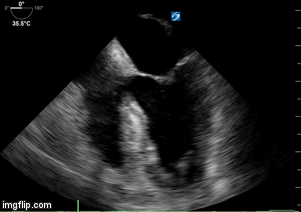
Chest X-ray

Figure 1. Admission chest x-ray.

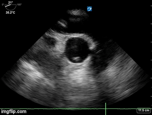
Thoracic CT (2 views)

Figure 2. Representative images from the thoracic CT scan in lung windows.

What is most likely etiology of CXR and thoracic CT findings? (Click on the correct answer to proceed to the second of seven pages)

  1. Coccidioidomycosis pneumonia
  2. Pulmonary edema
  3. Pulmonary embolism with infarcts
  4. Staphylococcus aureus pneumonia
  5. Streptococcus pneumoniae infection 

Cite as: Bijin B, Callaway J, Campion J. March 2018 critical care case of the month. Southwest J Pulm Crit Care. 2018;16(3):117-25. doi: https://doi.org/10.13175/swjpcc035-18 PDF 

Sunday
Feb042018

Ultrasound for Critical Care Physicians: Ghost in the Machine

Ross Davidson, DO

Michel Boivin, MD 

Division of Pulmonary, Critical Care and Sleep Medicine

University of New Mexico School of Medicine

Albuquerque, NM USA


A 53-year-old woman presented to the emergency department after a sudden cardiac arrest at home. The patient had a history of asthma and tracheal stenosis and had progressive shortness of breath over the previous days. The patient’s family noticed a “thump” sound from the patient’s room, and found her apneic. They called 911 and began cardiopulmonary resuscitation. Paramedics arrived on the scene, found an initial rhythm of pulseless electrical activity. The patient eventually achieved return of spontaneous circulation and was transported to the hospital. On arrival the patient was in normal sinus rhythm, with a heart rate of 110 beats per minute. Blood pressure was 80/45 mmHg, on an epinephrine infusion. The patient was afebrile, endotracheally intubated, unresponsive and ventilated at 30 breaths per minute. An initial chest radiograph was compatible with aspiration pneumonitis and a small pneumothorax. Initial electrocardiogram on arrival had 1mm ST-segment depressions in leads V4 to V6. Transthoracic echocardiography was unsuccessful due to patient’s habitus and mechanical ventilation. Because of the patient’s hemodynamic instability and unknown cause of cardiac arrest, an urgent trans-esophageal echocardiogram (TEE) was performed (Videos 1-3).


Video 1. Mid-esophageal 4-chamber view of the heart.


Video 2. Upper esophageal long-axis view of the pulmonary artery and short axis view of the ascending aorta.


Video 3. Upper esophageal short axis view of the pulmonary artery with the ascending aorta in long axis. 

Based on the images presented what do you suspect is the etiology of the patient’s cardiac arrest? (Click on the correct answer for an explanation-no penalty for guessing, you can go back and try again)

  1. Massive Pulmonary Embolism
  2. Myocardial infarction
  3. Pericardial Tamponade
  4. Unable to determine

Cite as: Davidson R, Boivin M. Ultrasound for critical care physicians: ghost in the machine. Southwest J Pulm Crit Care. 2018;16(2):76-80. doi: https://doi.org/10.13175/swjpcc027-18 PDF 

Friday
Feb022018

February 2018 Critical Care Case of the Month

Robert A. Raschke, MD

University of Arizona College of Medicine-Phoenix

Phoenix, AZ


History of Present Illness

A 25-year-old was admitted to an outside hospital with an acute episode of nausea and vomiting and chronic progressive weakness.  He smoked 2 cigarettes per day and drank a 12-pack of beer per month.  He had a history of undefined chronic liver disease.

Physician Examination

Physical examination was reported as showing a chronically ill appearing man who was “weak” using crutches to ambulate.

The patient was made NPO and was rehydrated with intravenous normal saline.

Which of the following are indicated at this time? (Click on the correct answer to proceed to the second of four pages)

  1. Creatinine phosphokinase (CPK)
  2. Serum potassium
  3. Thyroid studies
  4. 1 and 3
  5. All of the above

Cite as: Raschke RA. February 2018 critical care case of the month. Southwest J Pulm Crit Care. 2018;16(2):62-6. doi: https://doi.org/10.13175/swjpcc009-18 PDF

Monday
Jan012018

January 2018 Critical Care Case of the Month

Theodore Loftsgard, APRN, ACNP 

Department of Anesthesiology and Critical Care

Mayo Clinic Minnesota

Rochester, MN USA


History of Present Illness

The patient is a 51-year-old woman admitted with a long history of progressive shortness of breath.  She has a long history of “heart problems”. She uses supplemental oxygen at 1 LPM by nasal cannula.

Past Medical History, Social History and Family History

She also has several comorbidities including renal failure with two renal transplants and a history of relatively recent RSV and CMV pneumonia. She is a life-long nonsmoker. Her family history is noncontributory.

Physical Examination

  • Vital signs: Blood pressure 145/80 mm Hg, heart rate 59 beats/min, respiratory rate 18, T 37.0º C, SpO2 91% of 1 LPM.
  • Lungs: Clear.
  • Heart: Regular rhythm with G 3/6 systolic ejection murmur at the base.
  • Abdomen: unremarkable.
  • Extremities: no edema

Which of the following should be performed? (Click on the correct answer to proceed to the second of seven pages)

  1. Brain naturetic peptide (BNP)
  2. Chest x-ray
  3. Echocardiogram
  4. Electrocardiogram
  5. All of the above

Cite as: Loftsgard T. January 2018 critical care case of the month. Southwest J Pulm Crit Care. 2018;16(1):1-7. doi: https://doi.org/10.13175/swjpcc155-17 PDF

Saturday
Dec022017

December 2017 Critical Care Case of the Month

Michael B. Gotway, MD


Department of Radiology

Mayo Clinic Arizona

Scottsdale AZ USA


Clinical History: A 57-year-old man with no known previous medical history was brought to the emergency room via ambulance and admitted to the intensive care unit with a compliant of severe chest pain in the substernal region and epigastrium. The patient was awake and alert and did not complain of shortness of breath.

Physical examination was largely unremarkable and the patient’s oxygen saturation was 98% on room air. The patient’s vital signs revealed tachycardia (105 bpm) and his blood pressure was 108 mmHg / 60 mmHg.

Laboratory evaluation showed a slightly elevated white blood cell count (13 x 109 cells/L), but his hemoglobin and hematocrit values were with within normal limits, as was his platelet count. 

Which of the following diagnoses are appropriate considerations for this patient’s condition? (Click on the correct answer to proceed to the second of nine pages)

  1. Acute pericarditis
  2. Aortic dissection
  3. Community-acquired pneumonia
  4. Myocardial infarction
  5. All of the above

Cite as: Gotway MB. December 2017 critical care case of the month. Southwest J Pulm Crit Care. 2017;15(6):241-52. doi: https://doi.org/10.13175/swjpcc145-17 PDF